The Lerner Research Institute Notations - Cleveland Clinic Lerner ...
The Lerner Research Institute Notations - Cleveland Clinic Lerner ...
The Lerner Research Institute Notations - Cleveland Clinic Lerner ...
- No tags were found...

Create successful ePaper yourself
Turn your PDF publications into a flip-book with our unique Google optimized e-Paper software.
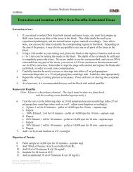
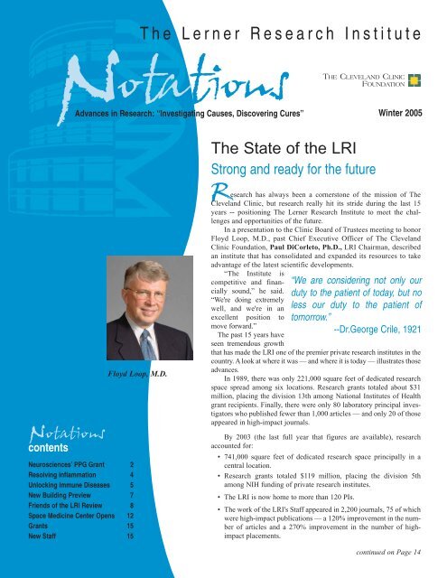
NEUROSCIENCES$6.2 million multiple sclerosis grantPPG renewal allows continued study of inflammation, tissue injury links<strong>The</strong> National <strong>Institute</strong>s of Health haveawarded a $6.2 million Program ProjectGrant renewal to a team of investigators at<strong>The</strong> <strong>Lerner</strong> <strong>Research</strong> <strong>Institute</strong> and <strong>The</strong>Mellen Center for MS Treatment and<strong>Research</strong> to continue investigating the interplaybetween inflammation and tissue injuryin multiple sclerosis (MS).Among the research objectives are toidentify molecular mechanisms responsiblefor inflammation and neurodegeneration inMS brains, to use magnetic resonance imagingto monitor and evaluate the physicalchanges to patients' brains during the courseof the disease, and to use cDNA macroarraysto identify biomarkers of the therapeuticresponse to interferon-beta (the most widelyused treatment for MS).Richard Ransohoff, M.D., Neurosciences,serves as Program Director forresearch that involves more than 30 physicians,bench scientists, support staff, statisticiansand research technologists throughoutthe <strong>Clinic</strong> and the LRI. In addition to Dr.Ransohoff, project leaders are Bruce Trapp,Ph.D., Chair, Neurosciences; RichardRudick, M.D., Chair, Division of <strong>Clinic</strong>al<strong>Research</strong>; and Elizabeth Fisher, Ph.D.,Biomedical Engineering.<strong>The</strong> pathology of multiple sclerosis isassociated with inflammation, scarring of themyelin (the tissue that covers and protectsnerve fibers) in the brain and spinal cord, anddegeneration of demyelinated nerve fibers.MS patients experience varying degrees ofneurological impairment depending in parton the location and extent of the scarring.MS patients in the early stage of the diseasetypically experience clearly defined flareupsand episodes of acute worsening of neurologicfunction, followed by partial or completerecovery periods. This stage of the diseaseis called relapsing-remitting MS. MostPart of LRI’s research into MS is to compare brain scans of people without MS (left)with scans of MS patients to measure brain atrophy (right).patients with relapsing-remitting diseaseeventually develop secondary-progressiveMS, in which patients steadily worsen withor without occasional flare-ups and minorrecoveries. Although MS is often describedas an autoimmune disease (one in which thebody's own immune system attacks normalbody tissue -- in this case, the cells that makemyelin), the “autoimmune hypothesis” is notcentral to the PPG-supported research program,termed “Tissue injury and inflammationin MS,” which is designed to obtain usefulinformation about how MS progresses,regardless of its underlying cause.<strong>The</strong> grant renewal allows the LRIresearch teams to continue a coordinatedresearch program initially funded in 1999.<strong>The</strong> initial program looked at three principalareas. Dr. Ransohoff investigated what signalingmight be involved to cause leukocytes(white blood cells) to invade the centralnervous system. Dr. Trapp focused on howlesions caused by the disease affect the axonalnetwork -- the long single-cell paths thatcarry signals from one nerve cell to anotheror to organs and muscles. Drs. Fisher andRudick directed their attention to relationshipsbetween conventional MRI imagesthought to show early signs of inflammationand subsequent atrophy of the brain.<strong>The</strong> LRI PPG is considered unique in itsexclusive focus on direct study of MS itself,rather than surrogate models of the disease.Part of the initial project was development ofa unique brain and spinal cord donation programin which MS patients and their familiesgave prior permission to use the materialafter the death of the patient. During the firstfive years, the studies provided tissues from36 patients, and 127 patients (including the36) have registered for the donation program.<strong>The</strong> efforts require collaborationamong the <strong>Clinic</strong>'s clinical, imaging, pathologyand administrative staffs.2 <strong>Notations</strong> · Winter 2005 <strong>The</strong> <strong>Lerner</strong> <strong>Research</strong> <strong>Institute</strong>
“I'm proud we study the human disease,”said Dr. Ransohoff. “It's the hardest thing todo (compared with studying models), but inessence it's the only thing that matters.”<strong>The</strong> grant renewal supportsfour primary projects:Chemokines and chemokine receptorsDr. Ransohoff's team has the overall objectiveto study chemokines, peptides that governthe migration and activation profiles ofleukocytes -- the major component of theinflammation that subsequently damages thetissues. <strong>The</strong> goal is to identify more rationaland effective treatment of inflammation,especially during the early stages of the diseasewhen inflammation seems to be moremanageable.Axonal pathologyDr. Trapp's previous project had two objectives:to determine the extent and nature ofthe disease and degeneration of the axonalpathways, and to study oligodendrocyte progenitorcells -- the cells of the central nervoussystem that are essential to the myelinsheath -- in the brains and lesions of MSpatients. <strong>The</strong> renewal allows him to investigatein greater detail the molecular mechanismsthat underlie neurodegeneration in thebrains and spinal cords of MS patients. Hiscontinued research is based on the premisethat reduced axonal energy metabolism inchronically demyelinated axons affects ionicfluxes across the axolemma, culminatingwith increased intraxonal Ca ++ and axonaldegeneration.“We are now at the point where we areconsidering adding neuroprotective therapiesto our arsenal of treatments for MS,” Dr.Trapp said. “If we can delay neurodegeneration,we can keep the MS patient out of thewheel chair.”Brain atrophy during the course of MS..Dr. Fisher will use advanced neuroimagingtechniques to study brain atrophy in patientswith relapsing-remitting and secondary progressiveMS. During the first phase of theresearch, a cohort of patients was identifiedand followed longitudinally over the courseABIOMEDICAL ENGINEERING14-year study of multiple sclerosispatients is providing a unique glimpseinto the significance of brain atrophy.Elizabeth Fisher, Ph.D., BiomedicalEngineering, said that for many years thestudy of brain tissue loss due to MS diseaseprocesses was not a high priority. Itwas only recently that the research communityhas started to examine brain atrophyin MS patients.“People knew brain atrophy happens,but many thought of the tissue loss assomething that only happens very late inthe disease in a subset of patients, perhapsas a consequence of repeated focal tissuedamage,” she said. “<strong>The</strong>re were also technologicallimitations at the time to preciselymeasuring the very slow process ofbrain atrophy.”Through <strong>The</strong> <strong>Lerner</strong> <strong>Research</strong><strong>Institute</strong>'s long-term dedication to MSresearch and landmark software developedby Dr. Fisher that provides accurate measurementsof minute changes in brain tissueeven early on in the disease, researchers havetracked brain atrophy -- an effort that willcontinue for another five years thanks to therenewal of a $6.2 million NIH grant.“Five years is a long time for an MSstudy,” Dr. Fisher said. “So a study thatwill eventually be 19 years is truly unique.Many people live with this disease for along time. It's a long-lasting and chronicillness, and this provides an opportunityto study how the disease changes andof four years. Analysis of MRIs from thesepatients has revealed that the pathologicprocesses that lead to overall brain tissue losscontinued on Page 13Brain atrophyGrant offers unique 19-year look at tissue lossaffects brain tissue over a long period oftime.”<strong>The</strong> precision of the imaging is essentialto the importance of the study.“On average, MS patients lose 0.5%to 1% of their brain tissue each year, soyou have to be able to detect very smallamounts of loss with a great deal of accuracyif you're going to monitor the benefitsof medications or other therapies,” Dr.Fisher said.About half of the 60 patients in thecurrent study were originally part of atwo-year trial started in 1990 to examinethe effects of a medication that was eventuallyapproved for use in treating MSpatients. With developments in technologyand Dr. Fisher's software, LRIresearchers were able to go back and reexaminethe images taken in 1990 -- andlocate and re-test the patients who werepart of the original clinical study.When the latest research is completedin five years, the LRI will have 19 yearsworth of images and quantitative informationon brain atrophy, in addition to clinicalfollow-up data on disability.“It's definitely the longest time to follow(brain atrophy) with a standardizedimaging protocol,” Dr. Fisher said.“Understanding atrophy is important.We're trying to determine what causes itand why it progresses faster in somepatients than in others. It also can be usedto help validate new treatments.”<strong>The</strong> <strong>Lerner</strong> <strong>Research</strong> <strong>Institute</strong> <strong>Notations</strong> · Winter 2005 3
CELL BIOLOGYMacrophage inflammationNovel mechanism holds promise of resolving many debilitating diseasesScientists at <strong>The</strong> <strong>Lerner</strong> <strong>Research</strong><strong>Institute</strong> have found a novel mechanismthat may contribute to resolvingmacrophage inflammation, the hallmarkof many debilitating diseases rangingfrom asthma to multiple sclerosis.<strong>Research</strong>ers in the laboratory ofPaul Fox, Ph.D., Cell Biology, reportedtheir findings in a Fall 2004 issue of thejournal Cell (www.cell.com/).<strong>The</strong> macrophage is a key cellular participantin inflammation because it helps todefend against pathogenic bacterial and viralmicroorganisms by the production of toxicagents. Macrophage activation can be triggeredby invasive microorganisms, as wellas by specific signaling cytokines released inthe local environment. Excessive or prolongedinflammation can lead to injury to thehost tissue, and immunologists and cell biologistsare now trying to unravel the cellularmechanisms that normally turn off theinflammatory response.Prabha Sampath, Ph.D., a formergraduate student from <strong>Cleveland</strong> StateUniversity and now a postdoctoral fellowat the University of Washington, conductedthe studies in Dr. Fox's laboratory. <strong>The</strong>laboratory had previously shown thatinterferon-gamma activates macrophagesto express the messenger RNA and proteinfor ceruloplasmin, a protein with oxidantand bactericidal activities.In the Cell article, the Fox laboratoryshows that glutamyl-prolyl-tRNA synthetase(GluProRS), a protein involved in normalprotein synthesis, becomes phosphorylatedand leaves its usual residence in the multisynthetasecomplex, a large complex containingabout half of the 20 tRNA synthetases.It then binds the ceruloplasminmessenger RNA in a complex containingthree additional proteins, and specificallysilences its translation into ceruloplasminprotein.<strong>The</strong> newly discovered mRNA silencingfunction of GluProRS may provide animportant endogenous mechanism for cellularresolution of inflammation. In addition,defects in this “off-switch” may be acause of tissue damage during chronicinflammatory diseases.At the more fundamental level, thework supports a new paradigm in whichordinary housekeeping proteins are mobilizedfrom parent complexes and assembledinto multi-subunit structures with extraordinaryfunctions. In a “Preview” accompanyingthe article in the same issue, PaulSchimmel, Ph.D., and Karla Ewalt, Ph.D., of<strong>The</strong> Skaggs <strong>Institute</strong> for Chemical Biologyat <strong>The</strong> Scripps <strong>Research</strong> <strong>Institute</strong> commentthat the work is the first example of a tRNAsynthetase acting on inflammation throughan intracellular pathway.<strong>The</strong> experiments were done by Dr.Sampath in collaboration with othermembers of the Department of CellBiology: Carrie Gerber, Ph.D., LaurentChavatte, Ph.D., Donna Driscoll, Ph.D.,Mike Kinter, Ph.D., BarsanjitMazumder, Ph.D. (now a faculty memberat <strong>Cleveland</strong> State University), andVasudevan Seshadri, Ph.D. (now at theNational Center for Cell Science, Pune,India).Dr. Fox's research program on translationalcontrol of inflammatory gene expressionis supported by the National Heart,Lung and Blood <strong>Institute</strong> of the National<strong>Institute</strong>s of Health as part of the VascularCell Function and Atherosclerosis ProgramProject headed by Paul DiCorleto, Ph.D.,LRI Chairman.A study by Tatiana V. Byzova, Ph.D., MolecularCardiology, about the mechanisms of blood vessel formationwas selected by the American Society of Hematologyfor presentation at the Society's annual meeting, heldDecember 4-7, 2004.<strong>The</strong> study identifies new factors that control the developmentand function of vasculature in tumors as well as in normaltissues. It was chosen from among more than 5,700abstracts submitted for the meeting.<strong>The</strong> study, "Akt-1 Regulates Angiogenesis in Skin," examinesthe molecular mechanisms of the development and maturationof blood vessels and identifies new pathways thatcontrol extracellular matrix composition of the vessel walland the blood vessel’s permeability. <strong>The</strong>se findings providea new insight on the regulation of blood vessel formation ingrowing tumors as well as in normal skin and have numerousimplications for cancer treatment, wound healing anddisorders associated with malfunctioning blood vessels.4 <strong>Notations</strong> · Winter 2005 <strong>The</strong> <strong>Lerner</strong> <strong>Research</strong> <strong>Institute</strong>
IMMUNOLOGYImmunity’s balanceGenetic key might unlock variety of human immune diseasesAn important genetic key that mayhelp to unlock some of the mysteriesunderlying a variety of humanimmune diseases has been identified.Xiaoxia Li, Ph.D., Immunology, andYoucun Qian, Ph.D., PostdoctoralFellow, Immunology, say the noveladapter molecule named Act1 plays a criticalrole in the survival of B-cells, whichare important immune cells much like T-cells. <strong>The</strong> team's findings were publishedin the October 2004 issue of Immunity(www.immunity.com/).B-cells are formed in bone marrow,then migrate to peripheral sites like thelymph nodes and spleen to mature. Whencalled upon, the B-cells are activated andjoin other immune cells to fightpathogens.“We know that two receptors --CD40 and BAFF -- are critical to B-cellmaturation and survival,” said Dr. Li.“<strong>The</strong>y provide the 'signal' to the cells tomature and produce. What we didn't knowwas how these two signaling pathwaysare regulated, and how that might affectB-cell survival.”Like any cell, B-cells must maintaina delicate balance of survival and death.When the CD40 and BAFF receptorswork properly together, the B-cell populationremains in balance -- just the rightamount of B-cells and only when they areneeded to fight pathogens.But if the CD40 and BAFF receptorstransmit too much or too little signaling,then an imbalance occurs. <strong>The</strong>re might betoo few B-cells (creating an immune deficiency)or too many B cells (resulting inhyperimmune responses and autoimmunedisease).<strong>The</strong> question became: What is influencingthe signaling by the two receptorsand causingthem to createtoo many or toofew B cells?Drs. Li andQian recentlydiscovered thata novel adaptormolecule, Act1,is one of theimportant regulatorsthat helpto adjust the signalingstrengthof the CD40 andBAFF receptorsXiaoxia Li, Ph.D., (left), Youcun Qian, Ph.D.to maintain the balanceof B-cell survival and death. <strong>The</strong>y CD40-Act1 or BAFF-Act1 genes aremade this discovery through a genetic simultaneously removed. <strong>The</strong> hyperapproach, by constructing a mouse in immune responses and autoimmune diseasedisplayed by the Act1 mutant mousewhich the Act1 gene is deleted. In thismutant mouse (lacking Act1), B-cell formationin the bone marrow remained knockout” mice. What they found strong-are greatly reduced in these “doubleunchanged and unaffected. However, in ly suggests that Act1 modulates immunethe absence of Act1, the peripheral B-cells responses through its interaction with(in the spleen, for example) are dramaticallyincreased. Too many B-cells caused “Our studies indicate that the signal-CD40 and BAFF pathways.the Act1 mutant mouse to develop hyper - ing molecule Act1 probably plays a criticalregulatory role in the survival ofimmune responses and autoimmune disease.<strong>The</strong> conclusion: Act1 is a necessary peripheral B-cells. It may be one of thecomponent for maintaining B-cell balance.there are too many, too few or the properfactors that determines whether or not<strong>The</strong> team previously found that the number of B cells to fight pathogens,”two key receptors necessary for B-cell said Dr. Li. “We see how delicately balancedour immune system has to be regu-function and immune responses -- CD40and BAFFR -- interacts with the Act1 proteinupon their activation. Does Act1 From this work, Drs. Li and Qianlated all the time.”work through its impact on the CD40- and know that Act1 “talks” to receptors --BAFFR-mediated pathways? To find out modulating the growth and survival of Bwhat specific regulatory role Act1 plays cells. Can this have implications in otherin the immune response system, Drs. Li areas?and Qian created CD40-Act1 and BAFF- <strong>The</strong> next step for Drs. Li and Qian isAct1 “double knockout” mice in whichcontinued on Page 14<strong>The</strong> <strong>Lerner</strong> <strong>Research</strong> <strong>Institute</strong> <strong>Notations</strong> · Winter 2005 5
RESEARCH DAYYoung Investigators ShineBasic Science, <strong>Clinic</strong>al <strong>Research</strong> work honored<strong>The</strong> F. Merlin Bumpus JuniorInvestigator Awards in both BasicScience and <strong>Clinic</strong>al <strong>Research</strong> were presentedduring the annual <strong>Lerner</strong> <strong>Research</strong><strong>Institute</strong> <strong>Research</strong> Day on Nov. 17, 2004.Paul DiCorleto, Ph.D., LRI Chair,presented the honors in the Basic Sciencecategory. Richard Rudick, M.D., Chair,Division of <strong>Clinic</strong>al <strong>Research</strong>, bestowedthe recognition in the <strong>Clinic</strong>al <strong>Research</strong>class.<strong>The</strong> following people were recognizedfor their work.Basic ScienceFirst Place:Laurent Chavatte,Ph.D., Cell Biology(sponsor: DonnaDriscoll, Ph.D., CellB i o l o g y ) ,“Mechanism ofselenoprotein biosynthesisin eukaryotes:Ribosomal protein L30 is a componentof UGA/selenocysteine recodingmachinery.”Second Place: Mark Whitmore,Ph.D., Cancer Biology (sponsor: BryanWilliams, Ph.D., Chair, Cancer Biology),“Synergistic activation of innate immunityby poly-IC and CPG DNA stimulatesenhanced antitumor activity against establishedB16-F10 pulmonary metastases.”Finalists: Mukesh Kumar Agarwal,Ph.D., Molecular Biology (sponsor:George Stark, Ph.D., F.R.S., MolecularBiology), “Placental transforming growthfactor beta protects p53-null ovarian cancercells from PALA-mediated death”;Hiroyuki Amano, M.D., Immunology(sponsor: Rob Fairchild, Ph.D.,Immunology), “Acute rejection of cardiacallografts in the absence of recipient CCR5is mediated by antibody”; and LisaMiddleton, Ph.D., Cell Biology (sponsor:Dr. Driscoll), “Translational recoding ofUGA as selenocysteine in selenoproteinsynthesis.”Neal Peachey, Ph.D., Ophthalmic<strong>Research</strong>, Cole Eye <strong>Institute</strong>, was Chair ofthe Basic Science Abstract SelectionCommittee.<strong>Clinic</strong>al <strong>Research</strong>First Place: MichaelB. Davidson, D.O.,Internal Medicine(sponsor: DanielBrotman, M.D.,General InternalMedicine), “Diurnalblood pressure variationand triglyceride to HDL ratio inpatients without diabetes “Second Place: David Anthony, M.D.,Anesthesiology and Critical CareMedicine (sponsor: C. Allen Bashour,M.D., Anesthesia), “Serial changes in creatinineto predict postoperative hemodialysis,extended length of stay, and mortalityafter cardiac surgery.”Finalists: Joseph Abdelmalak, M.D.,Urology (sponsor: Raymond Rackley,M.D., Glickman Urological Insttute),“Botulinum A toxin (BOTOX) injectionfor the treatment of refractory overactivebladder”; Ratna Sajja, M.D., RadiationOncology (sponsor: John Suh, M.D., BrainTumor <strong>Institute</strong>), “Gamma knife radiosurgeryfor newly diagnosed and recurrentintracranial meningiomas”; and HuseyinNail Aydin, M.D., Colorectal Surgery(sponsor: Feza Remzi, M.D., ColorectalSurgery), “Evaluation of operative morbidityand mortality in diverticular disease -analysis of predictive risk factors and theirimpact on medical and surgical outcome.”Maria Siemionow, M.D., Ph.D.,Plastic Surgery, was the Chair of the<strong>Clinic</strong>al <strong>Research</strong> Abstract SelectionCommittee.Frederick W. Alt, Ph.D., Departmentof Genetics, Harvard University, was thekeynote speaker. His presentation wastitled “<strong>The</strong> Role of the DNA Double StrandBreak Response Recombination andSuppression of Oncogenic Translocationsand Amplification.” <strong>Research</strong> presentationswere given by Michael Kattan, Ph.D.,Chair, Department of Biostatistics andEpidemiology; Marina Antoch, Ph.D.,Cancer Biology; Riqiang Yan, Ph.D.,Neurosciences; and Roy Silverstein,M.D., Chair, Cell Biology.6 <strong>Notations</strong> · Winter 2005 <strong>The</strong> <strong>Lerner</strong> <strong>Research</strong> <strong>Institute</strong>
Genetics & Stem Cell <strong>Research</strong> Building<strong>Research</strong>ers start to move into new state-of-the-art facilityIt's a marriage of form and function.Work nears completion on <strong>The</strong><strong>Lerner</strong> <strong>Research</strong> <strong>Institute</strong>'s new Geneticsand Stem Cell <strong>Research</strong> Building -- astate-of-the-art facility that blends theopenness and aesthetics of the NA, NBand ND buildings with the functionalityand efficiency of the NC building.Laboratories will start to move in by theend of March.Paul Fox, Ph.D., Cell Biology, and amember of the Laboratory Design Team,said the team looked at the features of allthe LRI buildings and toured otherresearch facilities to see what works bestfor scientists. Among the team memberswere Paul DiCorleto, Ph.D., LRI Chair,Clemencia Colmenares, Ph.D., Directorof LRI Core Services, and Chris Kaczmar,Kaczmar Architects, which designed thebuilding’s interior.“<strong>The</strong> new building definitely picksthe best from the NC and the NB buildings,”said Dr. Fox.About 3-1/2 floors are almost readyfor use. <strong>The</strong> remaining space in the sixfloor,151,000-square-foot building willbe furnished as the LRI adds Staff.Among the new building’s features:• An open lab design that incorporatesas much natural light as possible,with flexible and efficientuse of lab space. Modular furnishingsallow easy reconfiguration ofresearch modules, including modularwalls and doors if desired.• Shared facilities for tissue culture,fume hoods, and glassware andequipment areas for efficient use ofspace.• Easy indoor access to the NC building(with ramps) and shared LRI facilities(such as wireless and wired Ethernetnetworks and the glass wash services).“We set out to design research spacethat maximizes bench, desk and storagespace,” Dr. Fox said. “We also wanted toreduce ambient noise, so we included labdividers and an acoustic tile ceiling. Wealso designated space for low-temp freezersoutside of the lab areas.”<strong>Research</strong> modules can be easily andquickly expanded or reduced according toresearch needs, and the entire layout ofthe research floors allows for easy collaborationamong Staff and researchers.<strong>The</strong>re is a large classroom, conferencerooms, lunch break areas, darkrooms,and offices.Portions of two grants -- $10.9 millionfrom Ohio's Wright Capital Fund and$8.6 million from the state's Biomedical<strong>Research</strong> and Technology Transfer Fund-- were used for the $44 million buildingat East 96th Street and Cedar Avenue.Eventually it will house 500 people.<strong>The</strong> Ferchill Group, a <strong>Cleveland</strong>basedreal estate development firm, brokeground in October 2003 and leases it to<strong>The</strong> <strong>Cleveland</strong> <strong>Clinic</strong>.Eric Olson, Ph.D., one of the foremostauthorities on cardiovascular development,delivered the 20th Annual IrvineH. Page Lecture on October 6, 2004.Dr. Olson (right), Chairman of theDepartment of Molecular Biology at<strong>The</strong> University of Texas SouthwesternMedical Center in Dallas, spoke ongene regulatory mechanisms that areresponsible for cardiac growth andhow they relate to common diseases,including heart failure. His lecture also touched on novel molecularstrategies being developed in his laboratory for modifying cardiacremodeling and function through changes in gene expression.Dr. Olson spent nearly two days atCCF meeting with Staff researchers,students and postdoctoral fellows at<strong>The</strong> <strong>Lerner</strong> <strong>Research</strong> <strong>Institute</strong>. <strong>The</strong>Page Lecture was sponsored by theDepartment of Cell Biology and hostedby its chair, Roy Silverstein, M.D.(left).<strong>The</strong> lecture series honors the memoryof Irvine H. Page, M.D., the first directorof the Division of <strong>Research</strong> (nowthe LRI). Dr. Page was a pioneer in hypertension research and astrong proponent of translational and basic research as critical componentsof the mission of the <strong>Clinic</strong>.<strong>The</strong> <strong>Lerner</strong> <strong>Research</strong> <strong>Institute</strong> <strong>Notations</strong> · Winter 2005 7
An Inaugural EventFriends of theLERNER RESEARCH INSTITUTE<strong>The</strong> <strong>Lerner</strong> <strong>Research</strong> <strong>Institute</strong>'sintegral role in pediatric health was on fulldisplay during the inaugural Friends of theLRI speakers series, a new and ongoingopportunity for supporters and benefactorsto learn of the power and potential ofresearch undertaken at the <strong>Institute</strong>.<strong>The</strong> co-hosts for the event were NancyBeck, daughter of Al and Norma <strong>Lerner</strong>after whom the research institute is named,and Loyal Wilson, Managing Director ofPrimus Venture Partners, a private equityfirm in <strong>Cleveland</strong>. Both are members of<strong>The</strong> <strong>Cleveland</strong> <strong>Clinic</strong> Foundation's Boardof Trustees.<strong>The</strong> featured speakers were MichaelLevine, M.D., Chair, Pediatrics andPhysician-in-Chief at <strong>The</strong> Children'sHospital, Bryan Williams, Ph.D., Chair,LRI Cancer Biology, and Brian Duncan,M.D., Pediatric and Congenital HeartSurgery.Wilson welcomed the guests byexplaining that the series is an opportunityto educate and involve people in the communitywho share a passion for research.“<strong>The</strong> reputation of this <strong>Institute</strong> is based ona unique model of an intricate relationshipbetween clinical care and research and disseminatinginformation to help patientswith diseases,” he said.Beck invoked the care that her latefather received and the sense of hope thatpermeates the research at the LRI. “<strong>The</strong>scientists (at LRI) … have unbridled passion,”she said. “<strong>The</strong>y have dedicated theirlives to unraveling medical mysteries. <strong>The</strong>men and women in this building changelives; they change lives for the better. <strong>The</strong>irwork is the future and we all need to bepart of that future.”Nature and NurtureUnderstanding the intricacy of the humangenome, with its39,000 genes and 3billion bases, creates ablueprint for understandinggenetic risksand predispositionsMichael Levine, M.D. for disease in childrenand, by extension,adults, said Dr. Levine.For example, proteomics (the study ofproteins encoded by the genes) providesnew opportunities to understand proteinprofiling, which can then be used to developnovel tests that can diagnose cancer inchildren sooner than today's techniques.Science can unlock the very nature of aperson's biology at a young age.Prenatal genetic screening also caninfluence a child's lifestyle after birth, hesaid. Prenatal genetic screening might indicatethat an unborn child is likely to developType 2 diabetes or may be predisposedtoward obesity, so an appropriate diet andexercise program can be suggested prior tobirth and be used by the family earlier inthe child's life.Finally, there is the need to developtechnology that is smaller, more efficientand able to fit in a young child's smallerbody.Dr. Levine envisions a day whengenetic screening can help direct antibioticor anti-inflammatory therapies when achild is ill, or identify the type of diet,medications or testing the child shouldreceive starting early in life to preventconditions that are predicted to occur laterin life, such as hypertension.“We should start with genetic medicinebecause that's how we all begin,” Dr.Levine said. “Within the 21st century,medicine will focus on treatment of individualsand not diseases. Better geneticresearch will treat children, not diseases,and every child has the right to expect abetter life from us.”New Cancer TreatmentsFrom the most common malignant abdominaltumor among childrencomes insight —and hope — for new,more efficient cancertreatments, said Dr.Williams.Wilms TumorBryan Williams, Ph.D.is a cancer of the kidneythat occurs in 1 in 10,000 childrenunder the age of 15. Characterized by aswollen abdomen, the cure rate is 80% to85% — though 30% of the children inwhom the disease metastasizes will diefrom the cancer. In many cases, the sideeffects of treatment are almost as debilitatingas the disease.To better understand the nature ofWilms tumor, researchers looked at thegenetics of the disease and identified theWT1 gene, which is a primary factor in 5%to 10% of Wilms tumor patients, Dr.Williams said. Knowing which gene orgenes influence whether a cancer cell isturned on or off can lead to more targetedand productive treatments for those children.<strong>The</strong> human genome project is beingharnessed to better predict the course ofWilms tumor. “We're using molecularmarker studies to produce specific gene8 <strong>Notations</strong> · Winter 2005 <strong>The</strong> <strong>Lerner</strong> <strong>Research</strong> <strong>Institute</strong>
signatures of tumors,” Dr. Williams said.“This may mean that a more appropriatetherapy that targets a certain type of cancercell is given to a child, or with screeningwe can get the child to an oncologistearlier on and put the child on a programto prevent metastatic cancer.”“Biology is starting to allow us todetermine risk and to stratify patients as towho is likely to respond to certain therapiesas from those who don't,” he said.“This is very exciting, and we've verifiedthat this can work for Wilms tumors andcan be trialed on a larger scale throughoutthe country.”<strong>The</strong> challenge now is to make thesame progress on tumors with a muchpoorer prognosis, such as neuroblastoma.Here promising results are being obtainedby Jawhar Rawwas, M.D., CancerBiology, who also is a pediatric oncologistand molecular biologist working inthe Department of Pediatric Oncology.challenges not encountered in adults.“Heart disease is very different in childrencompared to adults: extreme abnormalitiesof heart structure are common inpediatric cardiology and cardiac surgery,”said Dr. Duncan. “For example, some of thechildren we encounter are born with a singlepumping chamber of the heart, while inothers the heart may actually be completelywork required to proceed from concept toapproval by the Federal DrugAdministration (FDA) may exceed $150million. <strong>The</strong> <strong>Clinic</strong> is one of five institutionsto receive government grants totaling$20 million for the development of apediatric VAD from the National Heart,Lung and Blood <strong>Institute</strong> of the National<strong>Institute</strong>s of Health. <strong>The</strong> next two yearsHelping the Smallest HeartsIn a sense, children have been left behind inefforts to developtechnology to helppatients with heartfailure. Ventricularassist devices (VADs)have proven to be lifesavingfor adults withBrian Duncan, M.D.end-stage heart failure.However, currently available devicesare too large for children. <strong>The</strong> relativelysmall number of children suffering fromheart failure compared to the adult populationmakes it difficult for device companiesto invest the time and expense necessary forthe development of VADs designed specificallyfor pediatrics.To bridge this gap in patient care, cliniciansat <strong>The</strong> Children's Hospital andresearchers in the LRI's Department ofBiomedical Engineering are working on thePediPump — a VAD designed specificallyfor children.In addition to the obvious difference insize between pediatric and adult patients,children with heart disease pose a variety ofNorma <strong>Lerner</strong>, wife of the late Al <strong>Lerner</strong>, chats with Bryan Williams, Chair,Cancer Biology (right), about cancer research at the LRI following the inauguralFriends of the LRI session.backward. <strong>The</strong> task we face during thedevelopment of a pediatric VAD is todesign a device that can support theseunique features of heart failure in childrenin addition to being much smaller in size.<strong>The</strong> PediPump achieves both of thesegoals. While it is much smaller than anycurrently available VAD, inherent in itsdesign is the ability to provide circulatorysupport for a wide range of conditionsunique to heart failure in children.”While currently available VADs areoften themselves larger than the entire bodyof the smallest newborn patients, thePediPump is about as big around as a penciland as long as a golf tee.“Because the technology is getting sosmall, we eventually envision a fullyimplantable system that could even supportnewborns, whose hearts are no larger than awalnut,” Dr. Duncan said.This type of research is expensive:will see development and engineering ofthe PediPump, followed by two years ofanimal testing, with early clinical studiescommencing after that. <strong>The</strong> FDA couldconsider the PediPump in 10 years.“<strong>The</strong> great strength of this project isthe uniquely close collaboration betweenclinicians at <strong>The</strong> Children's Hospital andengineers at the LRI,” Dr. Duncan said.Paul DiCorleto, Ph.D., LRI Chair,summed up the role of research for theattendees: “<strong>The</strong> patients we're thinkingabout are the patients who arrive here fiveyears from now, 10 years from now,” hesaid. “And they're going to expect <strong>The</strong><strong>Cleveland</strong> <strong>Clinic</strong> to be at the forefront.<strong>The</strong> kind of research we do at the LRI andthe communication we have with the clinicianswill place us at the forefront.”Future sessions will examineresearch into genetic, stem cell and neurodegenerativediseases.<strong>The</strong> <strong>Lerner</strong> <strong>Research</strong> <strong>Institute</strong> <strong>Notations</strong> · Winter 2005 9
IMMUNOLOGYImproving the oddsFive-year study will look to improve long-term survivability of transplants<strong>The</strong> <strong>Lerner</strong> <strong>Research</strong> <strong>Institute</strong> and theCase Western Reserve University School ofMedicine are guiding a new five-year studythat could improve the long-term survival oftransplanted organs while minimizing theside effects of anti-rejection medicationsgiven to transplant patients.Peter Heeger, M.D., Immunology, isthe lead investigator of a consortium thatalso includes University Hospitals in<strong>Cleveland</strong>, Emory University in Atlanta,Yale University, and the University ofManitoba in Winnipeg. Don Hricik, M.D.,Medical Director of UH's Renal TransplantProgram, will serve as the study's clinicaldirector. <strong>The</strong> LRI consortium recentlyreceived a $10 million grant from theNational <strong>Institute</strong> of Allergy and InfectiousDisease, a part of the National <strong>Institute</strong>s ofHealth. It is one of three consortia workingtogether on the national project.<strong>The</strong> LRI consortium is focusing on kidneyand heart transplantation, and possiblyliver and lung. Dr. Heeger expects to start toenroll patients by this summer.“<strong>The</strong>re is a good chance for landmarkinformation on how transplant patients arecared for,” Dr. Heeger said. “We want tofind out why a treatment works or does notwork. <strong>The</strong> goal is to find better ways to treatindividual patients to improve long-termsurvival of the transplanted organs.”<strong>The</strong> short-term success of organ transplantsis well documented. In 1980, the oneyearsurvival rate for a transplanted kidneyvaried from 50% to 75%; the rate improvedto better than 90% by 2004.But the long-term survivability oftransplanted grafts remains elusive, and thefactors that influence long-term acceptanceare not well understood. In the early 1980s,a kidney transplant would last an average ofslightly more than eight years. By the late1990s, that improved to only about 10 years.“We have not impacted long-term survivability,”Dr. Heeger said. “If you're 30years old and have a kidney transplant, youface the possibility of needing two or threemore during your lifetime - and there is ashortage of donors.”One of the keys of the LRI consortiumis a series of immune monitoring testsdeveloped by Dr. Heeger's laboratory. <strong>The</strong>non-invasive tests look for clues in thepatient's blood and urine as to the effectivenessof anti-rejection medications -- resultsthat could tell researchers how well a particulardrug is or is not working.“Our basic question is, 'Can we usethese tests to predict what will happen to apatient?'” he said. “<strong>The</strong>n we can makechanges that will have a positive impact onthe long-term survivability of the organ.”Other components of the LRI study aregenetic and RNA studies, proteomic (protein)analysis of patients' urine, and investigatingreactions of antibodies and T-cells tograft antigens.“If we can individualize treatment andrisk assessment, we can improve what drugswe give to transplant patients and in whatamounts,” Dr. Heeger said. “We can makeclinical decisions based on quantitativemeasurements of an individual patient'simmune system and degree of organ injury.And that will impact both the long-term survivalrate for transplants and the extent ofside effects of anti-rejection medications.”George Stark, Ph.D.,Distinguished Scientist,Molecular Genetics and formerchairman of the <strong>Lerner</strong><strong>Research</strong> <strong>Institute</strong>, was selectedfor <strong>Cleveland</strong> Magazine's inauguralAward for Excellence in<strong>Research</strong>.In profiling him in theNovember 2004 issue, themagazine said that during fourdecades of research Dr. Stark“has put all of his brainpowerinto biochemistry research. Ithas served him -- and the medicalcommunity -- well.”“But intelligence alonedoesn't createand sustain a scientistlikeGeorge Stark,”the magazinesaid. “Somethingelse is at work inthe endlesslyupward-archingpath of hiscareer: a tenacity.... It's his insatiable passion forthe field of science and themoment of discovery.”In addition to his researchinto signalingpathwaysamong cells andgenetics, themagazine highlightedDr.Stark's work tocreate more collaborativeefforts betweenhospitals andresearch laboratories.Ganes Sen, Ph.D.,Molecular Genetics, said in thearticle, “His heart is inresearch.. … He promotes avery casual, human and informalinteraction. He's not interestedin whether you're a student,a researcher, a professor -- no matter what your designation,you just talk 'finds.' He'salso very open-minded aboutthe kind of research you do;he's interested in the quality ofthe research because he enjoysgetting excited about any kindof science.”10 <strong>Notations</strong> · Winter 2005 <strong>The</strong> <strong>Lerner</strong> <strong>Research</strong> <strong>Institute</strong>
BIOMEDICAL ENGINEERINGGood things in small packagesSummit explored potential of nanotechnology for clinical, patient useLate October was cool, but the topicwas hot: Nanomedicine. A project 10months in the planning, theNanoMedicine Summit was designed toemphasize how to translate the best innanoscale science and technology to currentand as yet unmet needs of cliniciansand patients.On Oct. 25-26, 2004, <strong>The</strong> <strong>Cleveland</strong><strong>Clinic</strong> hosted the NanoMedicine Summit,designed to bring together leaders whonormally do not meet in the same venues.Prominent names in nanoscienceresearch, industry, engineering and medicinegathered to hear and consider strategiesfor translating recent advances innanotechnology research into clinicalpractice, biomedical investigation andnew products and systems.<strong>The</strong> summit, presented in collaborationwith Case Western ReserveUniversity, Cornell University and theMaple Fund, was formally opened byOhio Gov. Bob Taft. It was the cornerstoneof NanoWeek, a series of presentationsthat involved <strong>Clinic</strong> staff and othercolleagues in the summit and a NationalCancer <strong>Institute</strong> symposium. That meeting,cosponsored by the cancer centers of<strong>The</strong> <strong>Clinic</strong>, Case Western ReserveUniversity and University Hospitals of<strong>Cleveland</strong>, focused on nanotechnologyapplied to research in cancer diagnosisand treatment.<strong>The</strong> <strong>Clinic</strong> was represented with oneorganizer and 11 members on the summit'sparticipating faculty. Shuvo Roy,Ph.D., Co-Director of the BioMEMSLaboratory, Biomedical Engineering, ledthe summit's organizing committee. “This[summit was] a great show of the capabilitiesof nanotechnology at the <strong>Clinic</strong> andthroughout the biotech world,” he said.“We felt we succeeded in bridging thecommunication gap and filtering out thehype” about what nanotechnology is andcan do.Nanotechnology, broadly, combineschemistry, physics and engineering todevelop new materials and devices bymanipulating matter at the molecularscale (usually 1-100 nanometers, or nm)to produce the precise structures needed.Nanomaterials and devices have dimensionsthat are smaller than a few hundrednanometers (billionthsof a meter).<strong>The</strong> field is seen tohold promise forrevolutionary newopportunities inhealthcare technology,but the speakersalso gave theiropinions as a “realitycheck” to reinin unrealisticexpectations aboutthe new technology.As Dr. Roycautioned, for allits almost “sciencefiction” potential,“nanotechnologyis not a panacea.”Bringing multidisciplinary viewpointstogether for such weeding out offact from fancy was the underlying strategyof the gathering, and it was rewarded.More than 300 people filled theInterContinental Hotel and ConferenceCenter's facilities, an attendance that surpassedexpectations and resulted in uniformlyfavorable comments on the venue.<strong>The</strong> <strong>Clinic</strong> was in an international spotlight,with attendees coming from the FarEast, Europe and Canada, as well as 30U.S. states. <strong>Clinic</strong>al specialists, whoobtained continuing medical educationcredits, included those in cardiology, neurology,oncology, orthopedics and surgery-- areas the <strong>Clinic</strong> is well known for -- butthe audience was even more diverse.Physical scientists (physics, chemistry),life scientists (biology) and all manner ofengineers also attended. Entrepreneurswhose groups are pushing the bounds ofthis new technology were well represented.On the research side, many came fromdisciplines such as cell biology, genetics,molecular biology and virology, neuroscienceand structural biology.<strong>The</strong> range of topics (diagnostics,gene therapy, wound healing and roboticsurgery, among others) was deliberatelystructured to highlight nanotechnologyconcepts relevant to clinical medicine andbiomedical research. <strong>The</strong>se included thestate of the art in top-down and bottom-upcontinued on Page 14<strong>The</strong> <strong>Lerner</strong> <strong>Research</strong> <strong>Institute</strong> <strong>Notations</strong> · Winter 2005 11
BIOMEDICAL ENGINEERINGCenter for Space MedicineVenture opens horizons to leadership role in space health sciencesIn January 2004, the NationalAeronautics and Space Administration(NASA) underwent a sea change in outlook,goals and procedures as the agencyreoriented to plan to reach the moon andMars.By October, two creative minds at the<strong>Cleveland</strong> <strong>Clinic</strong> Foundation — PeterCavanagh, Ph.D., D.Sc. (Med.), Chair,Biomedical Engineering, and JamesThomas, M.D., Cardiovascular Imaging— took a leap of imagination to create theCenter for Space Medicine. <strong>The</strong> Centerpromises to yield a wealth of medical andscientific data, generate knowledgethrough educational forums and openopportunities for industrial and intellectualproperty ventures that could take the<strong>Clinic</strong> into a high-profile leadership role inspace health sciences.On October 21, against the backdropof the official opening of an art exhibittitled “<strong>The</strong> Art of Science for Space” in theH Building lobby, the <strong>Clinic</strong> announcedthe establishment of the Center. It providesa new focal point for biomedical collaborationsinvolving NASA Glenn <strong>Research</strong>Center (GRC) and the <strong>Clinic</strong>'s medical andscientific investigators. Located in <strong>The</strong><strong>Lerner</strong> <strong>Research</strong> <strong>Institute</strong>, it is co-directedby Drs. Cavanagh and Thomas. <strong>The</strong>Center provides a hub for space-relatedmedical research and gives researchersaccess to the network of more than 2,000<strong>Clinic</strong> physicians and scientists.<strong>The</strong> goal of the Center is to contributeto the solution of medical problems experiencedby humans during space flight.Many biological systems are adverselyaffected. Some problems, such as spacemotion sickness, usually resolve after afew days in space; others, such as boneloss, cardiovascularalterationsand therisk of injurydue to radiationexposure,continue forthe duration ofthe missionand may haveimplications inthe post-flightperiod.A thirdgroup of risks,including lossof consciousnessdue tolow bloodNASA Astronaut and <strong>Cleveland</strong>-area native Don Thomas visited AveryHoover, a patient at <strong>The</strong> Children's Hospital, as part of the opening of theCenter for Space Medicine. Biomedical Engineering arranged the visit bythe four-time Space Shuttle mission specialist with patients at the hospital.pressure,occur duringre-entry into the Earth's atmosphere. Ifhuman beings are to sustain a prolongedpresence in space, solutions to these andother medical problems resulting fromspace flight must be found.Like space exploration itself, theCenter holds great potential and functionsas a conduit for internal and external collaborations,as well as a nexus for industry-fundedresearch and development, providingfertile ground for the patent andlicensing opportunities that attend suchstudies. Many novel collaborations can berealized from the use of funding that theCenter receives, including grants andinfrastructure supported by the GRC, theNational <strong>Institute</strong>s of Health (NIH),monies competitively awarded fromNASA, the National Space Biomedical<strong>Research</strong> <strong>Institute</strong> (NSBRI), and privatefunding from the philanthropic and venturecapital arenas.Opportunities for teaching, for bothprofessionals and the lay public, are many.Among the initiatives in the early planningstages are invited lectures and symposia onspace-related health complications, includingthose of the cardiovascular andbone/muscle systems. Extending the ongoingresearch studies to young professionalscoming up in the medical and scientificpipelines, plans include eventual fellowshipsand graduate student mentoring programs.Under the aegis of the Center, professionalsmay be able to spend time withinthe <strong>Clinic</strong>'s clinical and research departmentsto learn or teach new techniques andwork side by side with <strong>Clinic</strong> colleagues.<strong>The</strong> Center is constructing a Web site andlater may establish such technologicalinnovations as telemedicine capability forarea school systems.<strong>The</strong> technology-in-space possibilities12 <strong>Notations</strong> · Winter 2005 <strong>The</strong> <strong>Lerner</strong> <strong>Research</strong> <strong>Institute</strong>
State of LRIContinued from page 1• <strong>The</strong> <strong>Institute</strong> has made more than adozen key appointments for research.• NIH program project grants supportwork in areas of atherosclerosis,interferon and cancer, multiple sclerosis,thrombosis, inflammation,bone marrow failure, ischemic heartdisease, and orthopaedic research.• <strong>The</strong> LRI now focuses on cardiovascular,cancer, neurologic, musculoskeletal,allergic/immunologic,eye, metabolic, and infectious areasof disease study.• New spin-off companies continue todevelop the research undertaken atthe LRI; for example, PrognostiX,for diagnostics; and <strong>Cleveland</strong>BioLabs, for anti-cancer and radioprotectiondrugs.Also, the LRI is opening the newGenetics and Stem Cell <strong>Research</strong>Building this winter (see story on page 7),and is currently searching for chairs tolead the two new departments of humangenetics and stem cell biology. This willplace the LRI among the elite in the newerfields of genetics and stem cell research.“<strong>The</strong> words of George Crile at theopening of the first <strong>Clinic</strong> building in1921 are as important today as they werethen,” said Dr. DiCorleto. “Dr. Crile said,'It is through the <strong>Cleveland</strong> <strong>Clinic</strong>Foundation ... that a continual policy ofactive investigation of disease will beassured. That is to say, we are consideringnot only our duty to the patient of today,but no less our duty to the patient oftomorrow.'”Li and QianContinued from page 5to study how Act1 may affect otherimmune cells, especially dendritic cells(these cells detect the invadingpathogens), since CD40- and BAFFRmediatedsignaling pathways also play acritical role for dendritic cell maturationand activation. Another avenue of investigationis to see if Act1 modulates or influencesother CD40- and BAFFR-relatedreceptors in the tumor necrosis factor(TNF) superfamily. <strong>The</strong>y will investigatewhether or not Act1 influences immuneresponses and development autoimmunediseases through its function in multiplepathways and multiple immune cells.“<strong>The</strong> molecular mechanisms ofautoimmunity are not well understood,”said Dr. Li. “This is important to the basicunderstanding of the autoimmune diseaseprocess as well.”Five years in length, the researchproject received its initial seed fundingfrom <strong>The</strong> <strong>Cleveland</strong> <strong>Clinic</strong>, and Drs. Liand Qian hope to attract funds from theNational <strong>Institute</strong>s of Health. Also, theremay be interest by some biotech companiesin their Act1 knockout mice.Nanomedicine SummitContinued from page 11fabrication strategies. Dr. Roy explainedthese concepts of making nanodevices orsystems this way: “top-down” methodstake an existing technology, like lithography,and refine it down to the 1-100 nmscale; “bottom-up” techniques, such asmolecular self-assembly, operate at the fundamentalchemical level and rely on the stabilityfrom molecular thermodynamics toachieve creations at the 1-100 nm scale.Technology on the nanoscale is rapidlydeveloping and encompasses microsensorsfor rapid and sensitive diagnosis,nanoparticles for highly targeted drugdelivery to tumors, and patterned chips(substrates of glass or specialty plastics)for investigation of biological phenomena.Leading off the summit were DerekRaghavan, M.D., chairman of <strong>The</strong><strong>Cleveland</strong> <strong>Clinic</strong> Taussig Cancer Center,and Harold Craighead, co-director of theNanotechnology Center at CornellUniversity, the event's plenary speakers,addressing cancer diagnosis and therapyand nanotechnology appraisal, respectively.Other experts described how theoreticalsolutions to medical problems and testingsystems are reduced to practice in devicemanufacturing and in relation to the pharmaceuticalindustry.Support for the summit came from anarray of government, industry and privatesources and totaled more than $100,000.Reflecting on the success of the summit,Dr. Roy said he found the intense levelof interest in general and the high level ofenthusiasm of <strong>Clinic</strong> participants especiallygratifying. “<strong>The</strong> key to making nanotechnologytechniques relevant to clinicalneeds -- and this is what we want -- is todiscern what is scientifically plausiblefrom what is actually impossible,” he said.<strong>The</strong> 12th International Workshop on HIV Dynamics andEvolution will be held April 23-26 at theInterContinental Hotel and Conference Center<strong>The</strong> workshop provides a regular forum for specialists inHIV variation and pathogenesis to discuss the latest developmentsand information. Attendance is kept to 140 peopleto encourage discussion.This year's event is planned by Miguel E. Quiñones-Mateu,Ph.D., Section of Virology, and Eric Arts, Ph.D., Department ofInfectious Disease, Case Western Reserve University.For more information, visithttp://hivdynamics.ucsd.edu/index.html14 <strong>Notations</strong> · Winter 2005 <strong>The</strong> <strong>Lerner</strong> <strong>Research</strong> <strong>Institute</strong>
GRANTSGrants awarded for the second half of 2004 include:Peter Chumakov, Ph.D., Molecular Genetics, received $1,568,250through June 2009 for “Potentials of reactivated mutant p53.” Funded bythe National <strong>Institute</strong>s of Health.Christopher Elco, Molecular Genetics, received $40,000 through August2006 for “Sendai Virus and dsRNA Induce a Shared Group of GenesThrough Differing Mechanisms.” Funded by United Negro College Fund.Serpil Erzurum. M.D., Chair, Pathobiology, received $1,560,000through June 2009 for “Nitric oxide production & reactions in the lung.”Funded by NIH.Maria Febbraio, Ph.D., Cell Biology, received $778,854 throughMarch 2006 for “Class B scavenger receptor, CD36, in atherosclerosis.”Funded by NIH.Linda Graham, M.D., Biomedical Engineering, received $1,752,670through November 2009 for “Effect of lipids on vascular graft healing,”Funded by NIH.Linda Graham, M.D., Biomedical Engineering, received $1,912,500through November 2009 for “Role of smooth muscle cells in graft intimalhyperplasia.” Funded by NIH.Nikki Harter, Ph.D., Molecular Genetics, received $1,509,989 throughMay 2009 for “Interplay between MyoD and chromatin-modifyingenzymes.” Funded by NIH.Stanley Hazen, M.D., Cell Biology, received $9,200,103 through July2009 for “PPG: Oxidation in inflammation & cardiovascular disease.”Funded by NIH.Stanley Hazen, M.D., Cell Biology, received $95,581 through September2004 for “Atorvastatin therapy in early MS, protocol ITN020AI.” Fundedby NIH and University of California.Stanley Hazen, M.D., Cell Biology, received $275,317 through June 2005for “Determinants of atherosclerotic plaque progression.” Funded by NIH.Stanley Hazen, M.D., Cell Biology, received $6,000 through August2005 for “Plasma redox-active iron and genetic susceptibility to diabeticcardiovascular disease.” Funded by United States-Israel BinationalScience Foundation.Ronald Hector, Ph.D, Molecular Genetics, received $99,476 throughAugust 2006 for “Does Tel2p act DNA double-strand breaks?” Fundedby NIH.Jane Hoover-Plow, Ph.D., Molecular Cardiology, received $1,221,880through August 2008 for “Regulation of Leukocyte Migration byPlasminogen.” Funded by NIH.Veronique Lefebvre, Ph.D., Biomedical Engineering, received$1,680,668 through April 2009 for “Genetic analysis of chondrogenesis.”Funded by NIH.Thomas McIntyre, Ph.D., Cell Biology, received $1,492,418 throughDecember 2008 for “Biological activity of oxidized phospholipids.”Funded by NIH.Thomas McIntyre, Ph.D., Cell Biology, received $714,214 throughNovember 2006 for “Endotoxin generated lipid second messengers.”Funded by NIH.Thomas McIntyre, Ph.D., Cell Biology, received $1,183,588 throughJuly 2008 for “Leukocyte responses to endotoxin.” Funded byNIH/NIGMS.George Muschler, M.D., Biomedical Engineering, received $1,517,916through July 2008 for “Optimizing assays of human stem cells in bonemarrow.” Funded by NIH.Richard Rudick, M.D., Chair, <strong>Clinic</strong>al <strong>Research</strong>, received $13,541,059through July 2009 for “CASE/CCF multidisciplinary clinical research program.”Funded by NIH.Roy Silverstein, M.D., Chair, Cell Biology, received $299,254 throughJune 2005 for “Regulation of Endothelial Cell Function in Angiogenesis.”Funded by NIH/Cornell-Weill Medical College.Jonathan Smith, Ph.D., Cell Biology, received $77,250 through June2005 for “Tracking Cellular Cholesterol Efflux in Real Time.” Funded byNIH/CWRU.Bryan Williams, Ph.D., Chair, Cancer Biology, received $1,657,499through December 20008 for “Interferon regulated DS RNA activatedKinase, PKR.” Funded by NIH.NEW STAFFMargot Damaser, Ph.D., joins theDepartment of Biomedical Engineering inJanuary. She will hold joint appointmentsin CCF's Glickman Urological <strong>Institute</strong>and at <strong>Cleveland</strong>'s VA Medical Center. Shewas recruited from Loyola UniversityMedical Center in Chicago. A graduate ofHarvard and Radcliffe, Dr. Damaserobtained her Ph.D. degree from theUniversity of California. Her specialty isfemale pelvic floor biomechanics. Shereceived the Presidential Early CareerAward for Scientists and Engineers (2000-2005) for outstanding research on thehuman urinary bladder using mathematicalmodeling along with physiological andneurological studies. This is the highesthonor bestowed by the U.S. Governmenton young professionals at the outset oftheir independent research careers.Booki Min, D.V.M., Ph.D., AssistantStaff, joined the Department ofImmunology in November 2004. He wasrecruited from the laboratory ofImmunology, NIAID, NIH. A graduate ofSeoul National University's College ofVeterinary Medicine, Dr. Min obtained hisPh.D. from the University of Tennessee.His interests include understanding themechanism of T cell homeostasis, CD4 Tcell memory generation and the role ofbasophils in adaptive immunity. He is amember of the American Association ofImmunologists (AAI).Saurav Misra, Ph.D., Assistant Staff,joined the Department of MolecularCardiology in September 2004. He wasrecruited from the NIH Laboratory of CellBiology in the National Cancer <strong>Institute</strong>,where his project was the structural investigationof the bacterial flagellar motorusing cryoelectron microscopy.<strong>The</strong> <strong>Lerner</strong> <strong>Research</strong> <strong>Institute</strong> <strong>Notations</strong> · Winter 2005 15
N otationsAdvances in <strong>Research</strong>: Investigating Causes, Discovering Cures<strong>Notations</strong> is a quarterly newsletter withinformation on research, staff activities,grants, and other pursuits that highlight the<strong>Institute</strong>’s continued excellence in investigatingcauses and discovering cures.Keith Jameson, Editorjamesok@ccf.orgis Published by<strong>The</strong> <strong>Lerner</strong> <strong>Research</strong> <strong>Institute</strong>Jim Lang, PhotographerChristine Kassuba, Editorial AssistantDavid Schumick, Designer<strong>The</strong> <strong>Lerner</strong> <strong>Research</strong> <strong>Institute</strong>9500 Euclid Avenue / ND 40<strong>Cleveland</strong>, OH 44195216 • 444 • 1798Forgive Antonie (Ton) van den Bogert, Ph.D., BiomedicalEngineering, if he has a bit of a Southern California attitude workingfor him. <strong>The</strong> Academy of Motion Pictures Arts and Sciences is honoring himwith an Academy Award® for his work with motion capture software.Specifically, it's a Technical Achievement Award for accomplishmentsthat contribute to the progress of the industry. He will receive a certificatedescribing the achievement and listing the names of all of the individuals whocontributed to its development at a gala black tie dinner on February 12 at theRitz-Carlton Huntington Hotel in Pasadena. He shares the award with threeother people.Motion capture is the creation of a 3D representation of a live performance.This is in contrast to animation which is created "by hand" through aprocess known as keyframing. Motion capture used to be considered a fairlycontroversial tool for creating animation. In the early days, the effort requiredto 'clean up' motion capture data often took as long as if the animation wascreated by an animator, from scratch. Thanks to hard work by the manufacturersof motion capture systems as well as numerous software developers,motion capture has become a feasible tool for the generation of animation.Software tools for working with motion-captured data have evolved tothe point where animators now have the means to edit and blend takes frommultiple capture sessions and mix and match them with keyframed animationtechniques, allowing great control of style and quality of final output for anythingranging from realistic to 'cartoony' motion.Dr. van den Bogert's software has been used in many products, such as thegames by Acclaim Entertainment and Electronic Arts, and various movies.THE CLEVELAND CLINICFOUNDATIONLERNER RESEARCH INSTITUTE9500 Euclid Avenue<strong>Cleveland</strong>, Ohio 44195Non-ProfitOrganizationUS Postage PaidBerea, OhioPermit #296