5 Cell Growth and Division
5 Cell Growth and Division
5 Cell Growth and Division

You also want an ePaper? Increase the reach of your titles
YUMPU automatically turns print PDFs into web optimized ePapers that Google loves.
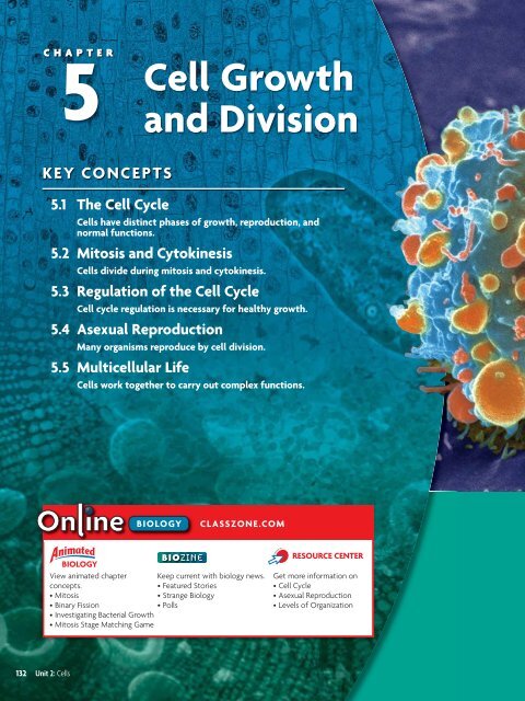
CHAPTER5<strong>Cell</strong> <strong>Growth</strong><strong>and</strong> <strong>Division</strong>KEY CONCEPTS5.1 The <strong>Cell</strong> Cycle<strong>Cell</strong>s have distinct phases of growth, reproduction, <strong>and</strong>normal functions.5.2 Mitosis <strong>and</strong> Cytokinesis<strong>Cell</strong>s divide during mitosis <strong>and</strong> cytokinesis.5.3 Regulation of the <strong>Cell</strong> Cycle<strong>Cell</strong> cycle regulation is necessary for healthy growth.5.4 Asexual ReproductionMany organisms reproduce by cell division.5.5 Multicellular Life<strong>Cell</strong>s work together to carry out complex functions.BIOLOGYCLASSZONE.COMBIOLOGYView animated chapter Keep current with biology news.concepts.• Featured Stories• Mitosis• Strange Biology• Binary Fission• Polls• Investigating Bacterial <strong>Growth</strong>• Mitosis Stage Matching GameRESOURCE CENTERGet more information on• <strong>Cell</strong> Cycle• Asexual Reproduction• Levels of Organization132 Unit 2: <strong>Cell</strong>s
5.1 The <strong>Cell</strong> CycleKEY CONCEPT <strong>Cell</strong>s have distinct phases of growth, reproduction, <strong>and</strong> normal functions.MAIN IDEAS• The cell cycle has four main stages.• <strong>Cell</strong>s divide at different rates.• <strong>Cell</strong> size is limited.VOCABULARYcell cycle, p. 134mitosis, p. 135cytokinesis, p. 135Connect Many of life’s little chores such as sweeping <strong>and</strong> dusting, are quietlysatisfying <strong>and</strong> rather fun. Washing dishes by h<strong>and</strong>, however, is never fun, whichis why some clever person made the dishwasher. This h<strong>and</strong>y invention soaks,washes, <strong>and</strong> rinses your dishes to a spot-free, sanitary sparkle. You unload thedishes, <strong>and</strong> the machine is ready to start the cycle all over again. A cell goesthrough a cycle, too. This cycle of growth, DNA synthesis, <strong>and</strong> division is essentialfor an organism to grow <strong>and</strong> heal. If it goes out of control, abnormal cellgrowth may occur, resulting in cancer cells like those shown on the previous page.FIGURE 5.1 <strong>Cell</strong>s grow <strong>and</strong> copytheir DNA during interphase.They also carry out cell-specificfunctions in G 1 <strong>and</strong> G 2 . DuringM stage, both the nucleus(in mitosis) <strong>and</strong> cytoplasm(in cytokinesis) are divided. MAIN IDEAThe cell cycle has four main stages.Just as all species have life cycles, from tiny chihuahuas to massive belugawhales, cells also have a life cycle. The cell cycle is the regular pattern ofgrowth, DNA duplication, <strong>and</strong> cell division that occurs in eukaryotic cells.FIGURE 5.1 shows its four main stages: gap 1, synthesis, gap 2, <strong>and</strong> mitosis. Gap1, synthesis, <strong>and</strong> gap 2 together make up what is called interphase.The stages of the cell cycle get their names from early studies of celldivision. Scientists’ observations were limited by the microscopes of the time.When a cell was not actively dividing, they could not seeactivity in it. Thus, they originally divided the cell cycleinto two parts: interphase, when the cell appeared tobe at rest, <strong>and</strong> mitosis, when the cell was dividing.Improved techniques <strong>and</strong> tools later allowedscientists to detect the copying of DNA (DNAsynthesis), <strong>and</strong> they changed their descriptionof the cell cycle to include the synthesis stage.Since they still could not see anythinghappening during the other parts of interphase,scientists named the periods betweenmitosis <strong>and</strong> synthesis “gap 1” <strong>and</strong> “gap 2.”Eventually, scientists learned that, duringinterphase, cells carry out their normalfunctions <strong>and</strong> undergo critical growth <strong>and</strong>preparation for cell division.134 Unit 2: <strong>Cell</strong>s
5.1 The <strong>Cell</strong> CycleKEY CONCEPT <strong>Cell</strong>s have distinct phases of growth, reproduction, <strong>and</strong> normal functions.MAIN IDEAS• The cell cycle has four main stages.• <strong>Cell</strong>s divide at different rates.• <strong>Cell</strong> size is limited.VOCABULARYcell cycle, p. 134mitosis, p. 135cytokinesis, p. 135Connect Many of life’s little chores such as sweeping <strong>and</strong> dusting, are quietlysatisfying <strong>and</strong> rather fun. Washing dishes by h<strong>and</strong>, however, is never fun, whichis why some clever person made the dishwasher. This h<strong>and</strong>y invention soaks,washes, <strong>and</strong> rinses your dishes to a spot-free, sanitary sparkle. You unload thedishes, <strong>and</strong> the machine is ready to start the cycle all over again. A cell goesthrough a cycle, too. This cycle of growth, DNA synthesis, <strong>and</strong> division is essentialfor an organism to grow <strong>and</strong> heal. If it goes out of control, abnormal cellgrowth may occur, resulting in cancer cells like those shown on the previous page.FIGURE 5.1 <strong>Cell</strong>s grow <strong>and</strong> copytheir DNA during interphase.They also carry out cell-specificfunctions in G 1 <strong>and</strong> G 2 . DuringM stage, both the nucleus(in mitosis) <strong>and</strong> cytoplasm(in cytokinesis) are divided. MAIN IDEAThe cell cycle has four main stages.Just as all species have life cycles, from tiny chihuahuas to massive belugawhales, cells also have a life cycle. The cell cycle is the regular pattern ofgrowth, DNA duplication, <strong>and</strong> cell division that occurs in eukaryotic cells.FIGURE 5.1 shows its four main stages: gap 1, synthesis, gap 2, <strong>and</strong> mitosis. Gap1, synthesis, <strong>and</strong> gap 2 together make up what is called interphase.The stages of the cell cycle get their names from early studies of celldivision. Scientists’ observations were limited by the microscopes of the time.When a cell was not actively dividing, they could not seeactivity in it. Thus, they originally divided the cell cycleinto two parts: interphase, when the cell appeared tobe at rest, <strong>and</strong> mitosis, when the cell was dividing.Improved techniques <strong>and</strong> tools later allowedscientists to detect the copying of DNA (DNAsynthesis), <strong>and</strong> they changed their descriptionof the cell cycle to include the synthesis stage.Since they still could not see anythinghappening during the other parts of interphase,scientists named the periods betweenmitosis <strong>and</strong> synthesis “gap 1” <strong>and</strong> “gap 2.”Eventually, scientists learned that, duringinterphase, cells carry out their normalfunctions <strong>and</strong> undergo critical growth <strong>and</strong>preparation for cell division.134 Unit 2: <strong>Cell</strong>s
Gap 1 (G 1 )The first stage of the cell cycle is gap 1 (G 1 ). During G 1 , a cell carries out itsnormal functions. If it is a skeletal muscle cell, it contracts to move joints. If itis an adrenal cell, it secretes hormones such as adrenaline. If it is an intestinalcell, it absorbs nutrients. During G 1 , cells also increase in size, <strong>and</strong> organellesincrease in number. A cell spends most of its time in the G 1 stage, although thelength of this stage varies by cell type.During G 1 , the cell must pass a critical checkpoint before it can proceed tothe synthesis stage. Just as it would be dangerous for you to run a marathon ifyou had not slept or eaten for several days, it would also be dangerous for yourcells to continue dividing if certain conditions were not met. For instance,most animal cells need enough nutrition, adequate size, <strong>and</strong> relatively undamagedDNA to divide successfully. They also need specific signals from othercells, telling them whether more cell division is needed.Synthesis (S)The second stage of the cell cycle is the synthesis (S) stage. Synthesis means“the combining of parts to make a whole.” During the S stage, the cell makes acopy of its nuclear DNA. In eukaryotes, DNA is located in the nucleus. Duringinterphase, it is loosely organized <strong>and</strong> appears grainy in photographs. By theend of the S stage, the cell nucleus contains two complete sets of DNA.Gap 2 (G 2 )Gap 2 (G 2 ) is the third stage of the cell cycle. During G 2 , cells continue to carryout their normal functions, <strong>and</strong> additional growth occurs. Like G 1 , this stageincludes a critical checkpoint. Everything must be in order—adequate cellsize, undamaged DNA—before the cell goes through mitosis <strong>and</strong> division.TAKING NOTESConstruct your own cycle diagramto take notes about processessuch as the cell cycle.G 1<strong>Growth</strong>ConnectingSSynthesisCONCEPTSDNA replication As you willlearn in Chapter 8, DNA synthesisis also called DNA replication.During this process, the DNAmolecule unzips <strong>and</strong> each str<strong>and</strong>is used as a pattern for a newDNA str<strong>and</strong>.Mitosis (M)Mitosis (M), the fourth stage of thecell cycle, includes two processes:mitosis <strong>and</strong> cytokinesis. Mitosis(my-TOH-sihs) is the division of thecell nucleus <strong>and</strong> its contents. Duringmitosis, the nuclear membranedissolves, the duplicated DNA condensesaround proteins <strong>and</strong> separates,<strong>and</strong> two new nuclei form. Lastly,VISUAL VOCABMitosis is the division of the cellnucleus <strong>and</strong> its contents.parent cellmitosiscytokinesisdaughter cellscytokinesis (SY-toh-kuh-NEE-sihs) isthe process that divides the cellCytokinesis divides the cell cytoplasm.cytoplasm. The result is two daughtercells that are genetically identical to the original cell.The stages of the cell cycle <strong>and</strong> the proteins that control it are similar inall eukaryotes. For example, scientists have demonstrated that some of themolecules that regulate checkpoints in the yeast cell cycle can work in humancells, too. Such similarities suggest that eukaryotes share a common ancestry.Predict What might happen if the G 2 checkpoint stopped working in cells?Chapter 5: <strong>Cell</strong> <strong>Growth</strong> <strong>and</strong> <strong>Division</strong> 135
FIGURE 5.2 CELL LIFE SPANCELL TYPESkin cellRed blood cellLiver cellIntestine—internal liningIntestine—muscle <strong>and</strong>other tissuesSource: Spaulding et al., <strong>Cell</strong> 122:1.ConnectingCONCEPTSMulticellular organisms use celldivision for growth <strong>and</strong> repair.APPROXIMATE LIFE SPAN2 weeks4 months300–500 days4–5 days16 yearsLymphocytes As you will learnin Chapter 31, lymphocytes are apart of your immune system.There are two major types oflymphocytes, B <strong>and</strong> T cells. Bothtypes recognize specific antigens.MAIN IDEA<strong>Cell</strong>s divide at different rates.Rates of cell division vary widely, as shown in FIGURE 5.2.The prokaryotic cell cycle is similar but not identical tothat of eukaryotic cells. Recall that prokaryotes do nothave the membrane-bound organelles <strong>and</strong> cytoskeletonfound in eukaryotes. Thus, prokaryotic cells typicallydivide much faster than do eukaryotic cells.The rate at which your cells divide is linked to yourbody’s need for those cells. In human cells, the S, G 2 , <strong>and</strong>M stages together usually take about 12 hours. The lengthof the G 1 stage differs most from cell type to cell type. Therate of cell division is greater in embryos <strong>and</strong> childrenthan it is in adults. Their cell cycle is shorter, <strong>and</strong> many oftheir organs are still developing. But the rate of celldivision also varies within different tissues of the adultbody. The internal lining of your digestive tract receives alot of wear <strong>and</strong> tear. As a result, cells that line yourstomach <strong>and</strong> intestine are replaced every few days. Incontrast, cells that make up the rest of your intestine(mainly smooth muscle) <strong>and</strong> many of your internal organs, such as lungs,kidney, <strong>and</strong> liver, divide only occasionally, in response to injury or cell death.<strong>Cell</strong>s that divide only rarely are thought to enter a stage that some scientistscall G 0 . In G 0 , cells are unlikely to divide, although they continue to carry outtheir normal functions. Some cells, such as neurons, appear to stay permanentlyin the G 0 stage. However, some data suggest that neurons actually c<strong>and</strong>ivide, <strong>and</strong> this question continues to be actively researched. Other cells, suchas lymphocytes, a type of white blood cell, may remain in G 0 for years untilthey recognize an invader. Once the invader binds to a lymphocyte receptor,the lymphocyte goes through rapid cell divisions to help fight infection.Infer Do you think a skin cell would have a long or short G 1 stage? Explain why.136 Unit 2: <strong>Cell</strong>sMAIN IDEA<strong>Cell</strong> size is limited.<strong>Cell</strong>s have upper <strong>and</strong> lower size limits. If cells were too small, they could notcontain all of the necessary organelles <strong>and</strong> molecules. For instance, a cell withtoo few mitochondria would not have enough energy to live. Nor can cellsgrow beyond a certain size, even if surrounded by plenty of nutrients. Theupper limit on cell size is due to the ratio of cell surface area to volume. Recallthat oxygen, nutrients, <strong>and</strong> wastes move across the cell membrane, or thesurface of the cell. These materials must be transported in adequate amounts<strong>and</strong> with adequate speed to keep the inside of the cell functioning. But as a cellincreases in size, its volume increases faster than its surface area, as shown inFIGURE 5.3. Therefore, a further increase in size could result in a surface area toosmall for the adequate exchange of materials.
FIGURE 5.3 Ratio of Surface Area to Volume in <strong>Cell</strong>sAs a cell grows, its volume increases more rapidly than its surface area. When thesurface area–to–volume ratio is too small, the cell cannot move materials into <strong>and</strong>out of the cell at a sufficient rate or in sufficient quantities.Relative size1 2 3Surface area(length width number of sides)Volume(length width height)Ratio of surface areato volume6 24 541 8 276_241= 6:1 __ 854= 3:1 __27 = 2:1Compare Which cell has the largest surface area? Which cell has the largestsurface area to volume ratio?Some cells, however, must be large. A neuron running down a giraffe’s neckto its legs may be several meters long, for instance. But it is not shaped like acube or a sphere. Instead, it is extremely long <strong>and</strong> thin. This structure gives theneuron a large surface area with a relatively small increase in volume.To maintain a suitable cell size, growth <strong>and</strong> division must be coordinated.If a cell more than doubled its size before dividing, the daughter cells would belarger than the original cell. If this happened each generation, cells wouldquickly become too large to live. Similarly, if a cell did not double its sizebefore dividing, the daughter cells would be smaller than the original cell. Ifthis happened each generation, cells would quickly become too small to live.Connect Which has the larger ratio of surface area to volume, a tennis ball or asoccer ball? Explain your reasoning.For more information aboutmitosis, go to scilinks.org.Keycode: MLB0055.1 ASSESSMENTONLINE QUIZClassZone.comREVIEWINGMAIN IDEAS1. During which stage of the cell cycleis the DNA copied?2. Which stages of the cell cyclegenerally require about the sameamount of time in all human cells?3. What limits the maximum size of acell?CRITICAL THINKING4. Infer Suppose you were to draw adiagram representing the cell cycleof a neuron. Explain where <strong>and</strong> howyou would represent G 0 .5. Predict Suppose you treatcells with chemicals that blockcytokinesis. Describe what youthink the cells would look like.Connecting CONCEPTS6. Scientific Process Predict howthe rate of cell division woulddiffer between single-celledalgae living in a sunny, nutrientrichpond versus algae living in ashady, nutrient-poor pond. Howcould you test your prediction?Chapter 5: <strong>Cell</strong> <strong>Growth</strong> <strong>and</strong> <strong>Division</strong> 137
5.2Mitosis <strong>and</strong> CytokinesisKEY CONCEPT <strong>Cell</strong>s divide during mitosis <strong>and</strong> cytokinesis.MAIN IDEAS• Chromosomes condense at the startof mitosis.• Mitosis <strong>and</strong> cytokinesis produce twogenetically identical daughter cells.VOCABULARYchromosome, p. 138histone, p. 139chromatin, p. 139chromatid, p. 139centromere, p. 139telomere, p. 139prophase, p. 140metaphase, p. 140anaphase, p. 140telophase, p. 140Connect When you were a child, perhaps you attended a birthday party wheregoody bags were h<strong>and</strong>ed out. Whoever stuffed the bags had to make sure thateach bag had exactly the same number of erasers, c<strong>and</strong>ies, <strong>and</strong> stickers. Otherwise,some ill-mannered child (not you, of course) might have raised a fuss if anitem was missing. In a similar way, your cells must receive a full set of DNA—nomore, no less—to work properly. Dividing DNA is a complicated task becausethe DNA is so long <strong>and</strong> stringy. Mitosis is an amazing process that efficientlysorts two sets of DNA <strong>and</strong> divides them between two nuclei.ConnectingCONCEPTSBiochemistry As you will learnin Chapter 8, a nucleotide ismade of three parts: a sugar, aphosphate group, <strong>and</strong> a nitrogencontainingmolecule called abase. When the sugars <strong>and</strong> phosphategroups bond, they formthe backbones of the longchains called nucleic acids.phosphate basesugarMAIN IDEAChromosomes condense at the start of mitosis.DNA is a double-str<strong>and</strong>ed molecule made of four different subunits callednucleotides. A chromosome is one long continuous thread of DNA thatconsists of numerous genes along with regulatory information. Your bodycells have 46 chromosomes each. If stretched out straight <strong>and</strong> laid end to end,the DNA in just one of your cells would be about 3 meters (10 feet) long.How does it fit inside the nucleus of a microscopic cell?DNA wraps around proteins that help organize<strong>and</strong> condense it. During interphase, or when a cellis not dividing, DNA is loosely organized—itlooks a bit like spaghetti. During mitosis, however,your chromosomes are tightly condensed, asshown in FIGURE 5.4. These changes in DNA’sorganization allow a cell to carry out its necessaryfunctions. During all of interphase, proteins mustaccess specific genes for a cell to make specificproteins or to copy the entire DNA sequence.During mitosis, the duplicated chromosomesmust condense to be divided between two nuclei.FIGURE 5.4 This duplicatedchromosome is tightlycondensed. (colored SEM;magnification about 5000)If chromosomes remained stringy during mitosis, they could become entangled.Perhaps a cell would get two copies of one chromosome <strong>and</strong> no copies ofa different one. FIGURE 5.5 shows the process that converts a chromosome froma linear str<strong>and</strong> of DNA to its highly condensed form. The key to this process isthe association between DNA <strong>and</strong> proteins.138 Unit 2: <strong>Cell</strong>s
FIGURE 5.5 Chromosome StructureDNA condenses tightly during the early stages of mitosis.chromatidtelomerehistonecentromereDNA double helixEach continuous,double-str<strong>and</strong>ed DNAmolecule makes onechromosome.DNA <strong>and</strong> histonesDNA wraps at regularintervals around proteinscalled histones,forming chromatin.ChromatinInteractions betweenparts of the histonesfurther compact theDNA.Supercoiled DNAThe chromatincoils more <strong>and</strong> moretightly around organizingproteins.Infer Overall, DNA has a negative charge. Look at the histone proteins in thefigure. What type of overall charge do you think they have? Explain.telomereCondensed, duplicated chromosomeThe condensed, duplicated chromosomescan be aligned <strong>and</strong> separated duringmitosis.At almost all times during the cell cycle, each of your chromosomes isassociated with a group of proteins called histones. DNA wraps aroundhistones at regular intervals, similar to beads on a string. Parts of the histonesinteract with each other, further compacting the DNA. At this stage—the“spaghetti” stage—the loose combination of DNA <strong>and</strong> proteins is calledchromatin. The word “loose” describes how much the DNA str<strong>and</strong> folds backon itself; it does not mean the DNA is loosely wrapped around the histones.As a cell progresses into mitosis, chromatin further condenses. It continuesto coil more <strong>and</strong> more tightly around organizing proteins, finally formingsmall thick rods. Recall that each chromosome has already been copied duringthe previous S stage. Thus, the chromosome looks similar to an “X” in whichthe left <strong>and</strong> right halves are two identical DNA double helixes. One half of aduplicated chromosome is called a chromatid (KROH-muh-tihd). Together,the two identical chromatids are called sister chromatids. Sister chromatidsare held together at the centromere (SEHN-truh-MEER), a region of thecondensed chromosome that looks pinched.In addition, the ends of DNA molecules form structures called telomeres(TEHL-uh-meers), which are made of repeating nucleotides that do not formgenes. They prevent the ends of chromosomes from accidentally attaching toeach other, <strong>and</strong> they help prevent the loss of genes. A short section of nucleotidesis lost from a new DNA molecule each time it is copied. It is importantthat these nucleotides are lost from telomeres, not from the genes themselves.Apply What is the relationship between a molecule of DNA <strong>and</strong>a chromosome?TAKING NOTESUse a main idea web to help youstudy the makeup <strong>and</strong> organizationof chromosomes.chromosomesChapter 5: <strong>Cell</strong> <strong>Growth</strong> <strong>and</strong> <strong>Division</strong> 139
MAIN IDEAMitosis <strong>and</strong> cytokinesis produce two geneticallyidentical daughter cells.The combined processes of mitosis <strong>and</strong> cytokinesis produce two geneticallyidentical daughter cells. Follow along in FIGURE 5.7 as you read about theprocess in more detail below.FIGURE 5.6 The nucleus <strong>and</strong>chromosomes go through dramaticchanges in a dividing cell.Parent cellcentriolesInterphaseInterphase plays an important role in preparing the cell todivide. It provides critical time for the duplication of organelles<strong>and</strong> for DNA replication. By the end of interphase, anindividual cell has two full sets of DNA, or chromosomes, <strong>and</strong>is large enough to divide.INTERPHASEnucleuswith DNAConnectingcentrosomespindlefibersCONCEPTS<strong>Cell</strong>s As you will learn in Chapter6, your body has two majorcell types. Germ cells developinto eggs or sperm. Somatic cellsmake up the rest of your body.MitosisMITOSISMitosis divides a cell’s nucleus into two genetically identicalnuclei, each with its own single, full set of DNA. This processoccurs in all of your body cells—except those that form eggsor sperm—<strong>and</strong> prepares them for cytokinesis. Althoughmitosis <strong>and</strong> cytokinesis are continuous processes, scientistshave divided them into phases to make them easier to underst<strong>and</strong> <strong>and</strong> discuss.The four main phases of mitosis are prophase, metaphase, anaphase, <strong>and</strong>telophase. Cytokinesis begins during late anaphase or telophase.1 During prophase, chromatin condenses into tightly coiled chromosomes.Each consists of two identical sister chromatids. The nuclear envelopebreaks down, the nucleolus disappears, <strong>and</strong> the centrosomes <strong>and</strong> centriolesbegin to migrate to opposite sides of the cell. Organized microtubulescalled spindle fibers grow from the centrioles <strong>and</strong> radiate towardthe center of the cell.2 In metaphase, the spindle fibers attach to a protein structure on thecentromere of each chromosome <strong>and</strong> align the chromosomes alongthe cell equator, around the middle of the cell.3 During anaphase, sister chromatids separate from each other. Thespindle fibers begin to shorten, which pulls the sister chromatidsaway from each other <strong>and</strong> toward opposite sides of the cell.4 In telophase, a complete set of identical chromosomes is positionedat each pole of the cell. The nuclear membranes start to form, thechromosomes begin to uncoil, <strong>and</strong> the spindle fibers fall apart.CytokinesisCytokinesis divides the cytoplasm into two cells <strong>and</strong> completesa full stage of the cell cycle. Cytokinesis differs in animal<strong>and</strong> plant cells. In animal cells, the membrane forms a furrow,or trench, that is pulled inward by tiny filaments, like adrawstring. Gradually, the membrane pinches closed, forminga separate cell around each nucleus.CYTOKINESIS140 Unit 2: <strong>Cell</strong>s
FIGURE 5.7 The <strong>Cell</strong> Cycle in DetailFollowing interphase, mitosis divides duplicated chromosomes between two nuclei.Cytokinesis divides the cytoplasm. In this diagram, the mitosis stage is greatlyexp<strong>and</strong>ed to highlight its four major phases. (micrographs; magnification about 100)BIOLOGYWatch mitosisin action atClassZone.com.INTERPHASEThe cell copies its DNA<strong>and</strong> grows in preparationfor division. The DNA isloosely organized duringinterphase.MITOSISMitosis divides a cell’s nucleus into twonuclei, each with an identical set of DNA.1Prophase DNA <strong>and</strong> proteins condenseinto tightly coiled chromosomes. Thenuclear envelope breaks down, centriolesbegin to move to opposite poles, <strong>and</strong>spindle fibers form.CYTOKINESISCytokinesis dividescytoplasm between twodaughter cells, each witha genetically identicalnucleus. The cells enterinterphase <strong>and</strong> beginthe cycle again.2Metaphase Spindle fibersattach to each chromosome.They align the chromosomesalong the cell equator.4Telophase Nuclear membranesstart to form, chromosomesbegin to uncoil, <strong>and</strong> the spindlefibers fall apart.3Anaphase Chromatids separateto opposite sides of the cell.Cytokinesis usually begins in lateanaphase or telophase.CRITICALVIEWINGHow many chromosomes does the cell have at the start ofmitosis? How many does it have after cytokinesis?Chapter 5: <strong>Cell</strong> <strong>Growth</strong> <strong>and</strong> <strong>Division</strong> 141
DATA ANALYSISCONSTRUCTING DATA TABLESScientists use data tables to recordtheir data. Data tables are organizedby the independent <strong>and</strong> dependentvariables. Usually the independentvariable is listed in the left column,<strong>and</strong> the other columns list thedependent variables. Each separateobservation is listed in its own row.When measurements are taken usingunits, they are listed in the columnheadings in parentheses. All tablesshould have numbers <strong>and</strong> titles.independentvariableTABLE 1. EFFECT OF HORMONES ONCELL DIVISIONConcentration ofHormone Solution (%)Size of <strong>Cell</strong> ClumpAfter 24 Hours (mm)0 325 450 875 9100 9observationsTable 1 shows data from a hypothetical experiment in which growth hormoneswere added to clumps of cells in a laboratory, <strong>and</strong> the growth of the cell clumpswas measured.dependentvariable1. Display Data Suppose a scientist decided to measure the effect of temperatureon cell division in chlorella, a type of green algae. Set up a table that could beused to record the results for the number of daily doublings of the cells: 20°C,3 doublings; 30°C, 7 doublings; 40°C, 12 doublings; 50°C, 0 doublings.2. Apply Label the independent <strong>and</strong> dependent variables on your table.During cytokinesis in plant cells, the membrane cannot pinch inwardbecause of the cell wall. Instead, a cell plate forms between the two nuclei. It ismade by the Golgi apparatus, which supplies the new plasma membrane. A newwall then grows as cellulose <strong>and</strong> other materials are laid down. Typically, cytoplasmis divided evenly between daughter cells in both plant <strong>and</strong> animal cells.The formation of new cells is critical in both multicellular <strong>and</strong> single-celledorganisms. Single-celled organisms use cell division to reproduce, whereasmulticellular organisms use it for growth, development, <strong>and</strong> repair.Contrast How does cytokinesis differ in animal <strong>and</strong> plant cells?5.2 ASSESSMENTONLINE QUIZClassZone.comREVIEWINGMAIN IDEAS1. Draw what a chromosomelooks like during metaphase.Identify the chromatids <strong>and</strong>the centromere.2. Briefly explain why the daughtercells resulting from mitosis aregenetically identical to each other<strong>and</strong> to the original cell.CRITICAL THINKING3. Contrast How do prophase <strong>and</strong>telophase differ?4. Apply Using a light microscope,you observe a cell that has nonucleus. What features would youlook for to determine whether it isa eukaryotic cell undergoingmitosis or a prokaryotic cell?Connecting CONCEPTS5. Protein Synthesis For a cell tomake proteins, enzymes mustaccess its genes. When histonesare modified with acetyl groups(-COCH 3 ), their positive charge isneutralized, so they wrap DNAless tightly. How might this affectthe rate of protein synthesis?142 Unit 2: <strong>Cell</strong>s
CHAPTER 5MATERIALS• slides of onionroot cells• microscopePROCESS SKILLS• Observing• Collecting Data• ConcludingINVESTIGATIONMitosis in Onion Root <strong>Cell</strong>sIn this lab, you will examine cells from onion root tissue under the microscope <strong>and</strong>identify the different stages of cell division. You will also determine how muchtime is spent in each stage of the cell cycle.PROBLEM How much time do cells spend in each part of the cell cycle?PROCEDURE1. Obtain a slide of onion root cells. Examine the slide under the microscope usingthe low-power lens.2. Find examples of cells in each stage of the cell cycle, including interphase <strong>and</strong> thestages of mitosis—prophase, metaphase, anaphase, <strong>and</strong> telophase. Draw <strong>and</strong> labeleach cell. Label structures within the cell.3. Select a r<strong>and</strong>om area of the slide to study using the high-power lens.4. Identify <strong>and</strong> record the stage of each cell in the view. Make a data table, like theone shown below, <strong>and</strong> record your data.5. Repeat step 3 two more times.6. Calculate the percentage of cells in each part of the cell cycle for each sample.TABLE 1. STAGES OF THE CELL CYCLESample Total <strong>Cell</strong>s Interphase Prophase Metaphase Anaphase TelophaseNo. No. % No. % No. % No. % No. %123ANALYZE AND CONCLUDE1. Analyze What patterns exist in your data? In which stage of the cell cycle aremost of the cells you examined? How do these data support what you knowabout the cell cycle?2. Calculate Find the average percentage of cells in each stage of the cell cycleamong the three samples. Assume that a cell takes 24 hours to complete one cellcycle. Calculate how much time is spent in each stage of the cell cycle. (Hint:Multiply the percentage of cells in each stage, as a decimal, by 24 hours.)3. Apply The cells in the root of an onion are actively dividing. How might thenumbers you count here be different than if you had examined cells from adifferent part of the plant?4. Predict A chemical company is testing a new product that it believes will increasethe growth rate of food plants. Suppose you are able to view the slides of onionroot tips that have been treated with the product. If the product is successful,how might the slides look different from the slides you viewed in this lab? Drawsome examples of what the treated slides might look like.This onion cell lays down acell plate (middle) that willform new cell membranes<strong>and</strong> the cell wall. (LM, magnificationabout 800)EXTEND YOUR INVESTIGATIONDesign an experiment that would test the product described in question 4. Assumethe product is a liquid that can be added to the soil in which the plant is growing.Chapter 5: <strong>Cell</strong> <strong>Growth</strong> <strong>and</strong> <strong>Division</strong> 143
5.3Regulation of the <strong>Cell</strong> CycleKEY CONCEPT <strong>Cell</strong> cycle regulation is necessary for healthy growth.MAIN IDEAS• Internal <strong>and</strong> external factors regulatecell division.• <strong>Cell</strong> division is uncontrolled in cancer.VOCABULARYgrowth factor, p. 144apoptosis, p. 145cancer, p. 146benign, p. 146malignant, p. 146metastasize, p. 146carcinogen, p. 146Connect Have you ever watched a movie in which people play with the elementsof nature? They might bring back dinosaurs or make a newfangled robot. Andhave you noticed that these movies are always scary? That’s because things go outof control. The robots take over, or the dinosaurs start eating humans. If cellgrowth goes out of control in your body, the result can be even scarier. Cancer isuncontrolled cell growth <strong>and</strong> results from many factors that affect the cell cycle.So how does your body regulate all the millions of cell divisions happening inyour body?MAIN IDEAInternal <strong>and</strong> external factors regulatecell division.Both external <strong>and</strong> internal factors regulate the cell cycle in eukaryotic cells.External factors come from outside the cell. They include messages fromnearby cells <strong>and</strong> from distant parts of the organism’s body.Internal factors come from inside the cell <strong>and</strong> include several types ofmolecules found in the cytoplasm. Both types of factors work together to helpyour body control the process of cell division.FIGURE 5.8 Normal animal cells(top) respond to external factors<strong>and</strong> stop dividing when they toucheach other. Cancer cells (bottom)fail to respond <strong>and</strong> form clumps.External FactorsExternal factors that help regulate the cell cycle include physical <strong>and</strong> chemicalsignals. One example of a physical signal is cell–cell contact. Most mammalcells grown in the laboratory form a single layer on the bottom of a culturedish, as shown in FIGURE 5.8. Once a cell touches other cells, it stops dividing.The exact reason for this phenomenon is unknown. One hypothesis is thatreceptors on neighboring cells bind to each other <strong>and</strong> cause the cells’ cytoskeletonsto form structures that may block the signals that trigger growth.Many cells also release chemical signals that tell other cells to grow. Forexample, growth factors are a broad group of proteins that stimulate celldivision. <strong>Growth</strong> factors bind to receptors that activate specific genes to triggercell growth. In general, cells grow <strong>and</strong> divide in response to a combination ofdifferent growth factors, not just one.144 Unit 2: <strong>Cell</strong>s
Some growth factors affect many different types of cells. For example,platelets are sticky fragments of bone marrow cells. They form clots thathelp stop bleeding. Platelets store a type of growth factor that helps yourbody repair wounds by triggering the growth of many different cell types.Other growth factors have more specific targets. For instance, erythropoietin(ih-RIHTH-roh-poy-EE-tihn) stimulates the production only of cells that willbecome red blood cells. Red blood cells carry oxygen. If you moved fromthe coast to the mountains, your blood oxygen levels would be lower becausethe air pressure is lower at higher altitudes. The decrease in blood oxygenlevels would cause your body to produce more erythropoietin. That factorwould increase the number of red blood cells <strong>and</strong> raise your blood oxygenlevels.Various hormones may also stimulate the growth of certain cell types. Inparticular, growth hormone results in bone growth <strong>and</strong> also affects yourprotein <strong>and</strong> fat metabolism.Internal FactorsWhen external factors bind to their receptors, they can trigger internal factorsthat affect the cell cycle. Two of the most important <strong>and</strong> well-studied internalfactors involved in the eukaryotic cell cycle are kinases <strong>and</strong> cyclins. A kinase isan enzyme that, when activated, transfers a phosphate group from one moleculeto a specific target molecule. This action typically increases the energy ofthe target molecule or changes its shape. Your cells have manytypes of kinases, <strong>and</strong> they are almost always present in the cell.Those kinases that help control the cell cycle are activated bycyclins. Cyclins are a group of proteins that are rapidly made<strong>and</strong> destroyed at certain points in the cell cycle. These twofactors help a cell advance to different stages of the cell cyclewhen cells bind to each other.ApoptosisJust as some cells need to grow <strong>and</strong> divide, other cells need todie. Apoptosis (AP-uhp-TOH-sihs) is programmed cell death. Itoccurs when internal or external signals activate genes that helpproduce self-destructive enzymes. Many questions remain aboutthis process. What is known is that the nucleus of an apoptoticcell tends to shrink <strong>and</strong> break apart, <strong>and</strong> the cell is recognized byspecialized cells in the immune system. These cells very tidilygobble up the apoptotic cell <strong>and</strong> recycle its chemical parts foruse in building other molecules. FIGURE 5.9 shows a classicexample of apoptosis. In the early stages of development, humanembryos have webbing between their fingers <strong>and</strong> toes, or digits.Before a baby is born, those cells typically go through apoptosis.Most babies are born with little unwebbed fingers <strong>and</strong> toes theylove to put in their mouths.Predict Suppose a child was born whose receptors for growthhormone did not work properly. How do you think this would affectthe child’s development?webbed fingersFIGURE 5.9 Human embryos havewebbed digits early in their development.The cells between thedigits undergo apoptosis duringlater stages of development. Asa result, the baby is born withunwebbed fingers <strong>and</strong> toes.Chapter 5: <strong>Cell</strong> <strong>Growth</strong> <strong>and</strong> <strong>Division</strong> 145
FIGURE 5.10 Cancer cells formtumors that may metastasize toother parts of the body.normal cellcancer cellbloodstream1. A healthy cell may become acancer cell if certain genes aredamaged.2. Cancer cells divide more oftenthan do healthy cells <strong>and</strong> mayform disorganized clumps calledtumors.3. Sometimes, cancer cells breakaway from the tumor. They canbe carried in the bloodstreamto other parts of the body,where they form new tumors.MAIN IDEA<strong>Cell</strong> division is uncontrolled in cancer.Cancer is the common name for a class of diseases characterized by uncontrolledcell division. It arises when regulation of the cell cycle breaks down.Unlike healthy cells, cancer cells grown in a culture dish continue to divide,even when surrounded by neighboring cells. Cancer cells can also continue todivide in the absence of many of the growth factors required for division inhealthy cells. As a result, they divide much more often than do healthy cells.Cancer cells form disorganized clumps called tumors. In a benign tumor,the cancer cells typically remain clustered together. This means the tumor maybe relatively harmless <strong>and</strong> can probably be cured by removing it. However, if atumor is malignant, some of the cancer cells can break away, or metastasize(mih-TAS-tuh-SYZ), from the tumor. These breakaway cells can be carried inthe bloodstream or lymph system to other parts of the body, as shown inFIGURE 5.10, where they can form more tumors, called metastases. Once a tumormetastasizes, it is much more difficult to entirely rid the body of tumors.But why are tumors harmful? Cancer cells do not perform the specializedfunctions needed by the body. In the lung, for example, cancer cells do notexchange oxygen <strong>and</strong> carbon dioxide. In the brain, they do not transmitthe carefully ordered electrical messages needed to interpret information.Therefore, the body has large clumps of rapidly dividing cells that require lotsof food <strong>and</strong> a hearty blood supply but that contribute nothing to the body’sfunction. In addition, a growing tumor can exert great pressure on surroundingorgans. For instance, a tumor growing inside the skull will cramp the brainfor space, <strong>and</strong> some regions will be unable to function properly. If cancer cellscontinue to grow unchecked, they will eventually kill the organism.Cancer cells come from normal cells that have suffered damage to thegenes that help make proteins involved in cell-cycle regulation. Most cancercells carry mutations, or errors, in two types of genes. One type, calledoncogenes, accelerate the cell cycle. The second type act as cell-cycle brakes.Mutations in these genes can be inherited. For instance, some breastcancers appear to be caused by inheritederrors in specific genes. Other mutationscan be caused by exposure to radiation orchemicals. For example, some skin cancersare due to DNA damage caused byultraviolet radiation from sunlight. Substancesknown to produce or promotethe development of cancer are calledcarcinogens (kahr-SIHN-uh-juhnz).These include tobacco smoke <strong>and</strong> certainair pollutants, which are both associatedwith lung cancer. Some mutated forms ofoncogenes are even carried by viruses; onesuch virus can cause cervical cancer.FIGURE 5.11 This cancerous mole isan example of a skin cancer, whichmay metastasize quickly.146 Unit 2: <strong>Cell</strong>s
QUICK LABOBSERVINGCancerIn this lab, you will compare normal cells with cancerous cells <strong>and</strong> observethe differences between them.PROBLEM How do normal <strong>and</strong> cancerous cells compare?PROCEDURE1. Examine the slides of normal cells under the microscope. Draw <strong>and</strong> describeyour observations.2. Repeat step 1 with slides of cancer cells.ANALYZE AND CONCLUDE1. Compare How does the structure of the normal cells compare with thestructure of the cancerous cells for each of the slides you viewed?2. Infer Cancer cells not only appear different from normal cells but they alsodivide more rapidly. Why do you think chemotherapy, a common treatment forcancer, results in the loss of hair?MATERIALS• microscope• slides of normal cells• slides of cancerous cellsSt<strong>and</strong>ard cancer treatment often involves both radiation <strong>and</strong> chemotherapy.Radiation therapy is the use of radiation to kill cancer cells <strong>and</strong> shrinktumors. It works by damaging a cell’s DNA so much that the cell cannotdivide. Radiation is usually localized—that is, its use is targeted to a specificregion—because it can also hurt healthy cells. Chemotherapy uses certaindrugs, often in combination, to kill actively dividing cells. Like radiation, itkills both cancerous <strong>and</strong> healthy cells. However, chemotherapy is systemic—drugs travel throughout the entire body.Medical researchers use laboratory-grown cancer cells in their search forcancer treatments. Much of what is known about the cell cycle has come fromstudies that use cancer cells. The most famous cancer cells used for researchare called HeLa cells. HeLa cells were originally obtained in 1951 from acervical tumor removed from a woman named Henrietta Lacks. This cell linecontinues to be grown <strong>and</strong> studied in laboratories all over the world.Analyze HeLa cells are also used to study cell signaling processes. What might be adisadvantage of using cancer cells to study processes occurring in healthy cells?5.3 ASSESSMENTONLINE QUIZClassZone.comREVIEWINGMAIN IDEAS1. Describe what a growth factoris <strong>and</strong> how it influences the cellcycle.2. Explain how cancer cells differfrom healthy cells.CRITICAL THINKING3. Contrast How do benign <strong>and</strong> malignanttumors differ?4. Hypothesize Suppose chromosomes in askin cell are damaged by ultraviolet radiation.If the damaged genes do not affectcell cycle regulation, do you think the cellwill become cancerous? Explain.Connecting CONCEPTS5. <strong>Cell</strong> Organelles Someanticancer drugs preventmicrotubules from formingspindle fibers. Why do youthink these drugs might beeffective treatments forcancer?Chapter 5: <strong>Cell</strong> <strong>Growth</strong> <strong>and</strong> <strong>Division</strong> 147
5.4Asexual ReproductionKEY CONCEPT Many organisms reproduce by cell division.MAIN IDEAS• Binary fission is similar in functionto mitosis.• Some eukaryotes reproducethrough mitosis.VOCABULARYasexual reproduction, p. 148binary fission, p. 148Connect In this flashy world of ours, you may think that the humble bacteriumwould have little chance of finding a mate. No dazzling smile, no fancy hairproducts, no shiny car, <strong>and</strong>—if we are brutally honest—not even a brain. Withall of these limitatio\ns, it may seem that our bacteria friends would be destinedto die out. And yet, bacteria are found in abundance <strong>and</strong> live just about everywhereon Earth. How can there be so many bacteria?MAIN IDEABinary fission is similar in function to mitosis.Reproduction is a process that makes new organisms from one or moreparent organisms. It happens in two ways—sexually <strong>and</strong> asexually. Sexualreproduction involves the joining of two specialized cells called gametes (eggs<strong>and</strong> sperm cells), one from each of two parents. The offspring that result aregenetically unique; they have a mixture of genes from both parents. In contrast,asexual reproduction is the creation of offspring from a single parent<strong>and</strong> does not involve the joining of gametes. The offspring that result are, forthe most part, genetically identical to each other <strong>and</strong> to the single parent.ConnectingCONCEPTS<strong>Cell</strong> Structure Recall fromChapter 3 that many scientistshypothesize that mitochondria<strong>and</strong> chloroplasts were originallyfree-living prokaryotes. Onepiece of evidence that supportsthis hypothesis is the factthat these two organellesreplicate much as bacteriado, through fission.Binary Fission <strong>and</strong> MitosisMost prokaryotes reproduce throughbinary fission. Binary fission (BY-nuhreeFIHSH-uhn) is the asexual reproductionof a single-celled organism bydivision into two roughly equal parts.Binary fission <strong>and</strong> mitosis havesimilar results. That is, both processesform two daughter cells that aregenetically identical to the parent cell.However, the actual processes aredifferent in several important ways.VISUAL VOCABBinary fission is the asexual reproductionof a single-celled organism bydivision into two roughly equal parts.parent cellDNA duplicatescell begins to dividedaughter cellsAs you already learned, prokaryotessuch as bacteria do not havenuclei. And although they do have DNA, they have much less of it than domost eukaryotes. Also, most of a bacterium’s DNA is in the form of onecircular chromosome, <strong>and</strong> bacteria have no spindle fibers.148 Unit 2: <strong>Cell</strong>s
123BIOLOGYSee binary fissionin action atClassZone.com.FIGURE 5.13 Binary fission isshown in this micrograph of threeindividual bacteria, each at adifferent stage of binary fission.First, a cell elongates (1), <strong>and</strong> theDNA is replicated. Next, the cellmembrane pinches inward (2).Finally, the membrane meets, <strong>and</strong>a new cell wall is laid down, formingtwo separate cells (3). (coloredTEM; magnification 60,000)Binary fission, shown in FIGURE 5.13, starts when the bacterialchromosome is copied. The two chromosomes are both attached to thecell membrane. As the cell grows <strong>and</strong> gets longer, the chromosomesmove away from each other. When the cell is about twice its originalsize, it undergoes cytokinesis. The membrane pinches inward, <strong>and</strong> anew cell wall is laid down between the two chromosomes, whichcompletes the separation into two daughter cells.Advantages <strong>and</strong> Disadvantages of Asexual ReproductionVery often, whether something is helpful or harmful depends on thesituation. In favorable environments that do not change much, asexualreproduction can be more efficient than sexual reproduction. Recallthat asexual reproduction results in genetically identical offspring. Ifthey are well suited to the environment, genetic variation could bemore harmful than helpful. In other words, if it ain’t broke, don’t fix it.However, asexual reproduction may be a disadvantage in changingconditions. Genetically identical offspring will respond to the environmentin the same way. If population members lack traits that enablethem to reproduce, the entire population could die off. In contrast, sexualreproduction increases genetic diversity, which raises the chance that at least afew individuals will survive or even thrive in changing conditions.Keep in mind, however, that the act of asexual reproduction itself is notmore efficient; rather, the associated costs of sexual reproduction are greater.For example, all asexually reproducing organisms can potentially reproduce.Suppose two organisms each have ten offspring. If one organism reproducesasexually, all ten offspring can have offspring of their own. If the other organismreproduces sexually, having five females <strong>and</strong> five males, only the fivefemales can bear offspring. In addition, sexually reproducing organisms mustattract a mate. This effort involves not only the time <strong>and</strong> energy needed tofind a mate but also many structures, signals, <strong>and</strong> behaviors that have evolvedto attract mates. Organisms that reproduce asexually do not have these costs.Summarize How is asexual reproduction an advantage in some conditions?FIGURE 5.14 BACTERIA GROWTH One bacterium can result in a totalof 1024 cells after only 10 rounds ofcell division.Connecting CONCEPTSEvolution As you will learn inChapter 18, the misuse of antibioticshas resulted in multidrugresistantbacteria. The bacterianot killed by antibiotics canreproduce quickly, passing thegenes for antibiotic resistanceon to their offspring.Chapter 5: <strong>Cell</strong> <strong>Growth</strong> <strong>and</strong> <strong>Division</strong> 149
FIGURE 5.15 Yeast <strong>and</strong> hydrascan reproduce by budding.(colored SEMs; magnifications: yeast2400; hydra about 30)YeastHydrabudMAIN IDEASome eukaryotes reproduce through mitosis.Some eukaryotes also reproduce asexually, through mitosis. Have you evergrown a new plant from a stem cutting? Or seen a new sea star growing fromthe arm of another one? These new organisms are the result of mitotic reproduction<strong>and</strong> are therefore genetically the same as the parent organism. Mitoticreproduction is especially common in simpler plants <strong>and</strong> animals. It occurs inboth multicellular <strong>and</strong> unicellular eukaryotes. It can take several forms,including budding, fragmentation, <strong>and</strong> vegetative reproduction.In budding, a small projection grows on the surface of the parent organism,forming a separate new individual. The new organism may live independentlyor attached as part of a colony. For instance, hydras <strong>and</strong> some types ofyeast reproduce by budding. Examples are shown in FIGURE 5.15.In fragmentation, a parent organism splits into pieces, each of which cangrow into a new organism. Flatworms <strong>and</strong> sea stars both reproduce by fragmentation.Many plants, including strawberries <strong>and</strong> potatoes, reproduce viavegetative reproduction. In general, vegetative reproduction involves themodification of a stem or underground structures of the parent organism.The offspring often stay connected to the original organism, through structurescalled runners, for example.Many organisms can reproduce both asexually <strong>and</strong> sexually. The form ofreproduction may depend on the current conditions. The sea anemone canreproduce in many ways. It can reproduce asexually bydividing in half, by breaking off small pieces from its base, orby budding. It can also reproduce sexually by making eggs<strong>and</strong> sperm. Some species of anemone have males <strong>and</strong> females.In other anemone species, the same organism can produceboth eggs <strong>and</strong> sperm cells.Synthesize How might the asexual reproduction of geneticallyidentical plants be useful to humans? How could it proveharmful to our food supply?5.4 ASSESSMENTONLINE QUIZClassZone.comREVIEWINGMAIN IDEAS1. Explain how mitosis differs frombinary fission.2. Briefly explain why cutting aflatworm into pieces would notkill it.CRITICAL THINKING3. Infer How does an organism benefitby being able to reproduce bothsexually <strong>and</strong> asexually?4. Apply Yeasts are growing in twodishes. You treat one dish with achemical that blocks DNA replicationbut forget to label it. How canyou identify the treated dish?Connecting CONCEPTS5. Ecology Two populations livein the same habitat <strong>and</strong>compete for food. The firstgroup is larger <strong>and</strong> usesasexual reproduction; thesecond reproduces sexually.What could happen to causethe second group to outnumberthe first?150 Unit 2: <strong>Cell</strong>s
5.5Multicellular LifeKEY CONCEPT <strong>Cell</strong>s work together to carry out complex functions.MAIN IDEAS• Multicellular organisms depend oninteractions among different cell types.• Specialized cells perform specific functions.• Stem cells can develop into differentcell types.VOCABULARYtissue, p. 151organ, p. 151organ system, p. 151cell differentiation, p. 152stem cell, p. 153ReviewhomeostasisConnect Each of us enters this world as a screaming infant. At first, the ability toeat solid foods or take a step draws forth great praise. These general skills rapidlylose their wonder, however, <strong>and</strong> by the time you reach the age of 18, everyonewants to know what you plan to do with yourself. Will you build houses or designclothing or treat patients? What will your specialty be? <strong>Cell</strong>s, too, undergospecialization to carry out the complex functions required by the body.ConnectingCONCEPTSHomeostasis As you learned inChapter 1, homeostasis is themaintenance of a stable internalenvironment. Both an organism’sphysiology <strong>and</strong> its behavior helpit achieve homeostasis.MAIN IDEAMulticellular organisms depend on interactionsamong different cell types.Within multicellular organisms, cells communicate <strong>and</strong> work together ingroups that form increasingly larger, more complex structures. This arrangementprogresses from cells to tissues to organs to organ systems, as shown inFIGURE 5.16. Tissues are groups of cells that work together to perform a similarfunction. Groups of tissues that work together to perform a specific functionor related functions are called organs. For instance, plants have photosynthetictissues made of chlorophyll-containing cells. Conductive tissues transportsugars, water, <strong>and</strong> minerals to <strong>and</strong> from other parts of the plant. Protectivetissues help prevent water loss. Together, these <strong>and</strong> other tissues form aleaf, the plant’s food-producing organ.Organs that carry out similar functions are further grouped intoorgan systems. In plants, the shoot system is above the ground. It includesstems that support the plant, leaves that capture radiant energy, <strong>and</strong> flowersthat aid reproduction. Beneath the ground, the root system has different typesof roots <strong>and</strong> root hairs that anchor the plant <strong>and</strong> absorb water <strong>and</strong> minerals.As organ systems work together, they help an organism maintain homeostasis.For example, plants need to maintain a certain level of water withintheir cells, or they will wilt <strong>and</strong> die. They absorb water through their roots <strong>and</strong>expel it as water vapor through openings in their leaves called stomata. Stomataare controlled by special cells called guard cells, which close the stomatawhen a plant’s water intake cannot keep up with its water loss.Apply Suppose your family goes out of town <strong>and</strong> forgets to ask your neighbor towater the plants. Do you think the plants’ stomata will be open or closed? Explain.Chapter 5: <strong>Cell</strong> <strong>Growth</strong> <strong>and</strong> <strong>Division</strong> 151
FIGURE 5.16 Levels of Organization<strong>Cell</strong>s work together in groups that form larger, specialized structures.CELL TISSUE ORGANSYSTEMSVessel elements aretube-shaped cells. (coloredSEM; magnification 200)Vessel elements <strong>and</strong> tracheidsform xylem. (coloredSEM; magnification 200)© Dr. Richard Kessel & Dr. Gene Shih / Visuals UnlimitedXylem <strong>and</strong> other tissuesform roots that absorbwater <strong>and</strong> nutrients.Apply Draw a simple sketch of the middle photograph.Label the vessel elements. Identify what you think arethe tracheids <strong>and</strong> label them.stemvasculartissuelateralrootsprimaryrootleafshoot systemroot systemMAIN IDEASpecialized cells perform specific functions.ConnectingCONCEPTSGametogenesis As you will learnin Chapter 6, the egg is stockedwith organelles <strong>and</strong> moleculesnecessary for an embryo to grow.Many of these molecules are notevenly distributed throughoutthe cell; they form gradients.It is easy to see that a skin cell can divide to make a new skin cell, or that asingle bacterium can generate another bacterium. But how does a complexorganism like you develop? Your body began as a single fertilized egg. If theegg simply divided to make lots of identical cells, it would not form a baby. Toform the intricate structures that make up your body <strong>and</strong> the bodies ofcountless organisms around you, cells must specialize.<strong>Cell</strong> differentiation is the process by which unspecialized cells develop intotheir mature forms <strong>and</strong> functions. While almost every cell in your body has afull set of DNA, each type of cell uses only the specific genes it needs to carryout its function. That is, a cell differentiates among the genes <strong>and</strong> uses onlycertain ones. You can think of your DNA as a cookbook. When you want tomake a specific dish, you select that recipe <strong>and</strong> carry out its instructions. If youneed to make a dessert, you might bake turtle brownies. If you need to make amain course, you might roast apple-stuffed pork chops or fix a hearty lentilstew. The dishes are very different, but they all come from the same cookbook.A cell’s location within the embryo helps determine how it will differentiate.In plant cells, the first division of a fertilized egg is unequal, or asymmetric,<strong>and</strong> produces two cells—the apical cell <strong>and</strong> the basal cell. The apical cellforms most of the embryo, including the growth point for stems <strong>and</strong> leaves.The major role of the basal cell is to provide nutrients to the embryo; it alsocreates the growth point for the roots. Plant cells cannot easily migrate becauseof the cell wall, but they adapt to changing conditions <strong>and</strong> continue todevelop throughout their lifetime. As the plant grows, new cells continue to152 Unit 2: <strong>Cell</strong>s
differentiate based on their location. For example, cells on the outer layer of aleaf may become epidermal cells that secrete a waxy substance that helpsprevent water loss. <strong>Cell</strong>s on the lower leaf surface may become guard cells thatcontrol the exchange of water, air, <strong>and</strong> carbon dioxide.In animals, an egg undergoes many rapid divisions after it is fertilized. Theresulting cells can migrate to a specific area, <strong>and</strong> the cells quickly begin todifferentiate. The early animal embryo generally takes the shape of a hollowball. As the embryo develops, part of the ball folds inward, forming an innerlayer <strong>and</strong> creating an opening in the outer cell layer. A middle layer of cellsthen forms between the other two.FIGURE 5.17 <strong>Cell</strong> Differentiation<strong>Cell</strong> differentiation in the developing animal embryo is based on location.Animal embryo cross sectionoutermiddleinnerOuter Skin cells help prevent infection <strong>and</strong>dehydration (colored SEM; magnification 130).Outer Skin cells help preventinfection <strong>and</strong> dehydration.(colored SEM; magnification 500)Middle Bone cells form a hardmatrix (shown) that supports <strong>and</strong>protects organs.(colored SEM; magnification 15)Inner Intestinal epithelia have alarge surface area that increasesabsorption.(colored SEM; magnification 25)As shown in FIGURE 5.17, in vertebrates, the outer cell layer differentiates toform the outer layer of skin <strong>and</strong> elements of the nervous system such as thebrain <strong>and</strong> spinal cord. The middle cell layer forms bones, muscles, kidneys,<strong>and</strong> the inner layer of skin. The inner cell layer forms internal organs such asthe pancreas, lungs, <strong>and</strong> digestive system lining.Analyze Why is regulation of the differentiation process during the early stages ofdevelopment so critical?MAIN IDEAStem cells can develop into different cell types.Stem cells are a unique type of body cell that have the ability to (1) divide<strong>and</strong> renew themselves for long periods of time, (2) remain undifferentiated inform, <strong>and</strong> (3) develop into a variety of specialized cell types. When a stem celldivides, it forms either two stem cells or one stem cell <strong>and</strong> one specialized cell.Chapter 5: <strong>Cell</strong> <strong>Growth</strong> <strong>and</strong> <strong>Division</strong> 153
VOCABULARYPotent comes from a Latinword meaning “to be able.”The addition of prefixes definesthe level of power or ability.toti- = allpluri- = more, severalmulti- = manyStem <strong>Cell</strong> ClassificationStem cells can be categorized by their ability, or potential, to develop into thedifferentiated cell types of different tissues, as shown in FIGURE 5.18. In general, themore differentiated a stem cell already is, the fewer the types of cells it can form.• Totipotent stem cells can grow into any other cell type. Only afertilized egg <strong>and</strong> the cells produced by the first few divisions ofan embryo are totipotent.• Pluripotent stem cells can grow into any cell type except fortotipotent stem cells.• Multipotent stem cells can only grow into cells of a closelyrelated cell family.FIGURE 5.18 STEM CELL CLASSIFICATIONClass totipotent pluripotent multipotentType of cell fertilized egg embryonic stem cell adult stem cell(example from blood)Can give rise to all cells almost any cell closely related cellsExampleneworganisminnercellmassneurons, skin, muscle,kidney, cartilage,bone, liver, pancreasred blood cells,platelets, whiteblood cellsStem cells are also classified bytheir origin, as either adult or embryonic.Adult stem cells have beenstudied for decades, but the ability togrow human embryonic stem cells wasnot developed until 1998. Since thattime, embryonic stem cells haveattracted great attention because oftheir potential to form almost any celltype. Both adult <strong>and</strong> embryonic stemcells offer unique advantages <strong>and</strong>challenges to researchers.Adult Stem <strong>Cell</strong>sAdult stem cells are partially undifferentiated cells located among the specializedcells of many organs <strong>and</strong> tissues. They are found all over the body, in thebrain, liver, bone marrow, skeletal muscle, dental pulp, <strong>and</strong> even fat. Thesestem cells are also found in children <strong>and</strong> in umbilical cord blood, so the termsomatic stem cell is more accurate although less frequently used.A major advantage of adult stem cells is that they can be taken from apatient, grown in culture, <strong>and</strong> put back into the patient. Thus, the risk oftransplant rejection by a patient’s immune system is very low. This methodalso avoids many ethical issues associated with using embryonic stem cells.Adult stem cells currently pose many disadvantages as well. They are few innumber, difficult to isolate, <strong>and</strong> sometimes tricky to grow. They may alsocontain more DNA abnormalities than do embryonic stem cells. For years,much evidence suggested that adult stem cells were multipotent. This wouldmean that a stem cell from fat would produce only fat cells, never muscle cells.Newer data suggest otherwise. Adult stem cells treated with the right combinationof molecules may give rise to a completely different type of tissue. Thisprocess, called transdifferentiation, remains an active area of research.Embryonic Stem <strong>Cell</strong>sMost embryonic stem cells come from donated embryos grown in a clinic.These embryos are the result of in vitro fertilization, a process by which eggsare fertilized outside a woman’s body. The stem cells are taken from a clusterof undifferentiated cells in the three-to-five-day-old embryo.154 Unit 2: <strong>Cell</strong>s
These cells, called the inner cell mass, do nothave the characteristics of any specific celltype. Because they are pluripotent, they canform any of the 200 cell types of the body.They can also be grown indefinitely inculture. These qualities have given manypeople hope that many now devastatingdiseases will be treatable or even curable inthe future.Embryonic stem cells also have a downside.If these cells are used in treatment, apatient’s body might reject them as foreignmaterial. The stem cells could potentiallygrow unchecked in a patient’s body <strong>and</strong> forma tumor. The use of embryonic stem cellsalso raises many ethical questions. The mostcommon method of getting embryonic stemcells, FIGURE 5.19, currently involves destructionof the embryo, which some peopleconsider ethically unacceptable.FIGURE 5.19 HARVESTING EMBRYONIC STEM CELLSResearch <strong>and</strong> Treatment HopeStem cells have long been used to treat patients with leukemia <strong>and</strong> lymphoma,<strong>and</strong> they offer hope for treating many other diseases as well. For instance,some patients might be cured of diabetes if nonworking cells in the pancreaswere replaced with healthy, growing cells. Similarly, damaged organs, such asthe heart, might be strengthened by an injection of healthy cells. Research,such as the testing of new drugs, might also benefit. The current researchsystem requires a lot of time <strong>and</strong> money. Many of the most innovative drugshave little chance of reaching the patient. Potentially, these new compoundscould be tested on large numbers of specific cell types grown from stem cells.Compare <strong>and</strong> Contrast List treatment benefits <strong>and</strong> risks of both types ofstem cells.First, an egg is fertilized by a sperm cell in a petri dish. The egg divides,forming an inner cell mass. These cells are then removed <strong>and</strong> grown withnutrients. Scientists try to control how the cells specialize by adding orremoving certain molecules.5.5 ASSESSMENTONLINE QUIZClassZone.comREVIEWINGMAIN IDEAS1. How does communication betweencells help maintain homeostasis?2. Explain why cell differentiationis an important part of thedevelopment of a multicellularorganism.3. What are the defining characteristicsof stem cells?CRITICAL THINKING4. Compare Describe how tissues,organs, <strong>and</strong> organ systems aresimilar.5. Evaluate Explain which factor youthink is most important in decidingwhether stem-cell research shouldbe legal <strong>and</strong> government-funded.Connecting CONCEPTS6. Animal Behavior Choose ananimal <strong>and</strong> give an example ofhow its behavior reflects itsneed to maintain homeostasis.Chapter 5: <strong>Cell</strong> <strong>Growth</strong> <strong>and</strong> <strong>Division</strong> 155
CHAPTER 5OPTIONS FOR INQUIRYUse these inquiry-based labs <strong>and</strong> online activities to deepen yourunderst<strong>and</strong>ing of cell growth <strong>and</strong> development.INVESTIGATIONModeling <strong>Cell</strong> SurfaceArea–to–Volume RatioA cell’s surface area–to–volume ratio affects theamount of material that can diffuse across themembrane <strong>and</strong> throughout the cell. You will make cellmodels to determine how this ratio changes as cellsize increases.SKILLS Modeling, InferringMATERIALS• plastic knife• phenolphthalein agar• metric ruler• 250-mL beaker• 100-mL graduated cylinder• 100 mL sodium hydroxidesolution• timer• plastic spoon• paper towelPROBLEM Which cell has the greatest surfacearea–to–volume ratio?PROCEDURE1. Make three model cells by using the knife to cut three cubes from the phenolphthaleinagar. <strong>Cell</strong> A should be 3 cm on each side, cell B should be 2 cm on each side, <strong>and</strong> cell Cshould be 1 cm on each side. Use the ruler to make exact measurements.2. Calculate the area of one side of each cell. Calculate the total surface area of each cell.Record your data in Table 1. (Hint: Multiply the area of one side by the number of sides.)3. Calculate the volume of each cell. Record your data in the table. (Hint: Multiply thelength by the width by the height of the cube.)4. Calculate the ratio of surface area to volume for each cell. For example, for cell A, theratio would be 54 cm 2 :27 cm 3 2:1 2. Record your data.5. Put the model cells in the beaker. Carefully cover them with sodium hydroxidesolution, which turns the agar pink. Soak the cells in solution for four minutes. Use aspoon to turn the cells repeatedly throughout that time.6. Remove the cells from solution <strong>and</strong> dry them on the paper towel.7. Use the knife to cut each cube in half. Measure the distance from the edge of the cellto the inner edge of the pink line. This shows how far the sodium hydroxide diffused.TABLE 1. CALCULATIONS OF CELL SIZE<strong>Cell</strong> Area of OneSide (cm 2 )Total SurfaceArea (cm 2 )ABCVolume of <strong>Cell</strong>(cm 3 )Surface Area–to–Volume RatioANALYZE AND CONCLUDE1. Analyze How does the surface area–to–volume ratio change as cell size increases? Howmight this affect the diffusion of materials throughout a cell?2. Apply Identify which cell turned pink in the greatest proportion, <strong>and</strong> explain how thisrelates to cell size.3. Apply How does a cell’s surface area–to–volume ratio affect its ability to stay alive?156 Unit 2: <strong>Cell</strong>s
INVESTIGATIONApoptosisIn this lab, you will research the role of apoptosis ina developmental process.SKILL CommunicatingPROBLEM What role does apoptosis play asorganisms develop?RESEARCH1. Use the Internet to research one of the followingprocesses:• the development of neural connections in thehuman brain• the development <strong>and</strong> maintenance of thehuman immune system• the metamorphosis of a tadpole into anadult frog2. Explain the role of apoptosis in your topic in atyped, one-page summary. In your answer, clearlyidentify the state of the organism before theapoptotic changes, the state of the organismfollowing apoptosis, <strong>and</strong> what would be the resultif apoptosis failed to occur.BIOLOGYCLASSZONE.COMVIRTUAL LABInvestigating Bacterial <strong>Growth</strong>Not all bacteria thrive in the same environmentalconditions. In this interactive lab, youwill determine which strains of bacteria growin an environment with oxygen <strong>and</strong> whichstrains grow without oxygen.ANIMATED BIOLOGYMitosis Stage Matching GameHow well can you recognize the stages ofmitosis? Test your skills by categorizingimages showing different phases of mitosis.The top image is a normal white blood cell. The bottomimage shows an apoptotic white blood cell with a darkenednucleus <strong>and</strong> disintegrating cytoplasm. (colored TEMs;magnification about 10,000)WEBQUESTCancer occurs when the cell cycle breaksdown. In this WebQuest, you will learnabout the most common cancer in theUnited States—skin cancer. Explore itscauses, how cells become cancerous, <strong>and</strong>why prevention is truly the best medicine.Chapter 5: <strong>Cell</strong> <strong>Growth</strong> <strong>and</strong> <strong>Division</strong> 157
CHAPTER5@ CLASSZONE.COMKEY CONCEPTS Vocabulary Games Concept Maps Animated Biology Online Quiz5.1 The <strong>Cell</strong> Cycle<strong>Cell</strong>s have distinct phases of growth, reproduction,<strong>and</strong> normal functions. The cell cycle hasfour main stages: G 1 , S, G 2 , <strong>and</strong> M. The length ofthe cell cycle can vary, resulting in different ratesof cell division. This variability isbased on the body’s need fordifferent cell types. <strong>Cell</strong>salso divide because theyneed a sufficient surfacearea–to–volume ratio tomove materials into <strong>and</strong>out of the cell.5.2 Mitosis <strong>and</strong>Cytokinesis<strong>Cell</strong>s divide during mitosis <strong>and</strong> cytokinesis.Mitosis divides the nucleus into two geneticallyidentical nuclei in a four-phase process: prophase,metaphase, anaphase, telophase. In prophase, theduplicated chromosomes condense tightly. Cytokinesisactually divides the cell cytoplasm.5.3 Regulation of the <strong>Cell</strong> Cycle<strong>Cell</strong> cycle regulation is necessary for healthygrowth. <strong>Cell</strong> growth <strong>and</strong> division are regulated byboth external factors, such as hormones <strong>and</strong>growth factors, <strong>and</strong> internal factors, such ascyclins <strong>and</strong> kinases. When proper regulation ofcell growth is disrupted, a cell may become cancerous.Cancer cells grow more rapidly than donormal cells <strong>and</strong> form clumps called tumors thatmay metastasize to other regions of the body. 5.4 Asexual ReproductionMany organisms reproduce by cell division.Most prokaryotes reproduce through a processcalled binary fission, in which a cell divides intotwo approximately equal parts. Some eukaryotesreproduce through mitosis. The offspring thatresult from asexual reproduction are geneticallyidentical to the parent organism, except whenmutations occur. Whether being identical isan advantage or a disadvantage depends onthe environment.5.5 Multicellular Life<strong>Cell</strong>s work together to carry out complex functions.Within multicellular organisms, cells formtissues, tissues form organs, <strong>and</strong> organs formorgan systems. The cells differentiate to performspecific functions. Much of this specialization isdetermined by a cell’s location within the developingembryo. Stem cells are a special type ofcell that continue to divide <strong>and</strong> renew themselvesfor long periods of time.Synthesize Your NotesConcept Map Use a concept map like the one below tosummarize what you know about mitosis.mitosis1stprophaseduringVenn Diagram Draw a Venn diagram like the one below tosummarize the similarities <strong>and</strong> differences betweenembryonic <strong>and</strong> adult stem cells.EMBRYONICADULThasfourphases2nd3rd4thmay berejectedremainundifferentiatedin formunlikely to berejected158 Unit 2: <strong>Cell</strong>s
Chapter AssessmentChapter Vocabulary5.1 cell cycle, p. 134mitosis, p. 135cytokinesis, p. 1355.2 chromosome, p. 138histone, p. 139chromatin, p. 139chromatid, p. 139centromere, p. 139telomere, p. 139prophase, p. 140metaphase, p. 140anaphase, p. 140telophase, p. 1405.3 growth factor, p. 144apoptosis, p. 145cancer , p. 146benign, p. 146malignant, p. 146metastasize, p. 146carcinogen, p. 1465.4 asexual reproduction, p. 148binary fission, p. 1485.5 tissue, p. 151organ, p. 151organ system, p. 151cell differentiation, p. 152stem cell, p. 153Reviewing VocabularyVisualize VocabularyFor each term below, draw a simple picture thatrepresents the meaning of the word. Here is anexample for mitosis.Reviewing MAIN IDEAS11. The cell cycle has four main stages—G 1 , S, G 2 , <strong>and</strong> M.What occurs in the cell during each stage?12. Compare the rates of cell division occurring in yourneurons <strong>and</strong> your hair follicles.13. What is the relationship between a cell’s surface area<strong>and</strong> its volume?14. You know that a chromosome is a very long, continuousstr<strong>and</strong> of DNA. How do proteins help condensechromosomes?1. prophase2. metaphase3. anaphase4. telophase5. cytokinesis6. centromere7. telomereWord Origins8. The prefix pro- means “earlier than” or “prior to.” Explainhow this meaning relates to the word prophase.9. The prefix telo- means “distant, far, or end.” How doesthis meaning relate to the words telophase <strong>and</strong>telomere?10. The term mitosis comes from the Greek root mitos,which means “thread.” How does this meaning relate tothe process of mitosis?15. Describe what happens in each main phase of mitosis—prophase, metaphase, anaphase, <strong>and</strong> telophase.16. How does the process of cytokinesis differ from theprocess of mitosis?17. Increased levels of cyclin help trigger a cell to divide. Doyou think a growth factor would increase or decreasecyclin levels? Explain.18. Describe how uncontrolled cell division is dangerousin organisms.19. List one similarity <strong>and</strong> one difference between binaryfission <strong>and</strong> mitosis.20. You pull a leaf from a plant <strong>and</strong> place it in a cup ofwater. After a week, roots start to grow from the leaf.What type of reproduction has occurred, <strong>and</strong> what roledoes mitosis play in it?21. Briefly describe how cell differentiation occurs in thedeveloping animal embryo.22. List three characteristics of all stem cells.Chapter 5: <strong>Cell</strong> <strong>Growth</strong> <strong>and</strong> <strong>Division</strong> 159
Critical Thinking23. Synthesize How do regulatory proteins of the cell cyclehelp maintain homeostasis?24. Hypothesize Plants often grow in the direction of asunny window, yet plant cells cannot easily migrate dueto their rigid cell walls. How do you think plants growtoward light?25. Analyze A scientist wants to use asexually reproducingvegetables to increase crop yields. He plans to distributebudding potatoes <strong>and</strong> teach farmers how to separatethem into new plants. What are some potential benefits<strong>and</strong> risks that could result from this situation?26. Analyze The rates of DNA mutations in bacteria areknown to increase when they are under stressedenvironmental conditions. Why do you think this isimportant for an organism that reproduces asexually?27. Apply Suppose an organism usually has 24chromosomes in its nucleus. How many chromatidswould it have just after the S phase of the cell cycle?28. Predict If a mutation made histone proteins bindless tightly to DNA, how might the cell cycle beaffected?Interpreting VisualsUse the picture of onion root cells shown below toanswer the next three questions.LM; magnification 100Analyzing DataThe graph below shows the five-year survival rate,expressed as percentages, of patients diagnosed withcancer from 1985 through 1997. This data is for all typesof invasive cancers <strong>and</strong> includes males <strong>and</strong> females ofall races. Use the graph to answer the next twoquestions.5-YEAR CANCER SURVIVAL RATE32. Analyze Which points do not follow the best-fit linefor the data?Source: The National Cancer Institute33. Interpret What is the trend in the data of cancersurvival during the span of time given?Connecting CONCEPTS29. Apply In what stage of the cell cycle are most of thesecells? Explain.30. Apply Which cells are newly formed <strong>and</strong> which areslightly older?31. Synthesize If this onion were immersed in salt water,how would the cells undergoing mitosis be affected?(Hint: Think about the process of osmosis.)34. Write a Narrative Imagine that you are a singlechromosome about to undergo replication <strong>and</strong> mitosis.Describe what will happen to you from the S phasethrough mitosis. Be creative. Use humor <strong>and</strong> first-personpoint of view. Come up with sounds or perspectivesthat illustrate what is happening. Be sure to include alldetails of the process <strong>and</strong> related terms.35. Design an Experiment Cancer cells, such as thoseshown on page 133, are frequently grown in labs forresearch uses. Suppose you wanted to determinewhether a certain substance was a carcinogen. Outlinean experimental plan to describe what questions youwould want to answer, what experiments you wouldperform, <strong>and</strong> what the different possible resultswould suggest.160 Unit 2: <strong>Cell</strong>s
For more test practice,go to ClassZone.com.1. Scientists searching for new anticancer drugstreat a cell culture with a certain compound.Following treatment, they notice that theculture has stopped growing. Untreated cellsfrom the same culture, however, have continuedto grow. These results could indicate that thecompound blocks the normal cell cycle. Whatelse could have caused these results?A The compound had degraded.B The compound prevented cells from mutating.C The compound killed the treated cells.D The compound had no effect.THINK THROUGH THE QUESTIONThe untreated cells serve as a control in thisexperiment. Therefore, differences between thetreated <strong>and</strong> untreated cells should be the result ofthe drug. If the drug has no effect, the two groupsof cells should be the same.2. After being scraped or a cut, the skin is able toheal. What biological process best accounts forthe replacement of skin cells?A mitosisB meiosisC asexual reproductionD cementation3.AIn this diagram, cell A is undergoing mitosis.If cell A has 6 chromosomes, how manychromosomes will the resulting cells B <strong>and</strong>C have?A noneB 3 eachC 6 eachD 12 eachCB4. What is the main function of messengerRNA?A to build proteins out of amino acidsB to transfer genetic code into the cytoplasmC to copy DNA during mitosisD to activate polymers that initiate fission5.organ systemsXtissuesThis figure represents some levels of structural<strong>and</strong> functional organization in multicellularorganisms. Which term fits in the box marked“X”?A cellsB organellesC organsD organisms6. Stem cells can form many kinds of cells. Incontrast, most body cells cannot form differenttypes of cells. For example, skin cells can onlymake skin cells, <strong>and</strong> nerve cells only make nervecells. Which statement best explains why skincells will never become nerve cells?A Each type of cell gets a different message fromthe central DNA, which is stored in DNA cells.B Each type of cell has only the part of the DNAnecessary for making that type of cell.C Each cell type is determined by messages sentfrom the brain, which directs development.D Both types of cells have the same DNA, buteach cell uses only part of the DNA message.Chapter 5: <strong>Cell</strong> <strong>Growth</strong> <strong>and</strong> <strong>Division</strong> 161
UNIT 2: CELLSBIOZINEatINTERNET MAGAZINECLASSZONE.COMGo online for thelatest biology news<strong>and</strong> updates on allBioZine articles.Exp<strong>and</strong>ing the TextbookNews FeedsScience DailyCNNBBCA group of embryonic stem cells(colored SEM, magnification 1000)CareersBio BytesOpinion PollStrange BiologyStem <strong>Cell</strong> Research—Potential Solutions,Practical ChallengesA news program asks viewers to vote online:“Should stem cell research be banned?Yes or no?” Some people claim that stemcell therapy will revolutionize medicine.Others believe that some types of stem cellresearch violate ethical st<strong>and</strong>ards <strong>and</strong> arenot justified by the potential benefits. Betweenthese two positions exists a widerange of ideas about what is or is not acceptable.Would you know how to vote?162 Unit 2: <strong>Cell</strong>s
Using Stem <strong>Cell</strong>sStem cells are undifferentiated cells that can regeneratethemselves <strong>and</strong> develop into specialized types ofcells. Stem cell research offers the hope of underst<strong>and</strong>ingbasic cell processes <strong>and</strong> treating or even curingmany dreaded diseases. However, a lot of technicalchallenges must be overcome before stem celltherapy is a realistic option, <strong>and</strong> ethical issues continueto surround stem cell research.Potential BenefitsStem cell research offers many potential benefits.• Studying adult stem cells may help scientists betterunderst<strong>and</strong> how tissues develop <strong>and</strong> what goeswrong when those tissues become diseased.• A better underst<strong>and</strong>ing of the properties of stemcells may give scientists more information abouthow cancer cells replace themselves <strong>and</strong> thus helpsscientists develop more targeted cancer therapies.• Stem cells could be used to grow human tissues totest the effects of drugs <strong>and</strong> chemicals.• Stem cells may be used to replace healthy cells thatare killed by radiation treatment for cancer.• Stem cells may be used to replace tissues. Forexample, chemotherapy kills blood-producing cellsin bone marrow. Many chemotherapy patients havemarrow taken before treatment that is later reinjectedinto the patient after treatment. However,the marrow could have cancer cells. The use ofblood-producing stem cells in place of bonemarrow transplants would remove this danger.TECHNOLOGYSomatic <strong>Cell</strong> Nuclear TransferSomatic cell nuclear transfer (SCNT), also called therapeutic cloning,is a method for obtaining stem cells that has been used to cloneanimals. The process is still under development, however, <strong>and</strong> it hasnot yet been used to produce human stem cells. SCNT offers thehope of using a patient’s own DNA to produce stem cells that canform many types of specialized cells. The use of stem cells that aregenetically identical to a patient would decrease that person’s riskof cell rejection <strong>and</strong> the need for drugs to suppress the immunesystem. Many SCNT studies have been done in mice; the diagramto the right shows how the SCNT process might be applied inhuman cells.1 An unfertilized egg is taken from a female’s body, <strong>and</strong> thenucleus—containing the DNA—is removed. A cell is thentaken from a patient’s body. The nucleus is removed <strong>and</strong>inserted into the egg.2 The egg is given a mild electrical stimulation, which makes itdivide. The DNA comes from the patient’s nucleus, <strong>and</strong> thematerials needed for division come from the egg.3 The stem cells could then be cultured <strong>and</strong> caused todifferentiate into any tissue or organ needed by the patient.Once a stem cell line is established, in theory it can continue to grow indefinitely.Researchers could use these cell lines without having to harvest more stem cells. Thecell lines also could be frozen <strong>and</strong> shipped to other researchers around the world. at CLASSZONE.COMhumanegg cellThe eggcell DNAis extracted<strong>and</strong> discarded.heart cells312DNAspinal cord neuronscells insulinproducingcellsbody cellfrompatientThe DNA from thebody cell is extractedto be used, <strong>and</strong> therest is discarded.red bloodcellsAn organ or tissue can be transplantedinto a patient without rejection.BioZine 163
UNIT 2 BIOZINETechnical ChallengesAdult stem cells have been used therapeutically foryears in the form of bone marrow transplants. Nevertheless,many technical challenges must still be overcomebefore stem cells can be used to treat a widerange of disorders. Examples are highlighted below.Supply Stem cells can be taken from a variety ofsources, including an embryo, a patient, a patient’srelative, or an established stem cell line. Each sourcepresents unique challenges. Established cell lines mayseem like the obvious choice, but some scientists areconcerned that these cells have built up a lot of mutationsas they have undergone thous<strong>and</strong>s of divisions.CAREERS<strong>Cell</strong> Biologist in ActionDR. GAIL MARTINTITLE Professor, Anatomy, University ofCalifornia, San FranciscoEDUCATION Ph. D., Molecular Biology,University of California, BerkeleyIn 1974 Dr. Gail Martin was working at the UniversityCollege in London when she made a huge advance. Shedeveloped a way to grow stem cells in a petri dish.These fragile cells were hard to work with, so Dr.Martin’s breakthrough removed a big obstacle to stemcell research. Seven years later, she made another keydiscovery while working in her own laboratory at theUniversity of California at San Francisco (UCSF)in her native United States—how to harvest stem cellsfrom mouse embryos. Her work has helped otherscientists develop ways to harvest stem cells fromhuman embryos <strong>and</strong> explore their use in treatingdisorders.Dr. Martin likes to point out that her work showshow small advances in basic biology can pay off yearslater in unexpected ways. She states that many peoplefocus on cures for specific diseases, not realizing thatthese cures “may come from basic research inseemingly unrelated areas. What is really going to beimportant 20 years from now isn’t clear.” at CLASSZONE.COMThis researcher is micro-injecting mouse stem cells intofertilized mouse eggs to be used in drug research.Transplantation into the target area The delivery ofstem cells to targeted tissues can be complex, especiallyif the tissues are deep inside the body. And oncedelivered, stem cells must “learn” to work with othercells. For instance, inserted cardiac cells must beat inunison with a patient’s heart cells to be effective.Prevention of rejection Stem cells may be rejected ifa patient’s body sees them as foreign. This problemcan remain even when certain identifying proteinsare removed from the cells’ membranes. The developmentof SCNT technology in humans could helpsolve this problem so that patients would not have totake drugs to suppress their immune system.Suppression of tumor formation By their verynature, stem cells remain undifferentiated <strong>and</strong>continue to divide for long periods of time. Whentransplanted into an organism, many embryonicstem cells tend to form tumors. This risk must beremoved before they can be used therapeutically.Unanswered QuestionsStem cell research <strong>and</strong> therapy do not only involvequestions of what we can do. They also involvequestions about what we should do, who shouldbenefit, <strong>and</strong> who should pay.• Should human embryos be a source of stem cells?• How can scientists protect their work when patentlaws vary from country to country?• How should stem cell research be funded?• How can the benefits of stem cell research best beshared by all people, regardless of income?• Should insurance cover costly stem cell procedures? at CLASSZONE.COM164 Unit 2: <strong>Cell</strong>s