Empirical Dual Energy Calibration (EDEC) for Cone-Beam ...
Empirical Dual Energy Calibration (EDEC) for Cone-Beam ...
Empirical Dual Energy Calibration (EDEC) for Cone-Beam ...

You also want an ePaper? Increase the reach of your titles
YUMPU automatically turns print PDFs into web optimized ePapers that Google loves.
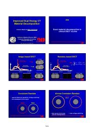
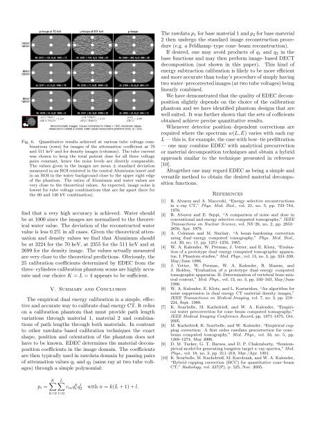
Fig. 6. Quantitative results achieved at various tube voltage combinations(rows) <strong>for</strong> images of the attenuation coefficient at 70and 511 keV and <strong>for</strong> density images (columns). The tube currentwas chosen to keep the total patient dose <strong>for</strong> all three voltagepairs constant, hence the noise levels are directly comparable.The values given in the images are mean ± standard deviationmeasured in an ROI centered in the central Aluminum insert andin an ROI in the water background close to the upper right edgeof the phantom. The ratios of Aluminum and water values arevery close to the theoretical values. As expected, image noise islowest <strong>for</strong> tube voltage combinations that are far apart (here <strong>for</strong>the 80 and 140 kV combination).find that a very high accuracy is achieved. Water shouldbe at 1000 since the images are normalized to the theoreticalwater value. The deviation of the reconstructed watervalue is less 0.2% in all cases. Given the theoretical attenuationand density values we find that Aluminum shouldbe at 3224 <strong>for</strong> the 70 keV, at 2355 <strong>for</strong> the 511 keV and at2699 <strong>for</strong> the density image. The values actually measuredare very close to the theoretical predictions. Obviously, the25 calibration coefficients determined by <strong>EDEC</strong> from thethree–cylinders calibration phantom scans are highly accurateand our choice K = L = 4 appears to be sufficient.V. Summary and ConclusionThe empirical dual energy calibration is a simple, effectiveand accurate way to calibrate dual energy CT. It relieson a calibration phantom that must provide path lengthvariations through material 1, material 2 and combinationsof path lengths through both materials. In contrastto other rawdata–based calibration techniques the exactshape, position and orientation of the phantom does nothave to be known. <strong>EDEC</strong> determines the material decompositioncoefficients in the image domain. The coefficientsare then typically used in rawdata domain by passing pairsof attenuation values q 1 and q 2 (same ray at two tube voltages)through a simple polynomial:The rawdata p 1 <strong>for</strong> base material 1 and p 2 <strong>for</strong> base material2 then undergo the standard image reconstruction procedure(e.g. a Feldkamp–type cone–beam reconstruction).If desired, one may avoid products of q 1 and q 2 in thebase functions and may then per<strong>for</strong>m image–based DECTdecomposition (not shown in this paper). This kind ofenergy subtraction calibration is likely to be more efficientand more accurate than today’s procedure of simply havingtwo water–precorrected images (at two tube voltages) beinglinearly combined.We have demonstrated that the quality of <strong>EDEC</strong> decompositionslightly depends on the choice of the calibrationphantom and we have identified phantom designs that arewell suited. It was further shown that the sets of cofficientsobtained achieve precise quantitative results.Whenever detector position–dependent corrections arerequired where the spectrum w(L, E) varies with each rayL — this is, <strong>for</strong> example, the case with bow–tie prefiltration— one may combine <strong>EDEC</strong> with analytical precorrectionor material decomposition techniques and obtain a hybridapproach similar to the technique presented in reference[10].Altogether one may regard <strong>EDEC</strong> as being a simple andversatile method to obtain the desired material decompositionfunctions.References[1] R. Alvarez and A. Macovski, “<strong>Energy</strong>–selective reconstructionsin x–ray CT,” Phys. Med. Biol., vol. 21, no. 5, pp. 733–744,1976.[2] R. Alvarez and E. Seppi, “A comparison of noise and dose inconventional and energy selective computed tomography,” IEEETransactions on Nuclear Science, vol. NS–26, no. 2, pp. 2853–2856, Apr. 1979.[3] A. Coleman and M. Sinclair, “A beam–hardening correctionusing dual–energy computed tomography,” Phys. Med. Biol.,vol. 30, no. 11, pp. 1251–1256, 1985.[4] W. A. Kalender, W. Perman, J. Vetter, and E. Klotz, “Evaluationof a prototype dual–energy computed tomographic apparatus.I. Phantom studies,” Med. Phys., vol. 13, no. 3, pp. 334–339,May/June 1986.[5] J. Vetter, W. Perman, W. A. Kalender, R. Mazess, andJ. Holden, “Evaluation of a prototype dual–energy computedtomographic apparatus. II. Determination of vertebral bone mineralcontent,” Med. Phys., vol. 13, no. 3, pp. 340–343, May/June1986.[6] W. A. Kalender, E. Klotz, and L. Kostaridou, “An algorithm <strong>for</strong>noise suppression in dual energy CT material density images,”IEEE Transactions on Medical Imaging, vol. 7, no. 3, pp. 218–224, Sept. 1988.[7] K. Sourbelle, M. Kachelrieß, and W. A. Kalender, “<strong>Empirical</strong>water precorrection <strong>for</strong> cone–beam computed tomography,”IEEE Medical Imaging Conference Record, pp. 1871–1875, Oct.2005.[8] M. Kachelrieß, K. Sourbelle, and W. Kalender, “<strong>Empirical</strong> cuppingcorrection: A first order rawdata precorrection <strong>for</strong> cone–beam computed tomography,” Med. Phys., vol. 33, no. 5, pp.1269–1274, May 2006.[9] D. M. Tucker, G. T. Barnes, and D. P. Chakraborty, “Semiempiricalmodel <strong>for</strong> generating tungsten target x–ray spectra,” Med.Phys., vol. 18, no. 3, pp. 211–218, Mar./Apr. 1991.[10] K. Sourbelle, M. Kachelrieß, M. Karolczak, and W. A. Kalender,“Hybrid cupping correction (HCC) <strong>for</strong> quantitative cone–beamCT,” Radiology, vol. 237(P), p. 525, Nov. 2005.K∑ L∑p i = c in q1q k 2 l with n = k(L + 1) + l.k=0 l=0