Fall 2008 - Cleveland Clinic Lerner Research Institute
Fall 2008 - Cleveland Clinic Lerner Research Institute
Fall 2008 - Cleveland Clinic Lerner Research Institute

You also want an ePaper? Increase the reach of your titles
YUMPU automatically turns print PDFs into web optimized ePapers that Google loves.
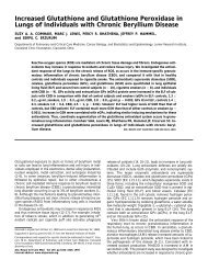
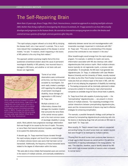
<strong>Lerner</strong> research instituteThe Self-Repairing BrainMore than 15 years ago, Bruce Trapp, PhD, Chair, Neurosciences, created an approach to studying multiple sclerosis(MS). Rather than being confined to investigating the disease in animals, Dr. Trapp wanted to see how MS actuallydevelops and progresses in the human brain. He started an innovative autopsy program to collect the brains andcentral nervous systems of multiple sclerosis patients after they die.“The rapid autopsy program allowed us to study MS by studyingthe disease itself, not a ‘near second’ in animals. This is muchmore relevant than investigating aspects of the disease in animalmodels,” he said. “In essence, what’s happening in the brainsof MS patients is the only thing that matters.”The approach yielded surprising insights that at the timequestioned conventional wisdom about the cause of permanentneurological disability in MS patients, how neuronal tissue isdamaged in MS brains, and whether or not brain cells andtissues can regenerate.White matter neurons are increased in a subset ofchronic multiple sclerosis lesions. Image shows a highmagnification view of post mortem tissue from amultiple sclerosis patient stained with an antibodythat recognizes neurons (microtubule associatedprotein 2, MAP2). The green line delimits a lesionwith increased neurons (top) from the surrounding,non-lesion white matter (bottom). (Photo: BruceTrapp, PhD, Chair, Neurosciences)“Some of our initialobservations were controversialat the time, but they’ve becomeaccepted by neurologists. Ourwork really created a paradigmshift regarding the pathogenesisof permanent neurologicaldisability, tissue damage andregeneration in MS brains,”Dr. Trapp said.Multiple sclerosis is aninflammatory disease thatdamages the insulation (myelin)that surrounds the nerve fibersof the central nervous systemand is the most common causeof neurologic disability in youngadults. Most patients have progressive neurologic deteriorationthat was thought to be caused by the loss of myelin. The axons,or nerve fibers that transmit electrical impulses, were thoughtto be spared this destructive process.A decade ago, Dr. Trapp examined tissues donated throughthe rapid autopsy program and found that a consistent featureof multiple sclerosis lesions was axons that had been cut (ortransected). Additionally, the frequency of these transected axonsrelated to the degree of inflammation within the lesion.“We were able to show initially that transected axons are commonin the lesions of multiple sclerosis, and eventually established arelationship between axonal loss and neurodegeneration withirreversible neurologic impairment in individuals with MS,”Dr. Trapp said. “This was an understanding of the diseaseprocess that contributed to new research in the area.”Other breakthroughs would emerge from the rapid autopsyprogram. For example, in addition to myelin and axons,inflammation associated with MS also destroys cells calledoligodendrocytes that produce myelin. Most multiple sclerosislesions typically do not regenerate myelin, a process calledremyelination. To remyelinate, the MS brain must generate newoligodendrocytes. Dr. Trapp, in collaboration with Case WesternReserve University and the University of Toledo, recently received$3 million by the Ohio Third Frontier Commission to develop smallmolecules that can enhance repair of the brain in MS, with thegoal of not only delaying the progression of disability but reversingit. The leading compounds will be chemically optimized to obtaincompounds suitable for licensing by major pharmaceuticalcompanies as candidate drugs for future clinical trials in patients.Dr. Trapp found that cells capable of producing myelin — the“premyelinating” oligodendrocytes — are present in chroniclesions of multiple sclerosis. “Our expanding knowledge of thecellular interactions between premyelinating oligodendrocytes,axons and the microenvironment of lesions may lead to effectivestrategies for enhancing remyelination.“For example, a possible therapeutic strategy could perhaps beachieved by transplanting oligodendrocyte-producing cells intothe lesions or developing drugs that will promote an MS brain torepair itself,” Dr. Trapp said.More recently, Dr. Trapp’s research team made an unexpectedand exciting finding: the adult human brain can replace neuronsthat were thought to be destroyed by multiple sclerosis.“Proving that the adult human brain produces new neurons isa tough egg to crack because we cannot perform the definitiveexperiments of injecting radioisotopes in the living patients,” hesaid. “The objective, therefore, was to identify areas of the MSbrain which had increased densities of neurons when comparedto normal brains.”8