Shell Morphology of the Egyptian Tortoise, Testudo kleinmanni ...
Shell Morphology of the Egyptian Tortoise, Testudo kleinmanni ...
Shell Morphology of the Egyptian Tortoise, Testudo kleinmanni ...

Create successful ePaper yourself
Turn your PDF publications into a flip-book with our unique Google optimized e-Paper software.
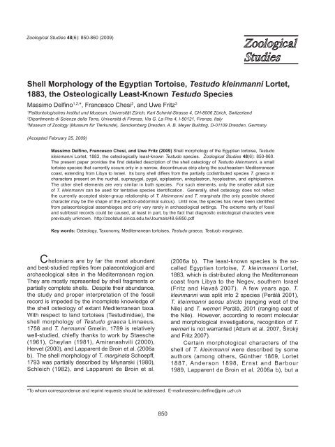
Delfino et al. – <strong>Shell</strong> <strong>Morphology</strong> <strong>of</strong> <strong>the</strong> <strong>Egyptian</strong> <strong>Tortoise</strong> 851comprehensive description was never published.Perhaps because <strong>of</strong> this, remains <strong>of</strong> T. <strong>kleinmanni</strong>have never been identified in palaeontologicalassemblages (Lapparent de Broin 2000) and arereported only very rarely in archaeological settings(von den Driesch and Boessneck 1985, Boessneck1988), even in sites within its current range.Interestingly, VM Chkhikvadze (in Gabashvili et al.2000) suggested that T. burtschaki Chkhikvadze,1975 from <strong>the</strong> Miocene <strong>of</strong> Georgia could bephylogenetically related to T. <strong>kleinmanni</strong>. Thisstatement, which was not supported by anyargument, is ra<strong>the</strong>r surprising because Gabashviliet al. (2000) proposed that T. burtschaki ischaracterized by <strong>the</strong> absence <strong>of</strong> a hypoxiphiplastralhinge, in contrast to T. <strong>kleinmanni</strong>.However, Danilov (2005) and Chkhikvadze (20062007) treated T. burtschaki as a representative <strong>of</strong><strong>the</strong> <strong>Testudo</strong> sensu stricto group, which suggests<strong>the</strong> presence <strong>of</strong> a plastral hinge. This contradictorysituation underlines <strong>the</strong> need for better knowledge<strong>of</strong> osteological features <strong>of</strong> <strong>the</strong> shells <strong>of</strong> all extant<strong>Testudo</strong> species in order to assess relationships <strong>of</strong>fossil taxa.The present study provides <strong>the</strong> first detailedosteological description <strong>of</strong> <strong>the</strong> shell <strong>of</strong> T.<strong>kleinmanni</strong>, which should allow <strong>the</strong> identification<strong>of</strong> palaeontological and archaeological remains.Particular emphasis is given to distinctions withT. graeca, <strong>the</strong> only codistributed extant tortoisespecies (Schleich et al. 1996, Bouskila and Amitai2001).MATERIALS AND METHODSOur osteological description <strong>of</strong> T. <strong>kleinmanni</strong>is based on 8 shells in <strong>the</strong> collection <strong>of</strong> <strong>the</strong>Museum <strong>of</strong> Zoology Dresden (Museum fürTierkunde Dresden = MTD: MTD D 26762, 32832,35692, 38650, 39221, 40289, 44284, and 44285;see Appendix for sex, shell lengths, and neuralformulae). With 1 exception (MTD D 26762; with asmall pleuro-peripheral fontanellae on 1 side only),all specimens were adults. In addition, <strong>the</strong> externalshell morphology as well as <strong>the</strong> position andrelationships <strong>of</strong> <strong>the</strong> scute sulci were studied in <strong>the</strong>following 41 alcohol-preserved specimens: MTDD 12469, 26016, 26017, 26850, 28286, 28287,29119, 30855, 31125, 31598, 31803, 32434,32435, 32710, 32742, 32798, 32831, 34857,35074, 35075, 36797, 36897-36907, 37473,39220, 40166, 42492, 45729, and 46938-46941.Osteological and alcohol-preserved specimens <strong>of</strong>T. graeca, T. hermanni, and T. marginata, housedin <strong>the</strong> same and o<strong>the</strong>r collections were used forcomparison. Anatomical nomenclature followedLapparent de Broin (2001), while <strong>the</strong> taxonomyfollowed Fritz and Havaš (2007).Description <strong>of</strong> <strong>the</strong> shell <strong>of</strong> T. <strong>kleinmanni</strong>In order to provide characters suitablefor <strong>the</strong> identification <strong>of</strong> palaeontological andarchaeological remains, <strong>the</strong> following descriptionis organized according to <strong>the</strong> bony shell elementsthat are usually preserved.Carapace(Figs. 1a, c, 2)The carapace is highly domed and reaches amaximum length <strong>of</strong> 144 mm (Farkas et al. 1997).Males are smaller than females, and reach amaximum length <strong>of</strong> only 100-106 mm accordingto Schleich et al. (1996) or 130 mm according toBaha el Din (2006). Our largest males and femalesdo not surpass <strong>the</strong>se lengths. In comparison too<strong>the</strong>r <strong>Testudo</strong> species, <strong>the</strong> carapacial and plastronelements <strong>of</strong> T. <strong>kleinmanni</strong> are quite thick; however,this character varies in different specimens.Nuchal: The nuchal is generally widerthan long and characteristically notched; <strong>the</strong>area covered by <strong>the</strong> cervical, usually triangularin shape, always distinctly protrudes from <strong>the</strong>anterior pr<strong>of</strong>ile <strong>of</strong> <strong>the</strong> nuchal; it is noteworthythat this character already occurs in our smallestspecimen (MTD D 26762; with a carapace length<strong>of</strong> 70 mm). Lapparent de Broin et al. (2006a: 292)stated that “<strong>the</strong> notch affects all <strong>the</strong> anterior border<strong>of</strong> <strong>the</strong> shell (nuchal and first peripherals) due to<strong>the</strong> apomorphic elongation <strong>of</strong> <strong>the</strong> peripheralsadjacent to <strong>the</strong> nuchal: <strong>the</strong> cervical is present,triangular, posteriorly wide, sometimes narrowedbut not absent … and protuberant in <strong>the</strong> middle<strong>of</strong> <strong>the</strong> notch”. The anterior edge <strong>of</strong> <strong>the</strong> nuchal is,<strong>the</strong>refore, W-shaped. The triple junction among<strong>the</strong> 1st marginal, vertebral, and pleural is alwayspresent on <strong>the</strong> nuchal and always relatively farfrom its lateral edge.Neural series: Seven or, more rarely, 8elements represent <strong>the</strong> neural series (Appendix).In a few specimens (MTD D 32832 and 40289),<strong>the</strong>y are alternatively rectangular and octagonalfrom <strong>the</strong> 1st to <strong>the</strong> 6th, whereas <strong>the</strong> 7th ishexagonal. Irregularities frequently occur, mostlyin <strong>the</strong> posterior sector <strong>of</strong> <strong>the</strong> series, and several
852Zoological Studies 48(6): 850-860 (2009)hexagonal elements with small posterolateraledges may be present (Appendix). The 1st,3rd, 5th, and 7th neurals are encroached by <strong>the</strong>intervertebral sulci. The ventral surface <strong>of</strong> allneural elements bears traces <strong>of</strong> <strong>the</strong> connectionwith <strong>the</strong> vertebrae.Suprapygal: All available specimens possessonly 1 suprapygal, which is approximatelyhexagonal (<strong>the</strong> posterolateral edges are veryshort), or in 1 case nearly trapezoidal (MTD D(a)ce(b)num3ne2pl3c3v4per10sppyg(c)scau(d)ceepiguaxentohumhyopechypoabdingxiphifemanFig. 1. Schematic drawings <strong>of</strong> <strong>the</strong> shell <strong>of</strong> <strong>Testudo</strong> <strong>kleinmanni</strong> Lortet, 1883. (a, b) male carapace in dorsal view and plastron in ventralview; (c, d) female carapace in dorsal view and plastron in ventral view. Thick lines represent scute sulci; thin lines, bony sutures. Bonyplates: ento, entoplastron; epi, epiplastron; hyo, hyoplastron; hypo, hypoplastron; ne2, 2nd neural; nu, nuchal; per10, 10th peripheral;pl3, 3rd pleural; pyg, pygal; sp, suprapygal; xiphi, xiphiplastron. Horny shields: abd, abdominal; an, anal; ax, axillary; c3, 3rd costal;ce, cervical; fem, femoral; gu, gular; hum, humeral; ing, inguinal; m3, 3rd marginal; pec, pectoral; scau, supracaudal; v4, 4th vertebral.Scale bar = 10 mm.pyg
Delfino et al. – <strong>Shell</strong> <strong>Morphology</strong> <strong>of</strong> <strong>the</strong> <strong>Egyptian</strong> <strong>Tortoise</strong> 85339221). The fusion <strong>of</strong> <strong>the</strong> suprapygals into a singleelement was also reported by Lapparent de Broinet al. (2006b).Pleural series: The 8 pleurals are trapezoidaland show <strong>the</strong> typical <strong>Testudo</strong> alternation <strong>of</strong> <strong>the</strong>pleurals with a wide proximal edge and a narrowdistal edge separated by pleurals in <strong>the</strong> oppositecondition. There is some variation, however, asin MTD D 38650. In this specimen, <strong>the</strong> 2nd to7th pleurals have approximately <strong>the</strong> same shape(note that <strong>the</strong> neural formula, given in Appendix,in this specimen is anomalous with 4 contiguoushexagonal elements). All even pleurals host<strong>the</strong> intercostal sulci. The visceral surface <strong>of</strong> <strong>the</strong>pleurals is characterized by an elongated convexity(transverse in strict anatomical terms, longitudinal if<strong>the</strong> main axis <strong>of</strong> <strong>the</strong> isolated pleural is considered).The proximal visceral area <strong>of</strong> <strong>the</strong> pleurals developsan irregular process contacting <strong>the</strong> vertebrae; in<strong>the</strong> 1st and last pleurals, <strong>the</strong> processes are moredeveloped than in <strong>the</strong> o<strong>the</strong>rs; <strong>the</strong> ones <strong>of</strong> <strong>the</strong> lastpleurals show a flattened facet which contacts <strong>the</strong>dorsal edge <strong>of</strong> <strong>the</strong> ilium.Peripheral series: The 3rd to 7th peripheralsform <strong>the</strong> bridge, <strong>the</strong> bony junction <strong>of</strong> <strong>the</strong> carapaceand plastron. In some cases, <strong>the</strong> external surface<strong>of</strong> <strong>the</strong> 1st (MTD D 39221) or <strong>the</strong> 1st 2 peripherals(MTD D 38650) is crossed by <strong>the</strong> costo-marginalsulcus which o<strong>the</strong>rwise corresponds (along <strong>the</strong>entire series, including <strong>the</strong> pygal-suprapygal) to<strong>the</strong> pleuro-peripheral suture. The 3rd peripheralhosts <strong>the</strong> sulcus which corresponds to <strong>the</strong> hornyaxillary shield, whereas <strong>the</strong> 7th hosts <strong>the</strong> onecorresponding to <strong>the</strong> inguinal shield (note thatexceptionally up to 2 inguinal shields can occur;Schleich et al. 1996). All peripherals are crossedby 1 intermarginal sulcus. The visceral surface<strong>of</strong> <strong>the</strong> 3rd and 7th peripherals is characterizedby <strong>the</strong> presence <strong>of</strong> an elongated scar <strong>of</strong> <strong>the</strong>dorsal projections <strong>of</strong> <strong>the</strong> hyo- and hypoplastron,respectively (<strong>the</strong> axillary and inguinal processes);<strong>the</strong> projection <strong>of</strong> <strong>the</strong> hyoplastron reaches <strong>the</strong>posterodorsal edge <strong>of</strong> <strong>the</strong> 2nd peripheral. The2nd and 3rd peripherals as well as <strong>the</strong> 7th to 11thperipherals can have a slight dorsally raised edge;at <strong>the</strong> same height, corresponding to this edge,<strong>the</strong> peripherals involved in <strong>the</strong> bridge can developa weak ridge; <strong>the</strong> entity <strong>of</strong> this raised edge andridge is slightly amplified by <strong>the</strong> horny shields.The edge <strong>of</strong> <strong>the</strong> 11th peripheral regularly showsa notch corresponding to <strong>the</strong> lateral margin <strong>of</strong> <strong>the</strong>supracaudal shield; this change in orientation <strong>of</strong><strong>the</strong> outline gives <strong>the</strong> ‘pointed’ appearance to <strong>the</strong>posterior edge <strong>of</strong> <strong>the</strong> carapace as described below.Pygal: The pygal plate is generally trapezoidal;in 1 case (MTD D 40289), it is hexagonalwith short anterolateral sides. Its dorsal surfaceis moderately convex to nearly flat, whereas <strong>the</strong>ventral surface is regularly concave in both sexeswith a gutter-like appearance (Fig. 2). On <strong>the</strong> pygalplate <strong>of</strong> all skeletonized specimens, no sagittalgroove is present which should be expected when<strong>the</strong> horny supracaudal scute is divided. However,divided supracaudals occur in 15 <strong>of</strong> <strong>the</strong> 41 alcoholpreservedspecimens (from both Egypt andLibya). In most cases, <strong>the</strong> division occurs only in<strong>the</strong> proximal sector <strong>of</strong> <strong>the</strong> external surface <strong>of</strong> <strong>the</strong>supracaudal, and very rarely on <strong>the</strong> internal one(as in MTD D 31125 and 32434, where just a hint<strong>of</strong> division is present). This character state waspreviously mentioned by Lortet (1887). Lapparentde Broin et al. (2006b) reported <strong>the</strong> presence <strong>of</strong>divided supracaudals in 14 <strong>of</strong> 67 specimens. Itseems that in <strong>the</strong> cases in which <strong>the</strong> supracaudalis divided, <strong>the</strong> sulcus between <strong>the</strong> last vertebraland supracaudal is not approximately straightbut anteriorly bi-convex. The lateral margins <strong>of</strong><strong>the</strong> pygal are generally straight (slightly concaveonly in MTD D 44285). The posterior edge <strong>of</strong> thiselement is variably pointed (see below for <strong>the</strong>general shape <strong>of</strong> <strong>the</strong> posterior region <strong>of</strong> this area),and this character tends to develop with age. Thepygal is not pointed in very small specimens, butweakly pointed in a specimen with a shell length<strong>of</strong> 70 mm (MTD D 26762), and markedly pointedin larger specimens (with <strong>the</strong> most pronouncedcondition in <strong>the</strong> largest specimen, MTD D 44285, afemale with a shell length <strong>of</strong> 133 mm).Plastron(Figs. 1b, d, 3)The plastral formula can be summarized asfollows: abd >> an > hum >< pect ≥ gul > fem.This slightly differs from <strong>the</strong> formulae reportedby Ernst and Barbour (1989; abd > an > pect > gul > fem) and Loveridge and Williams(1957; abd > [gu, hum, pec, an subequal] > fem)because our samples display some variability. Ina few cases, <strong>the</strong> gulars can be nearly as long as<strong>the</strong> pectorals. The anterior lobe <strong>of</strong> <strong>the</strong> plastron isapically curved in <strong>the</strong> dorsal direction. In adults, ahypo-xiphiplastral hinge is present.Epiplastron: This element is characteristicallyshort in <strong>the</strong> anteroposterior direction. In mostcases, <strong>the</strong> area covered by <strong>the</strong> gulars protrudesfrom <strong>the</strong> pr<strong>of</strong>ile <strong>of</strong> <strong>the</strong> anterior plastral lobe. The
854Zoological Studies 48(6): 850-860 (2009)epiplastral dorsal pad (corresponding to <strong>the</strong>dorsal fold <strong>of</strong> <strong>the</strong> gular; Fig. 3) is approximatelytrapezoidal, as long as wide, or slightly longerthan wide (as in MTD D 44285); it is moderatelydeveloped in <strong>the</strong> posterodorsal direction, sothat it barely reaches <strong>the</strong> anterior edge <strong>of</strong> <strong>the</strong>entoplastron (as in MTD D 32832) or slightlyoverhangs it (as in MTD D 44285). Due to suchmodest development, in most cases, a smallepiplastral pocket is present (particularly welldeveloped in MTD D 44285), but just a shallowand wide depression is visible in o<strong>the</strong>rs (as in MTDD 32832). The anterior edge <strong>of</strong> <strong>the</strong> pad generallydevelops a tubercle-like structure at its lateraledge, which is already developed in subadults (asin MTD D 26762). The dorsal surface between<strong>the</strong> pair <strong>of</strong> tubercles <strong>of</strong> <strong>the</strong> epiplastra is weaklyconcave, and its anterior margin is not rounded butacute (forming a ridge) and irregular in dorsal view.The ventral surface <strong>of</strong> <strong>the</strong> epiplastron (as well as<strong>of</strong> <strong>the</strong> entoplastron) covered by <strong>the</strong> gulars is not inrelief. As noted by Gün<strong>the</strong>r (1869), <strong>the</strong> sum <strong>of</strong> <strong>the</strong>posterior angles <strong>of</strong> <strong>the</strong> gulars, when seen in ventralview, is nearly equal to a right angle.Entoplastron: The entoplastron is roundedin external (ventral) view, but approximatelytriangular in visceral (dorsal) view. It is nearlyentirely located in <strong>the</strong> anterior lobe <strong>of</strong> <strong>the</strong> plastron.It is characterized by being partially covered by <strong>the</strong>gulars (generally extending to <strong>the</strong> anterior 1/3 <strong>of</strong><strong>the</strong> entoplastron). The entoplastron is not crossedby <strong>the</strong> humero-pectoral sulcus, but is slightly bentbecause it contributes, along with <strong>the</strong> hyoplastron,to <strong>the</strong> curvature to <strong>the</strong> anterior plastral lobe.Hyoplastron: The anterolateral region <strong>of</strong> <strong>the</strong>hyoplastron is markedly bent in <strong>the</strong> anterodorsaldirection. This paired element hosts <strong>the</strong> humeropectoraland <strong>the</strong> pectoro-abdominal sulci: <strong>the</strong>former is nearly straight whereas <strong>the</strong> latter isdistinctly curved (with a wide anterior convexity)so that in some cases (MTD D 38650), it nearlyreaches <strong>the</strong> hypo-hypoplastral suture, resembling<strong>the</strong> condition found in T. marginata. Theanterolateral projection <strong>of</strong> <strong>the</strong> hyoplastron hosts<strong>the</strong> sulci, which delimits <strong>the</strong> axillary shield.Hypoplastron: The hypoplastron is <strong>the</strong>largest plastral element. It is crossed by scutesulci only very close to its posterior border where<strong>the</strong> abdomino-femoral sulcus nearly coincideswith <strong>the</strong> hyo-xiphiplastral hinge. It is noteworthythat <strong>the</strong> abdomino-femoral sulcus is laterallyarched and <strong>the</strong>refore far from <strong>the</strong> hinge in <strong>the</strong>smallest skeleton available (MTD D 26762). TheepadsentohyoepihypoxiphiFig. 2. Posterior sector <strong>of</strong> <strong>the</strong> shell <strong>of</strong> <strong>Testudo</strong> <strong>kleinmanni</strong>Lortet, 1883 (MTD D 40289) showing <strong>the</strong> distinctly concaveventral surface <strong>of</strong> <strong>the</strong> pygal. Note <strong>the</strong> presence <strong>of</strong> horny shieldson <strong>the</strong> left 1/2 <strong>of</strong> shell (on <strong>the</strong> right in <strong>the</strong> picture). Scale bar =10 mm.Fig. 3. Schematic drawings <strong>of</strong> <strong>the</strong> morphology <strong>of</strong> <strong>the</strong> visceralsurface <strong>of</strong> <strong>the</strong> plastron <strong>of</strong> <strong>Testudo</strong> <strong>kleinmanni</strong> Lortet, 1883.Note that in this case, <strong>the</strong> epiplastral pads do not overhang<strong>the</strong> entoplaston. ento, entoplastron; epi, epiplastron; epads,epiplastral pads; hyo, hyoplastron; hypo, hypoplastron; xiphi,xiphiplastron. Scale bar = 10 mm.
Delfino et al. – <strong>Shell</strong> <strong>Morphology</strong> <strong>of</strong> <strong>the</strong> <strong>Egyptian</strong> <strong>Tortoise</strong> 855posterolateral corner <strong>of</strong> <strong>the</strong> hypoplastron hosts <strong>the</strong>sulci indicating <strong>the</strong> presence <strong>of</strong> <strong>the</strong> inguinal shield.Due to <strong>the</strong> development <strong>of</strong> <strong>the</strong> hinge and not asuture between <strong>the</strong> hypo- and <strong>the</strong> xiphiplastron,<strong>the</strong> posterior edge <strong>of</strong> <strong>the</strong> former and <strong>the</strong> anterioredge <strong>of</strong> <strong>the</strong> latter are smoo<strong>the</strong>r than <strong>the</strong> suturesbetween o<strong>the</strong>r plastral elements; a ventrallysloping surface develops on <strong>the</strong> visceral surfacealong <strong>the</strong> hinge, mostly in <strong>the</strong> lateral sector <strong>of</strong> <strong>the</strong>hypoplastron and xiphiplastron.Xiphiplastron: The ventral surface <strong>of</strong> <strong>the</strong>xiphiplastron is usually flat or nearly flat (seebelow under “Sexual dimorphism”). The dorsalarea covered by <strong>the</strong> horny shields is moderatelydeveloped. A deep anal notch occurs in bothsexes. The femoro-anal sulcus is generallystraight, and due to <strong>the</strong> fact that <strong>the</strong> femoralsare particularly short medially, it reaches <strong>the</strong>sagittal line quite near <strong>the</strong> anterior edge <strong>of</strong> <strong>the</strong>xiphiplastra, forming a markedly acute posteriorangle with that line. In our sample, <strong>the</strong> anal sutureusually is not “four times as long as <strong>the</strong> femoralsuture” (as stated by Anderson 1898: 28; see alsoLapparent de Broin et al. 2006a), but <strong>the</strong> areacovered by <strong>the</strong> femorals is generally wider thanthat covered by <strong>the</strong> anal shield. The lateral margin<strong>of</strong> <strong>the</strong> xiphiplastra frequently has a distinct stepcorresponding to <strong>the</strong> posterolateral end <strong>of</strong> <strong>the</strong>femoro-anal sulcus.Sexual dimorphismThe shape <strong>of</strong> <strong>the</strong> xiphiplastra is sexuallydimorphic being posteriorly wider in males thanin females. This difference is due to <strong>the</strong> medialedge <strong>of</strong> <strong>the</strong> xiphiplastra, which is longer in malesthan in females (compare Fig. 1b with 1d). Thewidth <strong>of</strong> <strong>the</strong> anal notch is similar in both sexes (butusually slightly larger in males than in females).The posterior tips <strong>of</strong> <strong>the</strong> xiphiplastra are slightlydirected downward in <strong>the</strong> male MTD D 38650. Asimilar, but weaker, character state occurs in <strong>the</strong>males MTD D 35692 and 39221, and perhaps isrelated to a weak, but evident, concavity locatedposteriorly to <strong>the</strong> femoro-anal sulcus along <strong>the</strong>lateral edge <strong>of</strong> <strong>the</strong> xiphiplastra (<strong>the</strong>refore between<strong>the</strong> sulcus and <strong>the</strong> posterior tip <strong>of</strong> <strong>the</strong> xiphiplastra).Le Berre (1989: 104) reported that “chez lefemelles âgées, le lobe postérieur du plastronprésente une charnière et est beaucoup plusmobile que chez les mâles”. This sentence wasapparently misinterpreted by Schleich et al. (1996:152) who, mentioning Le Berre (1989), wrote that<strong>the</strong> “plastral front lobe is movable” in very largefemales. The anterior plastral lobe <strong>of</strong> <strong>the</strong> largefemale MTD D 44285 (shell 133 mm) is firmlyattached to <strong>the</strong> rest <strong>of</strong> <strong>the</strong> plastron as in all o<strong>the</strong>rskeletonized and alcohol-preserved specimens atour disposal. Schleich et al. (1996: 152) reportedthat “males … show a prominently domed pygalwith a visceral trough” and that “<strong>the</strong> pygal isviscerally prominently curved in males”; but again,according to <strong>the</strong> sample at our disposal, both <strong>the</strong>external curvature and depth <strong>of</strong> <strong>the</strong> internal groove(= trough) do not significantly differ between <strong>the</strong> 2sexes (so that an isolated pygal cannot be referredto 1 sex or to <strong>the</strong> o<strong>the</strong>r). The plastron <strong>of</strong> male T.<strong>kleinmanni</strong> is quite flat and does not develop aconcavity like in many o<strong>the</strong>r testudinid species(including T. graeca).DISCUSSIONComparative morphologyAccording to our observations, neurals,pleurals, marginals, entoplastron, and hypoplastra<strong>of</strong> T. <strong>kleinmanni</strong> are not easily distinguishable from<strong>the</strong> only o<strong>the</strong>r species currently co-occurring withit, T. graeca. Extant specimens <strong>of</strong> both speciesdiffer in size in that T. graeca achieves a distinctlylarger shell length, so that it could be speculatedthat isolated large shell elements, belonging totortoises with shell lengths <strong>of</strong> > 14-15 cm, shouldrepresent T. graeca. However, except for <strong>the</strong>thickness and size, <strong>the</strong> following shell elementscan be identified at <strong>the</strong> species level with variousdegrees <strong>of</strong> precision: <strong>the</strong> nuchal, suprapygal,pygal, epiplastra, hyoplastra, and xiphiplastra (seeTable 1 for a summary <strong>of</strong> <strong>the</strong> major differencesbetween T. <strong>kleinmanni</strong> and T. graeca). In T.<strong>kleinmanni</strong>, <strong>the</strong> nuchal is deeply notched and withan anterior W-shaped edge (due to <strong>the</strong> protrudingcervical element); even if <strong>the</strong> anterior edge <strong>of</strong><strong>the</strong> carapace is concave in some T. graeca (asin MTD D 44856 from Tunisia and MTD D 11163from Georgia), a W-shaped notch (with <strong>the</strong> medialtriangular area covered by a distinctly protrudingcervical) never occurs. Moreover, <strong>the</strong> lateralcorner <strong>of</strong> <strong>the</strong> nuchal is largely covered by <strong>the</strong> 1stcostal shield in T. <strong>kleinmanni</strong> but not in T. graeca,where <strong>the</strong> triple junction among <strong>the</strong> 1st vertebral,costal, and marginal usually coincides (or nearlyso) with <strong>the</strong> edge <strong>of</strong> <strong>the</strong> nuchal (compare Figs. 1with 4a and 4b).The fact that <strong>the</strong> 2 suprapygals are alwaysfused into a single element in T. <strong>kleinmanni</strong> and
856Zoological Studies 48(6): 850-860 (2009)frequently separated in T. graeca (Lapparent deBroin et al. 2006a b; Fig. 4c) allows all separatedsuprapygals to be referred with certainty to <strong>the</strong>latter, but fused ones cannot unequivocally bereferred to <strong>the</strong> former.The pygal is always characteristicallyventrally concave in T. <strong>kleinmanni</strong>, whereas it isflat in T. graeca (and o<strong>the</strong>r <strong>Testudo</strong> species; for T.graeca see Fig. 4d). Moreover, in dorsal view, <strong>the</strong>pygal is vaguely V-shaped and pointed in adults<strong>of</strong> T. <strong>kleinmanni</strong> (and <strong>the</strong>refore all <strong>of</strong> <strong>the</strong> posteriorregion <strong>of</strong> <strong>the</strong> carapace is typically convex) butmore rectangular and never pointed in T. graeca(Figs. 4d, e). Due to <strong>the</strong> possible division <strong>of</strong> <strong>the</strong>supracaudal shield, a sagittal sulcus should bepresent in some cases, but such morphology wasnot seen in our sample.The epiplastra <strong>of</strong> T. <strong>kleinmanni</strong> and T. graecaseem to slightly differ in that <strong>the</strong> epiplastron <strong>of</strong> T.<strong>kleinmanni</strong> is anteroposteriorly shorter, and, aboveall, possesses pointed tubercles which delimita slightly concave dorsal surface; dorsal padsare only moderately developed. In comparativespecimens <strong>of</strong> T. graeca, <strong>the</strong> pads are moredeveloped (Fig. 4f) and <strong>the</strong> tubercles, whenpresent at all (as in MTD D 39616 and 44856),do not seem to be so pointed and do not delimit aconcave surface.The entoplastron <strong>of</strong> T. <strong>kleinmanni</strong> is notcrossed by <strong>the</strong> humero-pectoral sulcus, whereasthis may be <strong>the</strong> case in T. graeca (e.g., <strong>the</strong> juvenileMTD D 26763 from Libya).The hyoplastron <strong>of</strong> T. <strong>kleinmanni</strong> is characterizedby a nearly straight humero-pectoralsulcus and a deeply (anteriorly) convex pectoroabdominalsulcus; in T. graeca, <strong>the</strong> humeropectoralsulcus is anteriorly concave and <strong>the</strong>pectoro-abdominal sulcus is never so convex (Fig.Table 1. Comparison <strong>of</strong> major differences in shell morphology <strong>of</strong> adult <strong>Testudo</strong> <strong>kleinmanni</strong> Lortet, 1883 and T.graeca Linnaeus, 1758 (size differences not mentioned)T. <strong>kleinmanni</strong> T. graecaNuchal- deeply notched- anterior edge markedly W-shaped- covered by <strong>the</strong> 1st costal shield- usually not deeply notched- anterior edge not markedly W-shaped- not covered by <strong>the</strong> 1st costal shieldSuprapygals- always fused - frequently separatedPygal- V-shaped- posteriorly fairly pointed in dorsal view- always characteristically ventrally concaveEpiplastron- anteroposteriorly short- moderately developed dorsal pads not delimiting a deep pocketand not significantly overhanging <strong>the</strong> entoplastron in dorsal view- presence <strong>of</strong> pointed tubercles usually delimiting a dorsal slightlyconcave surfaceEntoplastron- nearly completely located in <strong>the</strong> anterior lobe <strong>of</strong> <strong>the</strong> plastron- covered only by gular and humeralHyoplastron- humero-pectoral sulcus nearly straight- deeply (anteriorly) convex pectoro-abdominal sulcusXiphiplastron- femoro-anal sulcus usually straight- femoral scutes medially short (so that <strong>the</strong> femoro-anal sulcusmedially approaches <strong>the</strong> anterior edge <strong>of</strong> <strong>the</strong> bone)- not V-shaped- never posteriorly pointed in dorsal view- fairly flat ventrally- anteroposteriorly long- well-developed dorsal pads delimiting a deep pocket and usuallyoverhanging <strong>the</strong> entoplastron in dorsal view- tubercles, if present, not so pointed and not delimiting a dorsalconcave surface- not completely located in <strong>the</strong> anterior lobe <strong>of</strong> <strong>the</strong> plastron- covered by gular, humeral, and sometimes also by pectoral- humero-pectoral sulcus anteriorly concave- pectoro-abdominal sulcus not deeply (anteriorly) convex- femoro-anal sulcus usually sinuous- femorals scutes medially long (femoro-anal sulcus not mediallyapproaching <strong>the</strong> anterior edge <strong>of</strong> <strong>the</strong> bone)
Delfino et al. – <strong>Shell</strong> <strong>Morphology</strong> <strong>of</strong> <strong>the</strong> <strong>Egyptian</strong> <strong>Tortoise</strong> 8574g).The xiphiplastron <strong>of</strong> T. <strong>kleinmanni</strong> is crossedby <strong>the</strong> femoro-anal sulcus which is nearly straightand more anteromedially directed than in T. graeca(Fig. 4d). The area covered by <strong>the</strong> femoral shieldis usually much shorter in T. <strong>kleinmanni</strong> than in T.graeca. It is noteworthy that <strong>the</strong> xiphiplastra are<strong>the</strong> only shell elements showing moderate sexualdimorphism.According to Lortet (1887), <strong>the</strong> thickness <strong>of</strong>(a)ce(b)cenunu(c)(d)sp Isp IIxiphifempyganpyg(e)(f)epad(g)humhyopecabdFig. 4. Details <strong>of</strong> <strong>the</strong> shell morphology <strong>of</strong> <strong>Testudo</strong> graeca Linnaeus, 1758 mentioned in <strong>the</strong> text: (a) anterior carapace <strong>of</strong> MTD D 3943in dorsal view, note <strong>the</strong> absence <strong>of</strong> a medial notch; (b) same region <strong>of</strong> MTD D 11163, note <strong>the</strong> wide notch which is not W-shaped (because<strong>the</strong> area corresponding to <strong>the</strong> cervical shield does not protrude anteriorly); (c) posterior carapace <strong>of</strong> MTD D 11163 in posterior view,note <strong>the</strong> 2 suprapygal elements (sutures marked with a black line) and approximately rectangular pygal; (d) <strong>the</strong> same area in ventralview, note that <strong>the</strong> visceral side <strong>of</strong> <strong>the</strong> pygal is nearly flat and that <strong>the</strong> sinuous femoro-anal suture (marked with a black line) does notapproach <strong>the</strong> anterior edge <strong>of</strong> <strong>the</strong> xiphiplastron (marked with a black line); (e) posterior carapace <strong>of</strong> MTD D 3943, note <strong>the</strong> absence <strong>of</strong><strong>the</strong> posteriorly protruding triangular pygal which characterizes T. <strong>kleinmanni</strong>; (f) left lateral view <strong>of</strong> <strong>the</strong> carapace <strong>of</strong> MTD D 11163, note<strong>the</strong> well-developed epiplastral pads (much better developed than in o<strong>the</strong>r specimens <strong>of</strong> <strong>the</strong> same species); (g) ventral view <strong>of</strong> anteriorplastron <strong>of</strong> same specimen, note <strong>the</strong> anteriorly concave humero-pectoral suture and weakly curved pectoro-abdominal suture (bothmarked with a black line). Bony plates and structures: epad, epiplastral pad; hyo, hyoplastron; nu, nuchal; pyg, pygal; sp I, suprapygalI; sp II, suprapygal II; xiphi, xiphiplastron. Horny shields: abd, abdominal; an, anal; ce, cervical; fem, femoral; hum, humeral; pec,pectoral. Specimens not to scale.
858Zoological Studies 48(6): 850-860 (2009)<strong>the</strong> shell <strong>of</strong> T. <strong>kleinmanni</strong>, if compared to its smallsize, is sufficiently diagnostic to distinguish it fromall o<strong>the</strong>r Mediterranean <strong>Testudo</strong> species. However,considering that <strong>the</strong> thickness <strong>of</strong> <strong>the</strong> shell is quitevariable in fossil populations (for T. hermanni, seefor example Lapparent de Broin et al. 2006b) andthat this character is not always easily assessableon <strong>the</strong> basis <strong>of</strong> isolated shell fragments, <strong>the</strong>identification <strong>of</strong> shell remains should not be basedon shell thickness alone.Notes on <strong>the</strong> phylogenetic relationships <strong>of</strong>T. <strong>kleinmanni</strong>Based mainly on craniological characters,Loveridge and Williams (1957) placed T.k l e i n m a n n i i n t h e m o n o t y p i c s u b g e n u sPseudotestudo, as opposed to <strong>the</strong> subgenus<strong>Testudo</strong> comprising T. graeca, T. hermanni, T.horsfieldii, and T. marginata. However, accordingto both morphological and molecular data, thisarrangement, which implies an isolated positionfor T. <strong>kleinmanni</strong>, is not warranted. Bour (1989)suggested that <strong>the</strong> taxon Pseudotestudo wasbased on ontogenetically variable morphologicalcharacters. A combined dataset <strong>of</strong> 3 mitochondrialand 2 nuclear genes provided evidence for a wellsupportedclade formed by (T. <strong>kleinmanni</strong> + T.marginata) + T. graeca, being sister to ano<strong>the</strong>rclade comprising T. hermanni and T. horsfieldii(Fritz and Bininda-Emonds 2007).With respect to shell osteology, <strong>the</strong> closerelationship <strong>of</strong> T. <strong>kleinmanni</strong> + T. marginata isnot confirmed by any significant synapomorphy.An exception could be <strong>the</strong> shape and position <strong>of</strong><strong>the</strong> pectoro-abdominal sulcus, which is distinctlyconvex in <strong>the</strong> anterior direction in both species,and in some cases medially approaches <strong>the</strong>hyo-hypoplastral suture. On <strong>the</strong> o<strong>the</strong>r hand, <strong>the</strong>plastral color patterns <strong>of</strong> T. <strong>kleinmanni</strong> and T.marginata are supportive <strong>of</strong> <strong>the</strong>ir close relationshipin that <strong>the</strong> plastra <strong>of</strong> both species bear triangulardark spots, a character which occurs in no o<strong>the</strong>r<strong>Testudo</strong> species (Fritz and Cheylan 2001).Remarkably, Lapparent de Broin et al.(2006b) stated that “<strong>the</strong> T. <strong>kleinmanni</strong>-wernerigroup is autapomorphic within <strong>the</strong> genus by itsanteriorly notched dorsal shell and, convergentlywith Eurotestudo [read <strong>Testudo</strong> hermanni], by itspotentially divided supracaudal and/or its fusedsuprapygal”. The nearly rectilinear humeropectoralsulcus, a character not known in anyo<strong>the</strong>r <strong>Testudo</strong> species, could represent a fur<strong>the</strong>rautapomorphy <strong>of</strong> T. <strong>kleinmanni</strong>.These few characters available on <strong>the</strong> shellshould be taken into consideration for a futurecomprehensive analysis <strong>of</strong> <strong>the</strong> phylogeneticrelationships <strong>of</strong> T. <strong>kleinmanni</strong>.CONCLUSIONS<strong>Testudo</strong> <strong>kleinmanni</strong> currently inhabits onlya narrow, discontinuous coastal strip from Libyato sou<strong>the</strong>rn Israel (Fritz and Havaš 2007), whereit is “mostly found in areas between <strong>the</strong> 50-120 mm isohyets” (Baha el Din 2006: 304).According to Bonin et al. (2006), T. <strong>kleinmanni</strong>occurs only 30-50 km inland. However, in <strong>the</strong>past, when aridification <strong>of</strong> nor<strong>the</strong>rn Africa wasless pronounced and anthropogenic impacts on<strong>the</strong> environment were less serious or absent, <strong>the</strong>species was more widely distributed: Perälä (2003ab) provided data for a formerly much-larger rangeextending up to 120 km inland. We suppose that<strong>the</strong> diagnostic osteological shell characters outlinedin <strong>the</strong> present study will result in <strong>the</strong> identification<strong>of</strong> more fossil or subfossil remains <strong>of</strong> T. <strong>kleinmanni</strong>that will shed new light on <strong>the</strong> range dynamics <strong>of</strong>this tortoise species. Such insights could havesignificant implications for conservation biologyand wildlife management (see Lyman 1996 andLyman et al. 2004 for <strong>the</strong> role <strong>of</strong> zooarchaeology inconservation biology and wildlife management).Acknowledgments: Thanks go to H. Heideckeand colleagues (Museum <strong>of</strong> Zoology Dresden)for skeletonizing <strong>the</strong> specimens. I. Danilov (SaintPetersburg) provided information about <strong>the</strong>morphology <strong>of</strong> T. burtschaki and G. Bar-Oz (Haifa),S. Ikram (Cairo), and V. Linseele (Leuven) aboutarchaeological remains <strong>of</strong> T. <strong>kleinmanni</strong>. S. Doglio(Roma), E. Razzetti (Pavia), and R. Sindaco(Torino) suggested or provided pertinent literature.T. Jashashvili (Zürich) translated Russian textsfor us. The manuscript <strong>of</strong> this study pr<strong>of</strong>ited fromhelpful comments by 2 anonymous reviewers.Francesco Chesi was partially supported by aDAAD scholarship.REFERENCESAmiranashvili NG. 2000. Differences in shell morphology <strong>of</strong><strong>Testudo</strong> graeca and <strong>Testudo</strong> hermanni, based on materialfrom Bulgaria. Amphib.-Reptil. 21: 67-81.Anderson J. 1898. Zoology <strong>of</strong> Egypt: Reptilia and Batrachia.London: Bernard Quaritch.Attum O, S Baha el Din, S Carranza, R Earley, EN Arnold, B
Delfino et al. – <strong>Shell</strong> <strong>Morphology</strong> <strong>of</strong> <strong>the</strong> <strong>Egyptian</strong> <strong>Tortoise</strong> 859Kingsbury. 2007. An evaluation <strong>of</strong> <strong>the</strong> taxonomic validity<strong>of</strong> <strong>Testudo</strong> werneri. Amphib.-Reptil. 28: 393-401Baha el Din S. 2006. A guide to <strong>the</strong> reptiles and amphibians <strong>of</strong>Egypt. New York: American Univ. in Cairo Press.Boessneck J. 1988. Die Tierwelt des Alten Ägypten untersuchtanhand kulturgeschichtlicher und zoologischer Quellen.München, Germany: CH Beck.Bonin F, B Devaux, A Dupré. 2006. Turtles <strong>of</strong> <strong>the</strong> world.London: A & C Black.Bour R. 1989. Caractères diagnostiques <strong>of</strong>ferts par le crânedes tortues terrestres du genre <strong>Testudo</strong>. Mésogée 48:13-19.Bouskila A, P Amitai. 2001. Handbook <strong>of</strong> amphibians andreptiles <strong>of</strong> Israel. Jerusalem, Israel: Keter PublishingHouse.Cheylan M. 1981. Biologie et écologie de la Tortue d'Hermann(<strong>Testudo</strong> hermanni Gmelin 1789). Contribution del'espéce à la connaissance des climats quaternaires de laFrance. Mém. Trav. EPHE Inst. Montpellier 13: 404.Chkhikvadze VM. 2006. Brief catalogue <strong>of</strong> recent and fossiltortoises <strong>of</strong> North Eurasia. Periodn. nauchn. j. “Prometei”7: 276-283. (in Russian)Chkhikvadze VM. 2007. A brief catalogue <strong>of</strong> recent andfossil tortoises <strong>of</strong> <strong>the</strong> North Eurasia. In Problems<strong>of</strong> paleobiology, vol. II. Tbilisi, Georgia: Institute <strong>of</strong>Paleobiology, Georgian National Museum, pp. 126-137. (inRussian)Danilov I. 2005. Die fossilen Schildkröten Europas. InU Fritz, ed. Handbuch der Reptilien und AmphibienEuropas. Schildkröten (Testudines) II. (Cheloniidae,Dermochelyidae, Fossile Schildkröten Europas).Wiebelsheim, Germany: AULA-Verlag, pp. 329-448.Ernst CH, RW Barbour. 1989. Turtles <strong>of</strong> <strong>the</strong> world.Washington DC: Smithsonian Institution Press.Farkas BL, L Sasvári, JR Buskirk. 1997. Maximum size <strong>of</strong> <strong>the</strong><strong>Egyptian</strong> tortoise, <strong>Testudo</strong> <strong>kleinmanni</strong>. Chelon. Conserv.Biol. 2: 415.Fritz U, ORP Bininda-Emonds. 2007. When genes meetnomenclature: tortoise phylogeny and <strong>the</strong> shifting genericconcepts <strong>of</strong> <strong>Testudo</strong> and Geochelone. Zoology 110:298-307.Fritz U, M Cheylan. 2001. <strong>Testudo</strong> Linnaeus, 1758 –Eigentliche Landschildkröten. In U Fritz, ed. Handbuchder Reptilien und Amphibien Europas. Schildkröten(Testudines) I. (Emydidae, Bataguridae, Testudinidae).Wiebelsheim, Germany: AULA-Verlag, pp. 113-124.Fritz U, P Havaš. 2007. Checklist <strong>of</strong> chelonians <strong>of</strong> <strong>the</strong> world.Vertebr. Zool. 57: 149-368.Gabashvili EG, NG Amiranashvili, VM Chkhikvadze. 2000.Fossil tortoises <strong>of</strong> Udabno locality (Late Miocene, EasternGeorgia). Transactions <strong>of</strong> <strong>the</strong> scientific session dedicatedto <strong>the</strong> 110th anniversary <strong>of</strong> <strong>the</strong> Academician A Janelidze.Georgian Acad. Sci. A Janelidze Geol. Inst. Proc. NewSer. 115: 177-180. (in Russian)Gün<strong>the</strong>r A. 1869. Report on two collections <strong>of</strong> Indian reptiles.London: Proceedings <strong>of</strong> <strong>the</strong> Zoological Society, pp.500-507.Hervet S. 2000. Tortues du Quaternaire de France: critères dedétermination, répartitions chronologique et géographique.Mésogée 58: 3-47.Lapparent de Broin F de. 2000. African chelonians from <strong>the</strong>Jurassic to <strong>the</strong> present: phases <strong>of</strong> development andpreliminary catalogue <strong>of</strong> <strong>the</strong> fossil record. Paleont. afr.36: 43-82.Lapparent de Broin F de. 2001. The European turtle faunafrom <strong>the</strong> Triassic to <strong>the</strong> present. Dumerilia 4: 155-217.Lapparent de Broin F de, R Bour, J Perälä. 2006a.Morphological definition <strong>of</strong> Eurotestudo (Testudinidae,Chelonii): first part. Ann. Paléontol. 92: 255-304.Lapparent de Broin F de, R Bour, J Perälä. 2006b.Morphological definition <strong>of</strong> Eurotestudo (Testudinidae,Chelonii): second part. Ann. Paléontol. 92: 325-357.Le Berre LM. 1989. Faune du Sahara 1 – Poisson,Amphibiens, Reptiles. Paris: R Chabaud.Lortet L. 1887. Observations sur les tortues terrestres etpaludines du bassin de la Méditerranée. Arch. Mus. Hist.Nat. Lyon 4: 1-26.Loveridge A, EE Williams. 1957. Revision <strong>of</strong> <strong>the</strong> Africantortoises and turtles <strong>of</strong> <strong>the</strong> suborder Cryptodira. Bull.Mus. Comp. Zool. 115: 163-557.Lyman RL. 1996. Applied zooarchaelogy: <strong>the</strong> relevance <strong>of</strong>faunal analysis to wildlife management. World Archaeol.28: 110-125.Lyman RL, K Cannon, R Gresswell. 2004. Zooarchaeologyand conservation biology. Salt Lake City, UT: Univ. <strong>of</strong>Utah Press.Młynarski M. 1980. Die pleistocänen Schildkröten Mittel- undOsteuropas (Bestimmung-schlüssel). Folia Quat. 52:1-44.Perälä J. 2001. A new species <strong>of</strong> <strong>Testudo</strong> (Testudines:Testudinidae) from <strong>the</strong> Middle East with implication forconservation. J. Herpetol. 35: 567-582.Perälä J. 2003a. <strong>Testudo</strong> <strong>kleinmanni</strong>. In IUCN 2008. 2008IUCN red list <strong>of</strong> threatened species. Available at www.iucnredlist.org; accessed on 27 Dec. 2008.Perälä J. 2003b. <strong>Testudo</strong> werneri. In IUCN 2008. 2008IUCN red list <strong>of</strong> threatened species. Available at www.iucnredlist.org; accessed on 27 Dec. 2008.Schleich HH. 1982. <strong>Testudo</strong> marginata Schoepff aus plio/pleistozänen Ablagerungen SE-Lakoniens (Peloponnes,Griechenlans). Paläont. Z. 56: 259-264.Schleich HH, W Kästle, K Kabisch. 1996. Amphibians andreptiles <strong>of</strong> North Africa. Koenigstein, Germany: KoeltzScientific Books.Široký P, U Fritz. 2007. Is <strong>Testudo</strong> werneri a distinct species?Biologia 62: 228-231.Staesche K. 1961. Beobachtungen am Panzer von <strong>Testudo</strong>graeca und <strong>Testudo</strong> hermanni. Stuttgarter. Beitr. Naturk.74: 1-16.von den Driesch A, J Boessneck. 1985. Die Knochenfundeaus der neolithischen Siedlung von Merimde-Benisalâmeam westlichen Nil-Delta. Munich, Germany: Institutfür Paläoanotomie, Domestikationsforschung undGeschichte der Tiermedizin, München and DeutschesArchäologisches Institut, Abteilung Kairo.
860Zoological Studies 48(6): 850-860 (2009)APPENDIXList <strong>of</strong> <strong>Testudo</strong> <strong>kleinmanni</strong> shells studied. For each specimen, <strong>the</strong> following data are reported: collection number,sex, total shell length, and neural formula. Note that letters ‘a’ (anterolateral) and ‘p’ (posterolateral) indicate<strong>the</strong> position <strong>of</strong> <strong>the</strong> smallest edges in <strong>the</strong> hexagonal neurals; <strong>the</strong> letters are not reported when <strong>the</strong> anterolateraland posterolateral edges <strong>of</strong> hexagonal neurals were equal or subequal in size. F, female; M, male.MTD D 26762 (?; 70 mm; 4-8-4-8-6p-4-4); MTD D 32832 (F; 114 mm; 4-8-4-8-4-8-6); MTD D 35692 (M; 84 mm;4-8-4-8-6p-4-8); MTD D 38650 (M; 102 mm; 4-8-6p-6p-6p-6p-4); MTD D 39221 (M; 93 mm; ?4-8-4-8-4-8-4-6);MTD D 40289 (F; 95 mm; 4-8-4-8-4-8-6); MTD D 44284 (F; 100 mm; 4-8-4-8-6p-6p-6p); MTD D 44285 (F; 133mm; 4-8-4-8-4-6p-4-6a).