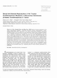
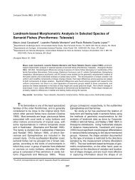
616<strong>Zoological</strong> <strong>Studies</strong> 50(5): 611-635 (2011)1 aes<strong>the</strong>tasc, and 8 + 1 aes<strong>the</strong>tasc. Parabasalprocess absent. Antenna (Fig. 3G) 2-segmented;corpus about twice as long as claw, former bearing1 broad basal seta on medial surface; claw bearingsimilar basal seta and terminal striations. Mandible(Fig. 3H) and maxillule (Fig. 3I) essentiallyas in previous species. Maxilla (Fig. 4A, B)2-segmented; lacertus unarmed; brachium bearing1 subterminal seta on medial margin, 1 blunt,terminal element, and large patch <strong>of</strong> denticles onouter surface; terminal claw fringed with rows <strong>of</strong>denticles along both edges. Maxilliped (Fig. 4C)2-segmented; corpus with fine denticles scatteredin myxal region; shaft longer than claw, with 1subterminal seta on medial margin; claw withstriations as in antenna.Ventral surface <strong>of</strong> leg 1 (Fig. 4D) with denticlesscattered on protopod and endopod;outer protopodal seta simple and thin, but innerprotopodal seta spiniform and arising from largepapilla; exopod 1-segmented and large, tipped with5 robust spines, inner 2 <strong>of</strong> which bear denticles onboth sides; endopod smaller than exopod, carrying1 long seta terminally. Leg 2 (Fig. 4E) protopodinconspicuous, without inner and outer setae;exopod armed as in leg 1, but seta on endopodbilaterally denticulate. Leg 3 with lamelliform ramicompletely fused to form a long plate completelycovering urosome ventrally (Fig. 3B) and leavinglarge gap dorsally (Fig. 3A); posterior edge <strong>of</strong>ventral lamella with 3 indentations (Fig. 3B). Leg 4a pair <strong>of</strong> long bilobate processes (Fig. 3A) arising(A)(B)(D)(C)(E)Fig. 4. Lernanthropodes trachinoti Pillai, 1962, female. (A) Maxilla, medial view; (B) tip <strong>of</strong> maxilla, medial view; (C) maxilliped, medialview; (D) leg 1, ventral view; (E) leg 2, ventral view. Scale bars: A = 50 μm; B = 10 μm; C = 0.1 mm; D = 30 μm; E = 20 μm.
Ho et al. – Copepods Parasitic on Marine Fishes <strong>of</strong> Taiwan 617from basal region <strong>of</strong> urosome (Fig. 3D). Leg 5absent.Male: Not collected.Remarks: Lernanthropodes trachinoti is s<strong>of</strong>ar known to occur on pompanos (Trachinotus)from India and Australia. In India, like in Taiwan, itwas taken from a snubnose pompano (Pillai 19621985), but in Australia, it was reported from ano<strong>the</strong>rspecies <strong>of</strong> pompano, Tra. botla (Shaw, 1803)(see Kabata 1979b). It was intriguing to note <strong>the</strong>specimen <strong>of</strong> Les. trachinoti in Pillai’s (1985) 2ndreport differed from his original report (Pillai 1962)in having a triangular head (cephalothorax) and alarge fused lamellae <strong>of</strong> leg 3 completely concealing<strong>the</strong> bifid leg 4 and urosome in ventral view <strong>of</strong> <strong>the</strong>animal. In o<strong>the</strong>r words, it may represent a differentspecies <strong>of</strong> Lernanthropodes. In fact, Pillai (1985)remarked in his 2nd report <strong>of</strong> Les. trachinoti that“This species closely resembles L. cuculus Bereand distinguished by only minor differences. Theymay turn out to be <strong>the</strong> same.” Inasmuch as <strong>the</strong>original description <strong>of</strong> Les. cuculus is sketchy, n<strong>of</strong>ur<strong>the</strong>r comment can be made at this point. Bere’s(1936) specimens <strong>of</strong> Les. cuculus were found onTra. carolinus (Linnaeus, 1766) and Tra. falcatus(Linnaeus, 1758) from <strong>the</strong> Gulf <strong>of</strong> Mexico.Specimens <strong>of</strong> Les. trachinoti from Taiwan fitwell with <strong>the</strong> original report <strong>of</strong> <strong>the</strong> species givenby Pillai (1962). The male <strong>of</strong> this species is notknown from India nor Taiwan, but Kabata (1979b)found it on Tra. botla from Australia.Genus Lernanthropus de Blainville, 1822Lernanthropus incilis sp. nov.(Figs. 5-7)Material examined: 4 and 2 found on gill filaments <strong>of</strong> Poey’s scabbardfish,Evoxymetopon poeyi Gün<strong>the</strong>r, 1887, landed atCheng-gong Fishing Port: 3 and 1 from 3(<strong>of</strong> 3) E. poeyi on 11 Feb. 2009, and 1 and 1 from 1 (<strong>of</strong> 1) E. poeyi on 25 Mar. 2009. Femaleholotype (USNM 1131890) and male allotype(USNM 1131891) were deposited in <strong>the</strong> NationalMuseum <strong>of</strong> Natural History, Smithsonian Institution,Washington, DC.Female: Body (Fig. 5A-C) large, 7.63 (7.50-7.76) mm long (from anterior rim <strong>of</strong> head to end <strong>of</strong>caudal ramus), divisible into head, neck, trunk, andurosome. Head nearly squarish, 1.95 (1.92-1.98)× 2.01 (1.80-2.22) mm, with narrowed antennalarea. Neck (1st pediger) short and wide, bearinglarge dorsal lobe. Remaining pedigers fused intotrunk, with pedigers 2 and 3 protruding out to forma lateral lobe and pediger 4 expanded posteriorlyinto a large subcircular dorsal plate that is deeplyemarginated in center. Genital complex andabdomen (Fig. 5D) wider than long, 0.40 (0.38-0.42) × 0.89 (0.84-0.94) and 0.48 (0.46-0.50) × 0.62(0.62-0.62) mm, respectively. Caudal ramus (Fig.5D) transformed into a long process, 2.21 (2.04-2.38) × 0.42 (0.40-0.44) mm, bearing 2 basal setaeon ventral surface (Fig. 5E), 1 subterminal seta onouter margin, and 2 small setae at tip. Egg saclong and straight.Antennule (Fig. 5F, G) stocky, indistinctly5-segmented; armature formula: 0, 0, 0, 0, and 9 +2 aes<strong>the</strong>tascs. Parabasal process (Fig. 5F) short.Antenna (Fig. 5H) robust, 2-segmented; corpusunarmed; claw armed with basal seta. Mandible(Fig. 6A) and maxillule (Fig. 6B) essentially as inprevious species. Maxilla (Fig. 6C) 2-segmented,with unarmed lacertus larger and longer thanbrachium; latter subterminally bearing 1 short,spiniform process and patch <strong>of</strong> denticles on medialsurface (usual terminal seta missing); terminalclaw (Fig. 6D) fringed with row <strong>of</strong> denticles onmedial surface. Maxilliped (Fig. 6E) 2-segmented;corpus robust and unarmed; subchela comprisingsmall, seta-bearing shaft and striated claw.Leg 1 (Fig. 6F) with protopod protruding outinto a process which carries an outer seta at itsbase; protopod also with inner conical process;exopod tipped with 5 stocky spines and endopodwith 1 blunt seta (Fig. 6G). Leg 2 (Fig. 6H) morereduced than leg 1, with inconspicuous protopodand weakly armed exopod (Fig. 6I). Leg 3 (Fig.5B) greatly modified, comprising large fleshy,folded lamella splayed ventrally at posterolateralcorners <strong>of</strong> trunk (Fig. 5C). Leg 4 (Fig. 5B) a pair<strong>of</strong> long, bifid processes with round, blunt tip. Leg5 (Fig. 5D) modified into a unilobate, long, obtuseprocess.Male: Body (Fig. 7A, B) smaller than femaleand without dorsal plate on trunk, measuring4.58 mm long (from tip <strong>of</strong> head to end <strong>of</strong> caudalramus). Head (cephalosome) wider than long, 1.64× 1.88 mm, with antennal region set apart fromrest <strong>of</strong> head. First 2 pedigers identifiable by <strong>the</strong>irlateral swellings, wider than long, measuring 0.24× 1.00 and 0.40 × 1.08 mm, respectively. Genitalcomplex indistinguishably fused to trunk. Caudalramus (Fig. 7A, B) long, slender, 745 × 186 μm,and armed as in female.Antennule (Fig. 7C) stocky as in female, butunsegmented and terminally armed with 3 moresetae (Fig. 7D). Parabasal process with basal