

7 - World Journal of Gastroenterology
7 - World Journal of Gastroenterology
7 - World Journal of Gastroenterology
- No tags were found...

Create successful ePaper yourself
Turn your PDF publications into a flip-book with our unique Google optimized e-Paper software.
ISSN 1948-5182 (online)<strong>World</strong> <strong>Journal</strong> <strong>of</strong>Hepatology<strong>World</strong> J Hepatol 2011 July 27; 3(7): 175-204www.wjgnet.com
Editorial Board2009-2013The <strong>World</strong> <strong>Journal</strong> <strong>of</strong> Hepatology Editorial Board consists <strong>of</strong> 573 members, representing a team <strong>of</strong> worldwide experts inhepatology. They are from 46 countries, including Argentina (4), Australia (7), Austria (2), Bangladesh (1), Belgium(3), Botswana (2), Brazil (8), Brunei Darussalam (1), Bulgaria (1), Canada (10), Chile (1), China (89), Denmark (1),Egypt (3), Finland (1), France (15), Gambia (1), Germany (28), Greece (8), Hungary (3), India (20), Ireland (1), Israel(7), Italy (65), Japan (45), Malaysia (1), Mexico (4), Netherlands (4), Pakistan (2), Poland (1), Portugal (1), Philippines(1), Romania (1), Saudi Arabia (1), Singapore (4), South Korea (17), Spain (22), Sri Lanka (1), Sudan (1), Switzerland(2), Thailand (6), Tunisia (2), Turkey (13), United Kingdom (17), United States (144), and Venezuela (1).PRESIDENT AND EDITOR-IN-CHIEFLian-Sheng Ma, BeijingSTRATEGY ASSOCIATEEDITORS-IN-CHIEFPaolo Cabassa, BresciaCheng-Shyong Chang, ChanghuaJing-Gung Chung, TaichungYi-Ming Chen, TaipeiAntonio Craxì, PalermoMoses S Elisaf, IoanninaFabio Grizzi, MilanMasatoshi Kudo, OsakaYasuhiro Kuramitsu, YamaguchiHuan-Yao Lei, TainanHsingjin Eugene Liu, TaipeiYasunobu Matsuda, Niigata CityChin-Hsiao Tseng, TaipeiYong Zeng, ChengduGUEST EDITORIAL BOARDMEMBERSYi-Chen Chen, TaichungTsung-Jung Lin, TaipeiYi-Wen Liu, ChiayiJen-Leih Wu, TaipeiSuh-Ching Yang, TaipeiMEMBERS OF THE EDITORIALBOARDArgentinaPatricia Cristina Baré, Buenos AiresMaria Cristina Carrillo, RosarioJuan Carlos Perazzo, Buenos AiresSilvia Cristina Sookoian, Buenos AiresAustraliaAnthony S-Y Leong, NewcastleDonald Peter McManus, QueenslandDes R Richardson, New South WalesMonica Robotin, SydneyNathan Subramaniam, BrisbaneNicholas Shackel, SydneyFiona J Warner, New South WalesAustriaWolfgang Mikulits, ViennaLothar Bernd Zimmerhackl, InnsbruckBangladeshMamun Al Mahta, BananiBelgiumFrederik C Berrevoet, GentOlivier Detry, LiègePhilip Meuleman, GhentBotswanaFrancesca Cainelli, GaboroneSandro Vento, GaboroneBrazilNiels OS Câmara, Sao PauloJoel Faintuch, Sao PauloRCS Ferreira, Santo AmaroRegina CS Godenberg, Rio de JaneiroCristina Miyazaki, Rio PretoCPMS Oliveira, Sao PauloMAF Ribeiro JR, ParnaibaMauricio Silva, Rio GrandeBrunei DarussalamVui Heng Chong, Bandar Seri BegawanBulgariaNikolai Vasilev Belev, PlovdivCanadaVasu D Appanna, OntarioElijah Dixon, AlbertaFernando Alvarez, QuebecSeyed Ali Gaskari, CalgarySerge Jothy, TorontoJennifer Linchee Kuk, TorontoQiang Liu, SaskatchewanEberhard L Renner, TorontoEldon A Shaffer, AlbertaGeorge Therapondos, OntarioChileLuis A Videla, SantiagoChinaPeng Bing, MD, ChengduWJH|www.wjgnet.comⅠJuly 27, 2011
Chiranjib Chakraborty, BeijingStephen Lam Chan, Hong KongGeorge G Chen, Hong KongMin-Shan Chen, GuangzhouYang Cheng, ShanghaiSiu Tim Cheung, Hong KongThomas YC Cheung, Hong KongYick-Pang Ching, Hong KangWilliam Chi-shing Cho, Hong KongChui Chung-hin, Hong KongShuang-Suo Dang, Xi'anYi-Tao Ding, NanjingJian-Gao Fan, ShanghaiYuen Man Fung, Hong KangZuo-Jiong Gong, WuhanTian-Quan Han, ShanghaiJin-Yang He, GuangzhouGarrett CL Ho, Hong KongJi-Ming Hu, WuhanCan-Hua Huang, ChengduZhi-Yong Huang, WuhanJian-Hui Jiang, ChangshaDong-Yan Jin, Hong KongHsiang-Fu Kung, Hong KongLai PBS Lai, Hong KongWan YJ Lau, Hong KongNancy WY Leung, Hong KongJin-Qing Li, GuangzhouLi-Ying Li, BeijingShu-Chen Li, HarbinXin-Wei Li, ShanghaiYu-Yuan Li, GuangzhouEn-Qi Liu, Xi’anYin-Kun Liu, ShanghaiChung-Mau Lo, Hong KongLun-Gen Lu, ShanghaiMing-De Lu, GuangzhouJohn M Luk, Hong KongGuang-Hua Luo, ChangzhouShuang Mei, ShanghaiKelvin KC Ng, Hong KongQin Ning, WuhanQin Pan, ShanghaiQi-Jun Qian, ShanghaiJian-Min Qin, ShanghaiXian-Jun Qu, JinanXue-Ying Sun, HarbinQin Su, BeijingWu-Yi Sun, HefeiHui-Ru Tang, WuhanPeng Tao, NanningEric WC Tse, Hong KongBin Wang, WeifangXiao-Zhong Wang, FuzhouXiu-Jie Wang, ChengduZhen-Xia Wang, HuhhotGrace LH Wong, Hong KongNathalie Wong, Hong KongXiong-Zhi Wu, TianjinDe-Xiang Xu, HefeiRui-An Xu, QuanzhouXun-Di Xu, ChangshaXiao Yang, BeijingZhen-Fan Yang, Hong KongBoon Hun Yong, Hong KongTing-He Yu, ChengduBenny CY Zee, Hong KongJia-Ning Zhang, DalianXiao-Dong Zhang, TianjinXiao-Lan Zhang, ShijiazhuangXiao-Yan Zhang, ShanghaiHong-Chuan Zhao, HefeiXiao-Ping Zhao, BeijingJiang-Fan Zhu, ShanghaiYi-Ping Zou, BeijingDenmarkHenning Grønbæk, AarhusEgyptNabil Mohie Abdel-Hamid, MiniaLaila AF Eissa, MansouraMona Mostafa Fahmy Nosseir, GizaFinlandThomas Kietzmann, OuluFranceAramando Abergel, Clenmont -FerrantHenri Bismuth, Villejuif CedexAna CFN Cardoso, ParisNicolas Chignard, ParisClaude C de Fromentel, LyonZdenko Herceg, LyonNathalie Janel, ParisVictor de Ledinghen, Pessac cedexAntoinette Lemoine, VillejuifMarcellin Patrick, ClichyRaoul Poupon, ParisRodrigue Rossignol, Bordeaux cedexChristian Trépo, LyonDominique A Vuitton, BesanconVirginie Wautot, Pierre BeniteGambiaMaimuna Ebirunkeh Mendy, BanjulGermanyThomas Bock, TuebingenAli Canbay, EssenEnrico Narciso De Toni, MünchenJoachim Drevs, FreiburgVolker Fendrich, MarburgPeter R Galle, MainzErich Gulbins, EssenRoland Kaufmann, JenaSebastian Hinz, KielPhilipp Kobbe, AachenMichael Kremer, HeidelbergChristian Liedtke, AachenMartin Loss, RegensburgArun Kumar Mankan, MunichLars Müller, MD, KielMichael D Menger, SaarbruckenAndreas K Nussler, MunichMargarete Odenthal, KoelnClaus Petersen, HannoverAndrej Potth<strong>of</strong>f, HannoverThomas Pusl, MünchenElke Roeb, GiessenFrank Tacke, AachenStefan Rose-John, KielAndreas Teufel, MainzLothar Thomas, FrankfurtJens JW Tischendorf, AachenArndt Vogel, HannoverGreeceAlex P Betrosian, AthensSpiros G Delis, AthensIoannis Diamantis, AthensPapandreou Dimitrios, MelaElias A Kouroumalis, CreteGeorge Papatheodoridis, AthensStamatios E. Theocharis, AthensHungaryGábor Bánhegyi, BudapestSubhamay Ghosh, PécsPeter Nagy, BudapestIndiaAnjali Deepak Amarapurkar, MumbaiDN Amararpurkar, MumbaiRunu Chakravarty, KolkataPronobesh Chattopadhyay, MoradabadPuneet Chopra, Gurgaon HaryanaTanya Das, KolkataRadha Krishan Dhiman, ChandigarhAjay Duseja, ChandigarhDevendra K Gupta, New DelhiP Kar, New DelhiSudhir Kumar, LucknowVijay Kumar, New DelhiAnoop Misra, New DelhiDevendra Parmar, LucknowRajendra Prasad, ChandigarhK Rajeshwari, New DelhiPallu Reddanna, HyderabadBarjesh Chander Sharma, New DelhiSarman Singh, New DelhiAjith TA, ThrissurIrelandMatthew William Lawless, DublinIsraelYaron Ilan, JerusalemWJH|www.wjgnet.com ⅡJuly 27, 2011
Yaakov Maor Kendler, Tel HashomerRan Oren, MD, Tel AvivAmir Shlomai, ModiinRifaat Safadi, JerusalemShira Zelber Sagi, Tel AvivYehuda Julius Shoenfeld, Tel HahsomerItalyLuca Aasaloni, BolognaGiovanni Addolorato, RomeLuigi E Adinolfi, NaplesPietro Andreone, BolognaM Appetecchia, RomeAntonio Ascione, NapoliFerruccio Bonino, MilanoBruno D Bruno, BeneventoSavino Bruno, MilanoMelchiorre Cervello, PalermoClaudio Chiesa, RomeStefano Colagrande, FirenzeMassimo G Colombo, MilanSamuele De Minicis, MontegranaroAlessandro Vitale, alessandroFabio Farinati, PadovaPaolo Feltracco, PadovaDomenico Ferri, BariAmalia Gastaldelli, PisaDomenico Girelli, VeronaFernando Goglia, BeneventoAlessandro Grasso, SavonaIgnazio Grattagliano, BariPietro Invernizzi, MilanFrancesco Izzo, NaplesAmedeo Lonardo, ModenaMalaguarnera Lucia, TrecastagniMassimo Di Maio, RossanoMelania Manco, RomeAndrea Mancuso, PalermoF Marotta, MilanoFabio Marra, FlorenceRoberto Mazzanti, FlorenceGiulia Morsica, MilanAntonio Moschetta, BariMassimo Negrini, FerraraAndrea Nicolini, PisaGiuseppe R Nigri, RomeValerio Nobili, RomeValentina Pallottini, RomeAdriano M Pellicelli, RomeMarcello Persico, NaplesMassimo Pinzani, FirenzeGiovanni Polimeni, MessinaCamillo Porta, PaviaPiero Portincasa, BariEmilio Quaia, TriesteGiuseppe Remuzzi, BergamoDomenico Ribatti, BariMassimo Roncalli, RozzanoCarlo Sabbà, BariOrazio Schillaci, RomeGaetano Serviddio, FoggiaAurelio Sonzogni, BergamoPaolo Sorrentino, SalernoEnea Spada, RomaGiovanni Tarantino, NaplesLuciano Tarantino, NaplesClaudio Tiribelli, TriestePierluigi Toniutto, UdinePietro Vajro, NaplesLuca Vigano, TorinoJapanYuichiro Eguchi, SagaMunechika Enjoji, FukuokaJiro Fujimoto, OsakaAtsushi Hosui, OsakaKazuo Ikeda, NagoyaToru Ishikawa, NiigataYoshiaki Iwasaki, OkayamaSatoru Kakizaki, GunmaNaoya Kato, TokyoTakumi Kawaguchi, KurumeKiminori Kimura, TokyoTsuneo Kitamura, ChibaKeiichi Kubota, TochigiSabina Mahmood, OkayamaHitoshi Maruyama, ChibaSachiko Matsuhashi, SagaToshihiro Mitaka, SapporoEiji Miyoshi, Yamada-oka SuitaZenichi Morise, Toyoake AichiRyuichi Morisihita, OsakaYoshiki Murakami, KyotoSatoru Murata, TokyoAtsushi Nakajima, KanagawaYasuni Nakanuma, KanazawaWaka Ohishi, HiroshimaMorikazu Onji, MatsuyamaToshiji Saibara, NankokuHiroaki Shiba, TokyoIkuo Shoji, HyogoRyo Sudo, YokohamaYoshio Sumida, NaraShinji Tanaka, TokyoTakuji Tanaka, GifuAkihiko Tsuchida, TokyoTakato Ueno, KurumeShinichi Ueno, KagoshimaKiyohito Yagi, OsakaYo-ichi Yamashita, HiroshimaTeruyoshi Yanagita, SagaShuang-Qin Yi, KanazawaHiroshi Yoshida, TokyoHitoshi Yoshiji, NaraMalaysiaKamsiah Jaarin, Kuala LumpurMexicoNorberto C Chavez-Tapia, TlalpanJavier Lizardi Cervera, Tlalpan CPSaúl Villa-Treviño, México DFFlorencia V Vorackova, México DFNetherlandsRobert Jacobus de Knegt, RotterdamTU Hoogenraad, HeidelberglaanMaarten E Tushuizen, MB AmsterdamRobert C Verdonk, RB GroningenPakistanSyed Hamid Ali, KarachiHuma IQ TI, IslamabadPolandMaria ES Lotowska, BialystokPortugalFelix Dias Carvalho, PortoPhilippinesJanus P Ong, ManilaRomaniaEugen Georgescu, CraiovaSaudi ArabiaAhmed Helmy, RiyadhSingaporeWei Ning Chen, SingaporeSi-Shen Feng, SingaporeLang Zhuo, SingaporeChun-Tao Wai, SingaporeSouth KoreaSang Hoon Ahn, SeoulSun Pyo Hong, YonginByung Ihn Choi, SeoulSeok Joo Han, SeoulKyung Lib Jang, BusanBum-Joon Kim, SeoulDong Goo Kim, SeoulKyung Sik Kim, SeoulMeehyein Kim, YonginYoung Chul Kim, SeoulMi-Kyung Lee, JeonnamYoung-Ik Lee, TaejonKwan-Kyu Park, DaeguHyunchul Rhim, SeoulIn Kyoung Lim, Gyunggi-doDae-Yeul Yu, DaejeonJong Won Yun, KyungbukWJH|www.wjgnet.com ⅢJuly 27, 2011
SpainJose AG Agundez, BadajozMaria Angeles, MadridAgustin Castiella, MendaroRuben Ciria, CordobaJoan Clari, BarcelonaMaria Buti Ferret, BarcelonaPuri Fortes, PamplonaJoan Genescà, BarcelonaMaría J Gómez-Lechón, ValenciaArias Jaime, MadridÁngeles Pajares María, MadridJordi Muntane, CordobaJose JG Marin, SalamancaJulia P Onsurbe, BarcelonaAlbert Parés, BarcelonaSonia Ramos, MadridCristina Ripoll, MadridIsabel F Romero, BarcelonaMarta R Romero, SalamancaJuan Macias Sanchez, SevillaJuan Sastre, ValenciaManuel Vázquez-Carrera, BarcelonaSri LankaEGD Shaman Rajindrajith, RagamaSudanHatim MY Mudawi, KhartoumSwitzerlandBeat Mullhaupt, ZurichMaurer A Christoph, LiestalThailandNattiya Hirankarn, BangkokSomchai Pinlaor, Khon KaenYong Poovorawan, BangkokAbhasnee Sobhonslidsuk, BangkokChanitra Thuwajit, BangkokSopit Wongkham, Khon KaenTunisiaOlfa Bahri, Tunis-BelvedereChadli Dziri, TunisTurkeyInci Alican, IstanbulAhmet Atessahin, ElazigYasemin Hatice Balaban, AnkaraHayrullah Derici, MD, IzmirCigdem Ulukaya Durakbasa, IstanbulMuhsin MM Harputluoglu, MalatyaAbdurrahman Kadayifci, GaziantepAdnan Kadayifci, AntalyaAli Sazci, KocaeliIlker Tasci, AnkaraMehmet Yalniz, ElazigSerkan Yener, IzmirYusuf Yilmaz, IstanbulUnited KingdomAlastair David Burt, NewcastleDavid O Cosgrove, LondonAnil Dhawan, LondonIndra Neil Guha, NottinghamPhillip M Harrison, LondonHübscher SG Hübscher, BirminghamLong R Jiao, LondonAT Koulaouzidis, EdinburghPatricia Lalor, BirminghamDavid A Lomas, CambridgeRajeshwar P Mookerjee, LondonGareth J Morris-Stiff, WalesKathryn L Nash, SouthamptonDerek Anthony O’Reilly,Christian P Selinge, BoltonKonstantinos Tziomalos, LondonFeng Wu, OxfordUnited StatesGary A Abrams, MontgomeryHassan H A-Kader, TucsonHans-Olov Adami, MassachusettsJoseph Ahn, MaywoodShannon Marie Bailey, AlabamaNuman Cem Balci, St Louis MOEdmund J Bini, New YorkVictor E Buckwold, FrederickRoniel Cabrera, GainesvilleGuoqing Cao, IndianaDisaya Chavalitdhamrong, New YorkChien-Shing Chen, Loma LindaFei Chen, MorgantownSu Chen, San AntonioYouhai H Chen, PhiladelphiaAnne M Covey, New YorkMark J Czaja, New YorkSrikanta Dash, New OrleansAnthony JB Demetris, PittsburghSridevi Devaraj, CaliforniaLisa Ross Dixon, GainesvilleTerrence M Donohue, OmahaQ Ping Dou, DetroitMurray N Ehrinpreis, DetroitMarwan Ghazi Fakih, BuffaloShengyun Fang, MarylandClaus J Fimmel, IllinoisRobert Anthony Fisher, VirginiaSamuel W French, TorrancePhillip A Furman, PrincetonM Eric Gershwin, CaliforniaJalal K Ghali, MichiganGrace Liejun Guo, Kansas CityDieter Haemmerich, CharlestonYoung S Hahn, CharlottesvilleStephen A Harrison, TexasDee Harrison-Findik, NebraskaSidhartha Hazari, LouisianaThomas S Helling, JacksonAlan W Hemming, FloridaIryna S Hepburn, EvansAi-Xuan L Holterman, ChicagoKe-Qin Hu, CaliforniaGuangcun Huang, OhioWendong Huang, CaliforniaRachel M Hudacko, New BrunswickMichael John Jacobs, MichiganHartmut W Jaeschke, Kansas CityRavi Jhaveri, North CarolinaLynt B Johnson, WashingtonNeil Louis Julie, BethesdaSanjay Kakar, San FranciscoSanjeeva P Kalva, BostonJing X Kang, MassachusettsHetal Karsan, GeorgiaEmmet B Keeffe, CaliforniaNancy Ellen Kemeny, New YorkAndrew Scott Kennedy, CaryKusum K Kharbanda, OmahaDavid H Kirn, CaliforniaHyam Lerner Leffert, La JollaStacee Marie Lerret, MilwaukeeFengzhi Li, New YorkWei Li, HoustonShuang Liu, IndianaSu Hao Lo, DavisDaniel G Maluf, RichmondJose E Manautou, StorrsRichard S Mangus, IndianaMary Ko Manibusan, VirginiaPaul Martin, Miami,Jochen Mattner, OhioJames A McCubrey, North CarolinaValentina Medici, SacramentoGeorge Michalopoulos, PittsburghSmruti R Mohanty, IllinoisJohn T Moore, GlaxoSmithKlineRavi Murthy, TexasLaura E Nagy, ClevelandSagar U Nigwekar, RochesterEileen M O‘Reilly, New YorkKevin FS O’Carroll, HersheyMelissa Kay Osborn, AtlantaHelieh Saatara Oz, KentuckyIgor P Pogribny, ArkansasNicholas C Popescu, Bethesda MarylandDaniel S Pratt, BostonRatna B Ray, LouisNancy Reau, ChicagoJanardan K Reddy, ChicagoMartin J Ronis, Little RockPhillip Ruiz, FloridaTanios B Saab, ColumbusAdnan Said, MadisonNeeraj Saxena, GeorgiaRaymund R Saxena, MinnesotaAnn Scheimann, BaltimoreTimothy M Schmitt, CharlottesvilleBernd Schnabl, La JollaKunwar Shailubhai, PennsylvaniaMuhammad Y Sheikh, CaliforniaPerry Shen, Winston-SalemViji Shridhar, RochesterShivendra D Shukla, MissouriAshwani K Singal, GalvestonKeshav K Singh, New YorkWJH|www.wjgnet.com ⅣJuly 27, 2011
Omar Skalli, ShreveportByoung-Joon Song, MarylandBranko Stefanovic, TallahasseeStephen Strom, PennsylvaniaXiao Su, San FranciscoWing-Kin Syn, North CarolinaGyongyi Szabo, MassachusettsShinako Takada, HoustonYueming Tang, ChicagoJohn M Taylor, PhiladelphiaSwee H The, SpringfieldChung-Jyi Tsai, LexingtonGeorge P Tuszynski, PennsylvaniaJean-Nicolas Vauthey, HoustonMichael E de Vera, PennsylvaniaYu-Jui Yvonne Wan, KansasJack R Wands, ProvidenceHanlin L Wang, Los AngelesXin Wei Wang, MarylandWahid Wassef, WorcesterRonald J Wong, CaliforniaGeorge YH Wu, FarmingtonHai-Shan Wu, New YorkVictor W Xia, CaliforniaXiming J Yang, ChicagoMatthew M Yeh, SeattleMei Po Yip, SeattleMin You, TampaZobair M Younossi, Falls ChurchXiao-Fang Yu, MarylandYong Yuan, PlainsboroJian X Zhang, CharlotteJian-Ying Zhang, El PasoKezhong Zhang, MichiganYu-Jing Zhang, New YorkYuao Zhu, DurhamSaša Živković, PittsburghWilliam A Zule, Research Triangle ParkVenezuelaFlor Pujol de Freychet, CaracasⅤWJH|www.wjgnet.com July 27, 2011
ContentsMonthly Volume 3 Number 7 July 27, 2011EDITORIAL175 A survey on herbal management <strong>of</strong> hepatocellular carcinomaAbdel-Hamid NM, Nazmy MH, Mahmoud AW, Fawzy MA, Youss<strong>of</strong> MORIGINAL ARTICLE184 Nonmuscle myosin Ⅱ regulates migration but not contraction in rat hepaticstellate cellsMoore CC, Lakner AM, Yengo CM, Schrum LWBRIEF ARTICLE198 An extended treatment protocol with pegylated interferon and ribavirin forhepatitis C recurrence after liver transplantationHashemi N, Araya V, Tufail K, Thummalakunta L,Feyssa E, Azhar A, Niazi M, Ortiz JWJH|www.wjgnet.comIJuly 27, 2011|Volume 3|Issue 7|
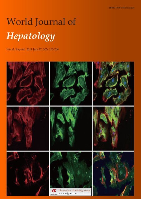
Contents<strong>World</strong> <strong>Journal</strong> <strong>of</strong> HepatologyVolume 3 Number 7 July 27, 2011ACKNOWLEDGMENTSIAcknowledgments to reviewers <strong>of</strong> <strong>World</strong> <strong>Journal</strong> <strong>of</strong> HepatologyAPPENDIXIMeetingsI-VInstructions to authorsABOUT COVERMoore CC, Lakner AM, Yengo CM, Schrum LW. Nonmuscle myosin Ⅱregulates migration but not contraction in rat hepatic stellate cells.<strong>World</strong> J Hepatol 2011; 3(7): 184-197http://www.wjgnet.com/1948-5182/full/v3/i7/184.htmAIM AND SCOPE<strong>World</strong> <strong>Journal</strong> <strong>of</strong> Hepatology (<strong>World</strong> J Hepatol, WJH, online ISSN 1948-5182, DOI:10.4254), is a monthly, open-access, peer-reviewed journal supported by an editorialboard <strong>of</strong> 573 experts in hepatology from 46 countries.The major task <strong>of</strong> WJH is to report rapidly the most recent results in basic andclinical research on hepatology, including: liver biology/pathology, cirrhosis and itscomplications, liver fibrosis, liver failure, portal hypertension, hepatitis B and C andinflammatory disorders, steatohepatitis and metabolic liver disease, hepatocellularcarcinoma, biliary tract disease, autoimmune disease, cholestatic and biliary disease,transplantation, genetics, epidemiology, microbiology, molecular and cell biology,nutrition, geriatric and pediatric hepatology, diagnosis and screening, endoscopy,imaging, and advanced technology.FLYLEAFI-VEditorial BoardEDITORS FORTHIS ISSUEResponsible Assistant Editor: Le ZhangResponsible Electronic Editor: Le ZhangPro<strong>of</strong>ing Editor-in-Chief: Lian-Sheng MaResponsible Science Editor: Hai-Ning ZhangPro<strong>of</strong>ing Editorial Office Director: Hai-Ning ZhangNAME OF JOURNAL<strong>World</strong> <strong>Journal</strong> <strong>of</strong> HepatologyLAUNCH DATEOctober 31, 2009SPONSORBeijing Baishideng BioMed Scientific Co., Ltd.,Room 903, Building D, Ocean International Center,No. 62 Dongsihuan Zhonglu, Chaoyang District,Beijing 100025, ChinaTelephone: +86-10-8538-1892Fax: +86-10-8538-1893E-mail: baishideng@wjgnet.comhttp://www.wjgnet.comEDITINGEditorial Board <strong>of</strong> <strong>World</strong> <strong>Journal</strong> <strong>of</strong> Hepatology,Room 903, Building D, Ocean International Center,No. 62 Dongsihuan Zhonglu, Chaoyang District,Beijing 100025, ChinaTelephone: +86-10-5908-0038Fax: +86-10-8538-1893E-mail: wjh@wjgnet.comhttp://www.wjgnet.comPUBLISHINGBaishideng Publishing Group Co., Limited,Room 1701, 17/F, Henan Building,No.90 Jaffe Road, Wanchai,Hong Kong, ChinaFax: +852-3115-8812Telephone: +852-5804-2046E-mail: baishideng@wjgnet.comhttp://www.wjgnet.comSUBSCRIPTIONBeijing Baishideng BioMed Scientific Co., Ltd.,Room 903, Building D, Ocean International Center,No. 62 Dongsihuan Zhonglu, Chaoyang District,Beijing 100025, ChinaTelephone: +86-10-8538-1892Fax: +86-10-8538-1893E-mail: baishideng@wjgnet.comhttp://www.wjgnet.comPUBLICATION DATEJuly 27, 2011ISSNISSN 1948-5182 (online)PRESIDENT AND EDITOR-IN-CHIEFLian-Sheng Ma, BeijingSTRATEGY ASSOCIATE EDITORS-IN-CHIEFPaolo Cabassa, BresciaCheng-Shyong Chang, ChanghuaJing-Gung Chung, TaichungYi-Ming Chen, TaipeiAntonio Craxì, PalermoMoses S Elisaf, IoanninaFabio Grizzi, MilanMasatoshi Kudo, OsakaYasuhiro Kuramitsu, YamaguchiHuan-Yao Lei, TainanHsingjin Eugene Liu, TaipeiYasunobu Matsuda, Niigata CityChin-Hsiao Tseng, TaipeiYong Zeng, ChengduEDITORIAL OFFICEHai-Ning Zhang, Director<strong>World</strong> <strong>Journal</strong> <strong>of</strong> HepatologyRoom 903, Building D, Ocean International Center,No. 62 Dongsihuan Zhonglu, Chaoyang District,Beijing 100025, ChinaTelephone: +86-10-5908-0038Fax: +86-10-8538-1893E-mail: wjh@wjgnet.comhttp://www.wjgnet.comCOPYRIGHT© 2011 Baishideng. Articles published by this Open-Access journal are distributed under the terms <strong>of</strong>the Creative Commons Attribution Non-commercialLicense, which permits use, distribution, and reproductionin any medium, provided the original work is properlycited, the use is non-commercial and is otherwisein compliance with the license.SPECIAL STATEMENTAll articles published in this journal represent theviewpoints <strong>of</strong> the authors except where indicatedotherwise.INSTRUCTIONS TO AUTHORSFull instructions are available online at http://www.wjgnet.com/1948-5182/g_info_20100316080002.htm.ONLINE SUBMISSIONhttp://www.wjgnet.com/1948-5182<strong>of</strong>ficeWJH|www.wjgnet.comIIJuly 27, 2011|Volume 3|Issue 7|
Online Submissions: http://www.wjgnet.com/1948-5182<strong>of</strong>ficewjh@wjgnet.comdoi:10.4254/wjh.v3.i7.175<strong>World</strong> J Hepatol 2011 July 27; 3(7): 175-183ISSN 1948-5182 (online)© 2011 Baishideng. All rights reserved.EDITORIALA survey on herbal management <strong>of</strong> hepatocellularcarcinomaNabil Mohie Abdel-Hamid, Maiiada Hasan Nazmy, Ahmed Wahid Mahmoud, Michael Atef Fawzy, MarcoYouss<strong>of</strong>Nabil Mohie Abdel-Hamid, Maiiada Hasan Nazmy, AhmedWahid Mahmoud, Michael Atef Fawzy, Marco Youss<strong>of</strong>, BiochemistryDepartment, Unit <strong>of</strong> Liver cancer research, Faulty <strong>of</strong>Pharmacy, Minia University, Minia 002086, EgyptAuthor contributions: Abdel-Hamid NM designed and revisedthe article; Nazmy MH collected the whole references; MahmoudAW cited the active constituents; and Fawzy MA and Youss<strong>of</strong>Mwere responsible for references management and editing.Correspondence to: Nabil Mohie Abdel-Hamid, PhD, Pr<strong>of</strong>essor,Diagnostic Laboratory, Abtal El-Faluga Street, Mit-Gomre,Dakahlia 002050, Egypt. nabilmohie@yahoo.comTelephone: +20-50-6913997 Fax: +20-86-2369075Received: January 5, 2011 Revised: May 6, 2011Accepted: May 13, 2011Published online: July 27, 2011AbstractIn this review we outline the different mechanisms mediatinghepatocarcinogenesis. We also discuss possibletargets <strong>of</strong> bioactive herbal agents at different stages <strong>of</strong>hepatocarcinogenesis and highlight their role at eachindividual stage. We gathered information on the mostcommon herbal prescriptions and extracts thought to beuseful in prevention or sensitization for chemotherapyin management <strong>of</strong> hepatocellular carcinoma (HCC). Thevalue <strong>of</strong> this topic may seem questionable compared tothe promise <strong>of</strong>fered for HCC management by chemotherapyand radiation. However, we would recommendthe use <strong>of</strong> herbal preparations not as alternatives tocommon chemo /and or radiotherapy, but rather for preventionamong at-risk individuals, given that drug/herbinteractions are still in need <strong>of</strong> extensive clarification.The bioactive constituents <strong>of</strong> various herbs seem to bepromising targets for isolation, cancer activity screeningand clinical evaluation. Finally, herbal preparations may<strong>of</strong>fer a cost effective protective alternative to individualsknown to have a high risk for HCC and possibly othercancers, through maintaining cell integrity, reversingoxidative stress and modulating different molecularpathways in preventing carcinogenesis.© 2011 Baishideng. All rights reserved.Key words: Active ingredients; Chemoprevention; Chemosensitization;Hepatocellular carcinoma; Herbs; MoleculartargetsPeer reviewers: Takuji Tanaka, MD, PhD, The Tohkai CytopathologyInstitute, Cancer Research and Prevention (TCI-CaRP), 4-33 Minami-Uzura, Gifu 500-8285, JapanAbdel-Hamid NM, Nazmy MH, Mahmoud AW, Fawzy MA,Youss<strong>of</strong> M. A Survey on herbal management <strong>of</strong> hepatocellularcarcinoma. <strong>World</strong> J Hepatol 2011; 3(7): 175-183 Available from:URL: http://www.wjgnet.com/1948-5182/full/v3/i7/175.htmDOI: http://dx.doi.org/10.4254/wjh.v3.i7.175INTRODUCTIONHepatocellular carcinoma (HCC) is the third deadliestand fifth most common malignancy worldwide [1-3] . It is ahighly malignant tumor having high morbidity and motality.HCC has a poor prognosis due to its rapid infiltratingpower which leads to complicating liver cirrhosis [4] . Therate <strong>of</strong> HCC is increasing worldwide between 3% and9% annually [5,6] . The incidence ranges from less than 10cases per 100 000 in North America and Western Europeto 50-150 cases per 100 000 in parts <strong>of</strong> Africa and Asia [7] .Hepatocarcinogenesis is associated with a background <strong>of</strong>chronic and persistent infection <strong>of</strong> hepatitis B virus (HBV)and hepatitis C virus (HCV) [8] . These infections along withalcohol and aflatoxin B1 exposure are widely recognizedetiological agents in HCC [9] .In Egypt, epidemiology <strong>of</strong> HCC is characterized bymarked demographic and geographic variations [10,11] . OverWJH|www.wjgnet.com 175July 27, 2011|Volume 3|Issue 7|
Abdel-Hamid NM et al . Herbal management <strong>of</strong> hepatocellular carcinomathe last decade, a remarkable increase, from 4.0% to 7.2%,was observed in the proportion <strong>of</strong> chronic liver disease(CLD) patients with HCC. The predominant age group(40-59 years) showed a slight increase compared witholder groups (> 60 years). A significant increase, from82.5% to 87.6%, was observed in the proportion <strong>of</strong> HCCamong males. The calculated risk <strong>of</strong> HCC developmentis nearly three times higher in men than in women [12] . Aunique invisible risk factor for development <strong>of</strong> HCC inEgypt could be Schistosomal infection and its injectiontherapy. Schistosomiasis induces immune suppression,which could result in increased persistence <strong>of</strong> viremia followingacute infection <strong>of</strong> both hepatitis B and C [13] .HCCs are phenotypically (morphology and microscopy)and genetically heterogenous tumors, possibly reflectingthe heterogeneity <strong>of</strong> etiological factors implicated inHCC development, the complexity <strong>of</strong> hepatocyte functionsand the late stage at which HCCs usually becomeclinically symptomatic and detectable [14,15] . Hepatocarcinogenesisis a multi-factor, multi-step and complex process [8] .It involves three distinguishable but closely connectedstages: initiation (normal cell → transformed or initiatedcell), promotion (initiated cell → preneoplastic cell),and progression (preneoplastic cell → neoplastic cell) [16] .Malignant transformation <strong>of</strong> hepatocytes may occur, regardless<strong>of</strong> the etiological agent, through a pathway <strong>of</strong> increasedliver cell turnover, induced by chronic liver injuryand regeneration in a context <strong>of</strong> inflammation, immuneresponse, and oxidative DNA damage [17-19] .MOLECULAR TARGETS FOR HERBALCOMPOUNDS DURING HCC PROGRESSIONSince ancient times, natural products, herbs and spiceshave been used as remedies for various diseases, incluingcancer (Table 1). The term chemoprevention was coinedin the late 1970s and referred to a pharmacological interventionaimed to arrest or reverse the process <strong>of</strong> carcinogenesis[20] . Previous attempts were made to identifyagents or combinations which could exhibit any <strong>of</strong> thefollowing characteristics: (1) prevention <strong>of</strong> tumor initiation;(2) delay or arrest <strong>of</strong> the development <strong>of</strong> tumors;(3) extention <strong>of</strong> cancer latency periods; (4) reduction incancer metastasis and mortality; and (5) prevention <strong>of</strong>recurrence <strong>of</strong> secondary tumors [21] . Recently, the focushas been directed towards molecular targeting <strong>of</strong> herbalcompounds to identify the mechanism(s) <strong>of</strong> action <strong>of</strong>these newly discovered bioactive compounds. Moreover,it has been recognized that single agents may not alwaysbe sufficient to provide chemopreventive efficacy andtherefore the new concept <strong>of</strong> combination chemopreventionby multiple agents or by the consumption <strong>of</strong> “wholefoods” has become an increasingly attractive area <strong>of</strong>study [22] . Steps in the development <strong>of</strong> cancer at cellularlevel are described below.InitiationInitiation involves gene mutation, carcinogen metabolismand aberrant DNA repair. In this initial stage, environmentalcarcinogens (e.g. dietary, tobacco, pollution) induceone or more simple mutations, including transitions orsmall deletions in genes which control the process <strong>of</strong> carcinogenesis.Activated carcinogens exert their effects byforming covalent adducts with individual molecules <strong>of</strong>DNA or RNA, causing deletions <strong>of</strong> genetic material ormistranslation <strong>of</strong> the DNA sequence which may producemutations in critical genes, such as tumor suppressors andoncogenes [23] . Reactive oxygen species (ROS) are generatednormally as part <strong>of</strong> the normal oxidative metabolismor may be end-products <strong>of</strong> the breakdown <strong>of</strong> xenobioticcompounds (Figure 1). Oxidative stress can result in extensiveDNA damage. Antioxidant herbs which scavengeactivated oxygen species are able to stimulate DNA repairpathways to prevent or overcome oxidative DNA damage.Vitamin C, genistein and compounds originating fromcruciferous vegetables are among the most well-studiedfor their scavenger properties [24] . In addition, chronic inflammationmay predispose individuals to certain cancers.Most precancerous and cancerous tissues show signs <strong>of</strong>inflammation involving the movement <strong>of</strong> innate immunecells into the tissue, the presence <strong>of</strong> specific inflammatorysignaling molecules (i.e. cytokines and chemokines),changes in tissue structure (remodeling) and the formation<strong>of</strong> new blood vessels (angiogenesis). Further studieshave found that cancer-associated inflammation actuallypromotes tumor growth and progression [25] . Several proinflammatorygene products (i.e. TNF-α , IL-6) have acritical role in regulation <strong>of</strong> apoptosis, proliferation, angiogenesis,invasion and metastasis. Their expression ismainly regulated by the transcription factor NF-kB, whichis constitutively active in most tumors and is induced bycarcinogens and chemotherapeutic agents. TNF-alphacan initiate signaling pathways which lead to the activation<strong>of</strong> NF-κB, the initiation <strong>of</strong> MAPK cascades, and celldeath [26] . These observations imply that anti-inflammatoryagents that suppress NF-κB or NF-κB-regulated productsshould have a potential in both the prevention and treatment<strong>of</strong> cancer [27] .Recently, diallyl sulphide (DAS) obtained from garlicand vitamin C were reported to decrease the levels <strong>of</strong>circulatory TNF-α and IL-6 in DENA-induced hepatocarcinogenesis[28] . Previous reports showed that vitamin Ccan inactivate nuclear factor kappa B in endothelial cellsduring the inflammation process, independently <strong>of</strong> itsatioxidant activity. Therefore, the anti-inflammatory activity<strong>of</strong> ascorbic acid (AA) may be mediated by multifactorialmechanisms, which are not necessarily associated withits intrinsic antioxidant activity [29] . DAS also was foundto promote an anti inflammatory environment by cytokinemodulation, leading to an overall inhibition <strong>of</strong> NFkBactivity in the surrounding tissue [30] . In addition, DASmay enhance antioxidants and suppresses inflammatorycytokines through the activation <strong>of</strong> Nrf2 transcriptionfactor [31] .WJH|www.wjgnet.com 176July 27, 2011|Volume 3|Issue 7|
Abdel-Hamid NM et al . Herbal management <strong>of</strong> hepatocellular carcinomaTable 1 Summary <strong>of</strong> the effects <strong>of</strong> some herbs and other natural compounds on hepatocellular carcinomaCompound Ref. Composition EffectHerbs with cancer chemotherapeutic effectGeiji-Bokryung-HwanGanfujian granulesMaharishi amritkalashScutellaria baicalensisand BupleurumscorzonerifolfiumwilldHuqi san(Qi-protectingpowder)[78,79][80][81][43][28,82]It is composed <strong>of</strong> five different herbs <strong>of</strong> CinnamomiRamulus, Poria Cocos Hoelen (Pachymae Fungus),Moutan Cortex Radicis, Paeoniae Radix, and PersicaeSemen. The active constituents are antioxidativephenolic compounds, trans-cinnamic acid, taxifolin,protocatechuic acid, trans-o-hydroxy cinnamicacid, protocatechuic aldehyde, benzoic acid, transo-methoxycinnamic acid, cis-o-methoxy cinnamicacid, 4-hydroxybenzoic acid, coumarin, daucosterol,Paeoniflorin, albiflorin and benzoylalbiflorin,paeonol and paeoniflorin.Ganfujian granules are an oral preparation consisting<strong>of</strong> dietary and medicinal Chinese herbs includingChinese yam (Rhizoma Dioscoreae), hawthorn fruit(Fructus Crataegi) and Chinese date (FructusZiziphiJujubae). The active constituents are flavonoidsincluding oligomeric procyanidins (OPCs), vitexin,vitexin 4'-O-rhamnoside, quercetin, and hyperosideMaharishi Amrit Kalash (MAK) is composed <strong>of</strong> amixture <strong>of</strong> two herbal mixtures, MAK-4 and MAK-5.The active constituents are multiple antioxidantsincluding alpha-tocopherol, beta-carotene, ascorbate,bi<strong>of</strong>lavonoid, catechin, polyphenols, rib<strong>of</strong>lavin andtannic acid.Chinese medicinal herbs. The active constituentsare antioxidant flavonoids, baicalein, wogonin, neobaicalein,and skullcapflavone.Huqi san is composed <strong>of</strong> eight medicinal herbs including(Ramulus Visci, Radix Astragali seu Hedysari,Radix Curcumae, Radix Salviae Miltiorrhizae). Theactive constituents are polysaccharides, flavonoids,alkaloids and tanshinones.The inhibitory effects <strong>of</strong> Geiji-Bokryung-Hwan (GBH) on the growth<strong>of</strong> cancer cell lines (HepG2 and Hep3B) and cancer chemopreventiveactivity were investigated. Tumor inhibition was found to bemediated via the inhibition <strong>of</strong> COX-1 activity.The herb was found to reduce and delay the incidence <strong>of</strong> diethylnitrosamine-inducedhepatocarcinoma by exerting director indirect effects on the cell cycle and inhibiting uncontrolledproliferation <strong>of</strong> rat hepatocytes.MAK was found to inhibit liver carcinogenesis when given assupplement to diet. The authors <strong>of</strong> this study suggested that themechanism <strong>of</strong> this inhibition involved the prevention <strong>of</strong> excessiveoxidative damage.The these herbs were found to enhance the chemopreventive effect <strong>of</strong>selenium on N-nitosobis (2-oxopropyl) amine-induced liver cancersin Syrian hamsters.The inhibitory effect <strong>of</strong> Huqi san on rat prehepatocarcinoma, whichwas induced via diethylinitrosamine (DEN), was investigated.It was found to inhibit the over-expression <strong>of</strong> c-jun, c-fos, andc-myc oncogenes, which were shown to play an important role inthe pathogenesis <strong>of</strong> hepatocellular carcinoma. Huqi san was alsoreported to inhibit DEN induced oxyradical formation in culturedhepatocytes, leading to suppression <strong>of</strong> oxidative DNA damage.Milk thistle[83,84]Herbs with cancer chemotherapeutic effect[85]Songyou YinMillettia reticulatabenth[86]Milk thistle, commonly known as silymarin, is extractedfrom Silybum marianum. The active constituentsare flavonoids from which silibinin and silymarin arethe biologically most active compound.This herbal extract is composed <strong>of</strong> a mixture <strong>of</strong> 5Chinese medicinal herbs (Salvia miltiorrhiza, Astragalusmembranaceus, lycium borbarum crataeguspinnatifida and trionyx sinensis). The active constituentsare diterpenoid tanshinones, flavonoids andsaponins.Millettia reticulata Benth is one <strong>of</strong> the oldest tonicherbs in traditional Chinese medicine. The activeconstituents are flavonoid derivatives: (-)-epicatechin,naringenin, 5,7,3',5'tetrahydroxyflavanone, formononetin,isoliquiritigenin, and genistein.It has been shown that a topical application <strong>of</strong> silymarin on miceresults in complete inhibition <strong>of</strong> an epidermal carcinogen andprevents the formation <strong>of</strong> pyrimidine dimers, which are consideredto be potential skin cancer agents."Songyou Yin" attenuates tumor proliferation and prolongs survival<strong>of</strong> nude mice bearing hepatocellular tumors without distinct toxicity.These findings suggest that "Songyou Yin" has some potential in thetreatment <strong>of</strong> hepatocellular carcinoma.It was demonstrated that Millettia reticulata Benth flavonoid derivativeshave a positive inhibitory effect on the viability <strong>of</strong> humancancer cells (including HepG2, SK-Hep-1, Huh7, PLC5, COLO 205,HT-29, and SW 872 cells). This Chinese herb also induces apoptosisin hepatocellular carcinoma cells via both Fas- and mitochondriamediatedpathways.Bushen huayu jiedurecipe[87]"bushen huayu jiedu recipe" (BSHYJDR) is a mixture<strong>of</strong> several herbs including Chinese Cassia Bark,Psoralea, Zedoary, Rhubarb. The active constituentsare alkaloids, flavonoid, arsenic trioxide, cinnamicacid, rhubarb and rhubarb substance.BSHYJDR was found to inhibit transplanted hepatocarcinoma inmice. This effect is improved in combination with chemotherapy(cisplatin (DDP)).WJH|www.wjgnet.com 177July 27, 2011|Volume 3|Issue 7|
Abdel-Hamid NM et al . Herbal management <strong>of</strong> hepatocellular carcinomaStar 99[88]Chinese herbal compoundHuman hepatocellular carcinoma was transplanted in nude miceand treated with Star 99 (intratumoral injection 10 days followingto cancer transplantation). The herbal compound was shown toinhibit and destruct liver cancer cells, in particular the membrane,cytoplasm and nucleus <strong>of</strong> the caner hepatocyte.Daesungki-TangLycium barbarumand rehmanniaglutinosaSemen coicisPaeoniae radixQingrejiedu,huoxuehuayu, andfuzhenggubenDelisheng[89][90][91][58][92][93]This is a preparation consisting <strong>of</strong> four herbs: Rheiradix et rhizoma (the roots <strong>of</strong> Rheum coreanumNakai), Aurantiii frutus immaturus (immature fruits<strong>of</strong> Poncirus trifolita Rafin), Magnoliae cortex (thestem bark <strong>of</strong> Magnolia <strong>of</strong>ficinalis Rehd. Et Wils), andMirabilite (Matrii sulfas). The active constituents aremagnolol, honokiol, physcion, chrysophanol, emodin,rhein, and aloe-emodin, naringenin glucuronideand hesperetin glucuronide.Lycium barbarum (LBE) and Rehmannia glutinosa(RGE) are traditionally used as chinese medicinesand herbal foods in China. The active constituentsare beta-carotene, vitamin C, vitamins B1 and B2,beta-sitosterol, linoleic acid, immunologically activepolysaccharides, sesquiterpenoids (cyperone, solavetivone),tetraterpenoids (zeaxanthin, physalin), andbetaine .Semen Coicis is a traditional chinese herbal medicinewhich yields the extract Kang-Lai-Te (KLT). Theactive constituents are protein, fat, carbohydrate,vitamin B1, amino acids (leucine, lysine, arginine,tyrosine), Coix factors, Coix esters, triterpenoids.This crude drug from the root <strong>of</strong> Paeonia lactifloraPallas is used in many traditional prescriptions inChina and Japan. The active constituents are Paeoniflorin,albiflorin and benzoylalbiflorin.Qingrejiedu, Huoxuehuayu, and Fuzhengguben(QHF) medicinal herbs. The active constituents arechlorogenic acid, geniposide, baicalin, forsythin,indirubin. ligustrazine chuanxiong, saponins, andis<strong>of</strong>lavonoids.Delisheng is a n atural medicinal compound composed<strong>of</strong> ginseng, milk vetch root, secretion bufonisand cantharidium.This herb is widely used in the treatment <strong>of</strong> cancer metastasis. DSTextracts were shown to inhib the invasion <strong>of</strong> the human hepatocellularcarcinoma cell line, Hep 3B. On this basis, DST may be apromising antitumor agent.Hot water-extracted Lycium barbarum (LBE) and Rehmanniaglutinosa (RGE) were found to inhibit cell proliferation andinduce p53 mediated apoptosis in hepatocellular carcinoma andinhibit oxidative DNA cleavage induced by various DNA damagechemicals. It also has immunological functions which lead tosuppression <strong>of</strong> malignant cell growth.KLT was found to inhibit HepG2 cell growth via a mechanisminvolving induction <strong>of</strong> apoptosis through activation <strong>of</strong> the Fas/FasLpathway.Paeoniae Radix was found to inhibit the growth <strong>of</strong> hepatoma celllines HepG2 and Hep3B via induction <strong>of</strong> apoptosis in a p53 independentpathway.The QHF mixture was found to be more efficient in combatingcancer than its separate ingredients. It was also reported to relievesymptoms that appear in patients with hepatocellular carcinomaand to decrease tumor growth by increasing the antitumor effect <strong>of</strong>cisplatin (DDP).The activity <strong>of</strong> Delishng on the human hepatocellular carcinoma cellline HepG2 was investigated using the MTT assay, and compared tothat <strong>of</strong> the chemotherapeutic drugs 5-fluorouracil and adriamycin.Delisheng was proved to have a positive anti-tumor activity,comparable to that <strong>of</strong> the chemotherapeutic drugs used.AstragalusmembranaceusMorarah and khaltita[94][95]This herb, also known as aka huang chi, is one <strong>of</strong> thefundamental herbs used in traditional Chinesemedicine. The active constituents are polysaccharides,saponins, flavonoids, amino acids.Medicinal herbs. The active constituents is KahalalideF.The herb was found to improve the function <strong>of</strong> T lymphocytes incancer patients compared with untreated cells.Morarah and Khaltita were found to induce cell death in a heptoma(Huh-7) cell line, suggesting that these herbs could have a promisinganti-cancer effect.Possible molecular targets <strong>of</strong> herbal agents in different stages <strong>of</strong> hepatocarcinogenesis.PromotionThis stage is characterized by dysregulation <strong>of</strong> signalingpathways which normally control cell proliferation andapoptosis (Figure 1). Apoptotic signaling within the cellis transduced mainly via two molecular pathways: thedeath receptor pathway (also called the extrinsic pathway)and the mitochondrial pathway (also called the intrinsicpathway) [32] . Both pathways activate a variety <strong>of</strong> proteases,mainly caspases (cysteinyl aspartate-specific proteases),and endonucleases, which finally degrade cellular components.Caspases are constitutively expressed as inactiveproenzy-mes, generally require proteolytic processing fortheir acti-vation, and are capable <strong>of</strong> self-activation as wellas activa-ting each other in a cascade-like process [33] . Theextrinsic and the intrinsic pathways are not mutually exclusiveand hepatocytes require mitochondrial involvementto amplify the apoptotic signal initiated by death receptors.The intrinsic pathway is triggered by various extra- orintracellular signals that induce mitochondrial dysfunction,resulting in altered membrane permeability and releaseinto the cytosol <strong>of</strong> mitochondrial proteins, including proapoptogenicfactors such as cytochrome c [34] . The Bcl-2WJH|www.wjgnet.com 178July 27, 2011|Volume 3|Issue 7|
Abdel-Hamid NM et al . Herbal management <strong>of</strong> hepatocellular carcinomacan stop or reverse the process <strong>of</strong> promotion is <strong>of</strong> a greatimportance [55] .This stage is characterized by invasion, angiogenesis,metastatic growth, and genetic alterations within thekaryotype <strong>of</strong> the cells due to accumulation <strong>of</strong> mutatedgenes, resulting in chromosomal abnormalities (see Figure1). Angiogenesis, the development <strong>of</strong> new blood vesselsfrom endothelial cells, is a crucial process which allows themalignant cells to get the nutrients and oxygen, which areessential for cancer progression [56] . Tumors that outgrowtheir oxygen supply cannot form masses greater than 1-2mm in diameter without developing central necrosis. Neoplasmsare genetically plastic and <strong>of</strong>ten adapt by switchingon genes that increase their ability to invade and metastasize.Tumours do not grow progressively unless theyinduce a blood supply from the surrounding stroma. Thetumour angiogenic switch seems to be activated when thebalance shifts from angiogenic inhibitors to angiogenicstimulators [57] . During angiogenesis, endothelial cells arestimulated by various growth factors, including vascularendothelial growth factor (VEGF) and fibroblast growthfactor (FGF). Thus, blocking the growth <strong>of</strong> new bloodvessels, and thereby reducing nutrients and oxygen supplyto tumour cells seems to be a successful strategy to preventcancer metastasis [58] .The process <strong>of</strong> cancer metastasis consists <strong>of</strong> a series<strong>of</strong> interrelated sequential steps, each <strong>of</strong> which is rate-limitingand may be a target for therapy. The outcome <strong>of</strong> theprocess depends on both the intrinsic properties <strong>of</strong> thetumour cells and the responses <strong>of</strong> the host. These stepsare summarized as follows: (1) Transformation <strong>of</strong> normalcells into tumour cells; (2) Extensive vascularization (angiogenesis)involving production and secretion <strong>of</strong> pro-angiogenicfactors by tumour cells and host cells to establisha capillary network from the surrounding host tissue; (3)Local invasion to the host stroma via thin-walled venules,fragmented arterioles, and lymphatic channels which <strong>of</strong>ferlittle resistance to penetration and entry <strong>of</strong> tumour cellsinto the circulation; (4) Detachment and embolization, inwhich most circulating tumour cells are rapidly destroyed,but those that survive arrest in the capillary beds <strong>of</strong> distantorgans by adhering either to capillary endothelial cellsor to the exposed subendothelial basement membrane;(5) Extravasation into a new host organ or tissue; and (6)Proliferation within the new host organ or tissue withthe micrometastasis developing a vascular network andevading destruction by host defenses. The cells can thencontinue to invade blood vessels, enter the circulation, andproduce additional metastases [59-61] .Recently, there has been significant interest in developingagents which can delay cancer cell progressionto metastasis. Many anti-angiogenic herbs, such as curcumin[62] , grape seed extract [63,64] , and green tea, have beenidentified [65,66] . These phytochemicals interact at multiplelevels to suppress the inflammatory, hyperproliferativeand transformative processes that promote angiogenesis.They inhibit aminopeptidase-N (CD13), a member <strong>of</strong>the matrix metalloproteinase family that is implicated inthe angiogenic switch process. They can also interferewith the expression <strong>of</strong> VEGF by suppressing a series<strong>of</strong> angiogenic pathways including production <strong>of</strong> transforminggrowth factor beta (TGF-Β), amplification <strong>of</strong>cyclooxygenase-2 (COX-2) and epidermal growth factorreceptor (EGFR), and amplification <strong>of</strong> nuclear factorkappa-B (NF-κB) signaling. They may also interfere withendothelial cell function by inhibiting the engagement <strong>of</strong>specific integrins. Other anti-angiogenic herbs includeChinese wormwood, Chinese skullcap, resveratrol andChinese magnolia tree, ginkgo biloba, quercetin, ginger,panax ginseng [67,68] .Most anti-cancer herbs can exert both chemopreventiveand chemotherapeutic actions. Taking into considerationthe sequence <strong>of</strong> events in carcinogenesis (i.e. initiation,promotion and progression), the boundary betweenthe two actions <strong>of</strong> herbal agents during progression <strong>of</strong>cancer is unclear. In other words, the same herbal agentcan both act as a chemopreventive agent for healthy orhigh risk patients, and can be used as a therapeutic agentor chemotherapy adjuvant to increase efficacy, decreaseside effects <strong>of</strong> conventional cytotoxic drugs, and preventtumour metastasis and recurrence in cancer patients. Thisdual action <strong>of</strong> herbal medicines combined with their abilityto target multiple biochemical and physiologic pathwaysinvolved in tumour development and to minimize normaltissuetoxicity emphasize their importance as an attractivealternative means <strong>of</strong> controlling malignancy [19] .HERB-DRUG INTERACTIONSAlthough herbal medicine has become a popular complementaryand alternative strategy for cancer, doubts concerninginterference with the action <strong>of</strong> conventional chemotherapeuticdrugs have been raised recently. Consideringthe narrow therapeutic borders <strong>of</strong> oncolytic drugs, theusef <strong>of</strong> herbs could increase the risk <strong>of</strong> clinically relevantherb-anticancer drug interactions. In addition, the lack<strong>of</strong> sufficient information about possible mechanismsfor such interactions makes it very difficult to accuratelyevaluate their possible adverse effects [69] . We have triedto highlight the negative side <strong>of</strong> random use <strong>of</strong> herbaltreatments without medical supervision and the extent towhich they can affect the safety and efficacy <strong>of</strong> chemotherapyin cancer patients.Herb-drug interactions can occur at different levels(pharmaceutical, pharmacodynamic or pharmacokinetic),but pharmacokinetic interactions are the most likely to occurand can result in changes in absorption, distribution,metabolism, or excretion <strong>of</strong> chemotherapeutic drugs [70] .Drug-metabolizing systems are among the main targetsfor such interactions. Phase I enzymes, mainly cytochromeP450, detoxify a variety <strong>of</strong> endogenous and exogenouschemicals and activate many carcinogens [71] . PhaseⅡenzyme systems, which include glutathione S-transferase(GST), 3-quinone reductase, sulfotransferases, and UDPglucuronosyl-transferase,catalyze the reduction or conjugation<strong>of</strong> phase I metabolites to various watersolubleWJH|www.wjgnet.com 180July 27, 2011|Volume 3|Issue 7|
Abdel-Hamid NM et al . Herbal management <strong>of</strong> hepatocellular carcinomamolecules and accelerate the rate <strong>of</strong> metabolite excretion[72,73] . Herbs can either inhibit or induce these systems,thus modulating the action <strong>of</strong> oncolytic drugs. Inhibitionoccurs when a herbal agent reduces the normal activitylevel <strong>of</strong> a certain metabolic enzyme or drug transporterinvolved in the disposition <strong>of</strong> the chemotherapeutic agentvia a competitive or noncompetitive mechanism, therebyleading to higher plasma levels <strong>of</strong> the cytotoxic drug [74,75] .On the other hand, induction is a much slower process, inwhich herbs increase the mRNA and protein levels <strong>of</strong> therelevant metabolizing enzyme or drug transporter, resultingin lower plasma levels <strong>of</strong> chemotherapeutic agent. Ineither case, significant clinical interactions can occur whichmay cause greater toxicity or therapeutic failure [70,76,77] .REFERENCES1 El-Serag HB. Epidemiology <strong>of</strong> hepatocellular carcinoma. ClinLiver Dis 2001; 5: 87-1072 El-Serag HB. Hepatocellular carcinoma: an epidemiologicview. J Clin Gastroenterol 2002; 35: S72-S783 Srivatanakul P, Sriplung H, Deerasamee S. Epidemiology<strong>of</strong> liver cancer: an overview. Asian Pac J Cancer Prev 2004; 5:118-1254 Llovet JM, Bruix J. Systematic review <strong>of</strong> randomized trialsfor unresectable hepatocellular carcinoma: Chemoembolizationimproves survival. Hepatology 2003; 37: 429-4425 Johnson PJ. Hepatocellular carcinoma: is current therapy reallyaltering outcome? Gut 2002; 51: 459-4626 Velázquez RF, Rodríguez M, Navascués CA, Linares A,Pérez R, Sotorríos NG, Martínez I, Rodrigo L. Prospectiveanalysis <strong>of</strong> risk factors for hepatocellular carcinoma in patientswith liver cirrhosis. Hepatology 2003; 37: 520-5277 Bosch FX, Ribes J, Díaz M, Cléries R. Primary liver cancer:worldwide incidence and trends. <strong>Gastroenterology</strong> 2004; 127:S5-S168 Yu AS, Keeffe EB. Management <strong>of</strong> hepatocellular carcinoma.Rev Gastroenterol Disord 2003; 3: 8-249 Tang ZY. Hepatocellular carcinoma--cause, treatment andmetastasis. <strong>World</strong> J Gastroenterol 2001; 7: 445-45410 Rahman El-Zayadi A, Abaza H, Shawky S, Mohamed MK,Selim OE, Badran HM. Prevalence and epidemiological features<strong>of</strong> hepatocellular carcinoma in Egypt-a single centerexperience. Hepatol Res 2001; 19: 170-17911 el-Zayadi AR, Badran HM, Barakat EM, Attia Mel-D,Shawky S, Mohamed MK, Selim O, Saeid A. Hepatocellularcarcinoma in Egypt: a single center study over a decade.<strong>World</strong> J Gastroenterol 2005; 11: 5193-519812 Kasahara A, Hayashi N, Mochizuki K, Takayanagi M, YoshiokaK, Kakumu S, Iijima A, Urushihara A, Kiyosawa K,Okuda M, Hino K, Okita K. Risk factors for hepatocellularcarcinoma and its incidence after interferon treatment inpatients with chronic hepatitis C. Osaka Liver Disease StudyGroup. Hepatology 1998; 27: 1394-140213 Ghaffar YA, Fattah SA, Kamel M, Badr RM, Mahomed FF,Strickland GT. The impact <strong>of</strong> endemic schistosomiasis onacute viral hepatitis. Am J Trop Med Hyg 1991; 45: 743-75014 Block TM, Mehta AS, Fimmel CJ, Jordan R. Molecular viraloncology <strong>of</strong> hepatocellular carcinoma. Oncogene 2003; 22:5093-510715 Suriawinata A, Xu R. An update on the molecular genetics <strong>of</strong>hepatocellular carcinoma. Semin Liver Dis 2004; 24: 77-8816 Kinghorn AD, Su BN, Jang DS, Chang LC, Lee D, Gu JQ,Carcache-Blanco EJ, Pawlus AD, Lee SK, Park EJ, Cuendet M,Gills JJ, Bhat K, Park HS, Mata-Greenwood E, Song LL, Jang M,Pezzuto JM. Natural inhibitors <strong>of</strong> carcinogenesis. Planta Med2004; 70: 691-70517 Bréchot C. Pathogenesis <strong>of</strong> hepatitis B virus-related hepatocellularcarcinoma: old and new paradigms. <strong>Gastroenterology</strong>2004; 127: S56-S6118 Yu MC, Yuan JM. Environmental factors and risk for hepatocellularcarcinoma. <strong>Gastroenterology</strong> 2004; 127: S72-S7819 Kwon KH, Barve A, Yu S, Huang MT, Kong AN. Cancerchemoprevention by phytochemicals: potential molecular targets,biomarkers and animal models. Acta Pharmacol Sin 2007;28: 1409-142120 Kapadia GJ, Azuine MA, Takayasu J, Konoshima T, TakasakiM, Nishino H, Tokuda H. Inhibition <strong>of</strong> epstein-barr virus earlyantigen activation promoted by 12-O-tetradecanoylphorbol-13-acetateby the non-steroidal anti-inflammatory drugs.Cancer Lett 2000; 161: 221-22921 Kell<strong>of</strong>f GJ. Perspectives on cancer chemoprevention researchand drug development. Adv Cancer Res 2000; 78: 199-33422 Mehta RG, Murillo G, Naithani R, Peng X. Cancer chemopreventionby natural products: how far have we come? PharmRes 2010; 27: 950-96123 Andreassen PR, Ho GP, D’Andrea AD. DNA damage responsesand their many interactions with the replication fork.Carcinogenesis 2006; 27: 883-89224 Guilford JM, Pezzuto JM. Natural products as inhibitors <strong>of</strong>carcinogenesis. Expert Opin Investig Drugs 2008; 17: 1341-135225 Balkwill F, Charles KA, Mantovani A. Smoldering and polarizedinflammation in the initiation and promotion <strong>of</strong> malignantdisease. Cancer Cell 2005; 7: 211-21726 Aggarwal BB. Signalling pathways <strong>of</strong> the TNF superfamily: adouble-edged sword. Nat Rev Immunol 2003; 3: 745-75627 Aggarwal BB, Shishodia S, Sandur SK, Pandey MK, Sethi G.Inflammation and cancer: how hot is the link? Biochem Pharmacol2006; 72: 1605-162128 Abdel-Hamid NM, Nazmy MH, Abdel-Bakey AI. Polyolpr<strong>of</strong>ile as an early diagnostic and prognostic marker in naturalproduct chemoprevention <strong>of</strong> hepatocellular carcinoma indiabetic rats. Diabetes Res Clin Pract 2011; 92: 228-23729 Bowie AG, O’Neill LA. Vitamin C inhibits NF-kappa Β activationby TNF via the activation <strong>of</strong> p38 mitogen-activatedprotein kinase. J Immunol 2000; 165: 7180-718830 Keiss HP, Dirsch VM, Hartung T, Haffner T, Trueman L,Auger J, Kahane R, Vollmar AM. Garlic (Allium sativum L.)modulates cytokine expression in lipopolysaccharide-activatedhuman blood thereby inhibiting NF-kappaB activity. JNutr 2003; 133: 2171-217531 Kalayarasan S, Prabhu PN, Sriram N, Manikandan R, ArumugamM, Sudhandiran G. Diallyl sulfide enhances antioxidantsand inhibits inflammation through the activation <strong>of</strong>Nrf2 against gentamicin-induced nephrotoxicity in Wistarrats. Eur J Pharmacol 2009; 606: 162-17132 Guicciardi ME, Gores GJ. Apoptosis: a mechanism <strong>of</strong> acuteand chronic liver injury. Gut 2005; 54: 1024-103333 Schuchmann M, Galle PR. Apoptosis in liver disease. Eur JGastroenterol Hepatol 2001; 13: 785-79034 Schafer ZT, Kornbluth S. The apoptosome: physiological, developmental,and pathological modes <strong>of</strong> regulation. Dev Cell2006; 10: 549-56135 Hockenbery D, Nuñez G, Milliman C, Schreiber RD, KorsmeyerSJ. Bcl-2 is an inner mitochondrial membrane proteinthat blocks programmed cell death. Nature 1990; 348: 334-33636 Luo D, Cheng SC, Xie H, Xie Y. Effects <strong>of</strong> Bcl-2 and Bcl-XLprotein levels on chemoresistance <strong>of</strong> hepatoblastoma HepG2cell line. Biochem Cell Biol 2000; 78: 119-12637 Zhao Y, Li S, Childs EE, Kuharsky DK, Yin XM. Activation <strong>of</strong>pro-death Bcl-2 family proteins and mitochondria apoptosispathway in tumor necrosis factor-alpha-induced liver injury.J Biol Chem 2001; 276: 27432-2744038 Volkmann M, Schiff JH, Hajjar Y, Otto G, Stilgenbauer F,Fiehn W, Galle PR, H<strong>of</strong>mann WJ. Loss <strong>of</strong> CD95 expression isWJH|www.wjgnet.com 181July 27, 2011|Volume 3|Issue 7|
Abdel-Hamid NM et al . Herbal management <strong>of</strong> hepatocellular carcinomalinked to most but not all p53 mutants in European hepatocellularcarcinoma. J Mol Med (Berl) 2001; 79: 594-60039 Takahashi M, Saito H, Okuyama T, Miyashita T, Kosuga M,Sumisa F, Yamada M, Ebinuma H, Ishii H. Overexpression<strong>of</strong> Bcl-2 protects human hepatoma cells from Fas-antibodymediatedapoptosis. J Hepatol 1999; 31: 315-32240 Sun SY, Hail N, Lotan R. Apoptosis as a novel target for cancerchemoprevention. J Natl Cancer Inst 2004; 96: 662-67241 Bruix J, Hessheimer AJ, Forner A, Boix L, Vilana R, LlovetJM. New aspects <strong>of</strong> diagnosis and therapy <strong>of</strong> hepatocellularcarcinoma. Oncogene 2006; 25: 3848-385642 Khan N, Afaq F, Mukhtar H. Apoptosis by dietary factors:the suicide solution for delaying cancer growth. Carcinogenesis2007; 28: 233-23943 Lee CY, Hsu YC, Wang JY, Chen CC, Chiu JH. Chemopreventiveeffect <strong>of</strong> selenium and Chinese medicinal herbs onN-nitrosobis(2-oxopropyl)amine-induced hepatocellular carcinomain Syrian hamsters. Liver Int 2008; 28: 841-85544 Wu LQ, Lu Y, Lu HJ, Zhao ZG, Yang M. Efficacy <strong>of</strong> intratumorinjection <strong>of</strong> Kang-Lai-Te in treating transplanted hepatomain rats. Hepatobiliary Pancreat Dis Int 2004; 3: 580-58445 Xiao D, Pinto JT, Gundersen GG, Weinstein IB. Effects <strong>of</strong>a series <strong>of</strong> organosulfur compounds on mitotic arrest andinduction <strong>of</strong> apoptosis in colon cancer cells. Mol Cancer Ther2005; 4: 1388-139846 Rotem R, Heyfets A, Fingrut O, Blickstein D, Shaklai M, FlescherE. Jasmonates: novel anticancer agents acting directlyand selectively on human cancer cell mitochondria. CancerRes 2005; 65: 1984-199347 You KR, Wen J, Lee ST, Kim DG. Cytochrome c oxidasesubunit III: a molecular marker for N-(4-hydroxyphenyl)retinamise-induced oxidative stress in hepatoma cells. J BiolChem 2002; 277: 3870-387748 Iwasaka K, Koyama N, Nogaki A, Maruyama S, Tamura A,Takano H, Takahama M, Kochi M, Satoh K, Sakagami H.Role <strong>of</strong> hydrogen peroxide in cytotoxicity induction by ascorbatesand other redox compounds. Anticancer Res 1998; 18:4333-433749 Kang JS, Cho D, Kim YI, Hahm E, Kim YS, Jin SN, Kim HN,Kim D, Hur D, Park H, Hwang YI, Lee WJ. Sodium ascorbate(vitamin C) induces apoptosis in melanoma cells via thedown-regulation <strong>of</strong> transferrin receptor dependent iron uptake.J Cell Physiol 2005; 204: 192-19750 Baader SL, Bruchelt G, Carmine TC, Lode HN, Rieth AG, NiethammerD. Ascorbic-acid-mediated iron release from cellularferritin and its relation to the formation <strong>of</strong> DNA strandbreaks in neuroblastoma cells. J Cancer Res Clin Oncol 1994;120: 415-42151 Menon M, Maramag C, Malhotra RK, Seethalakshmi L. Effect<strong>of</strong> vitamin C on androgen independent prostate cancercells (PC3 and Mat-Ly-Lu) in vitro: involvement <strong>of</strong> reactiveoxygen species-effect on cell number, viability and DNA synthesis.Cancer Biochem Biophys 1998; 16: 17-3052 Head KA. Ascorbic acid in the prevention and treatment <strong>of</strong>cancer. Altern Med Rev 1998; 3: 174-18653 Manivasagam T, Subramanian P, Suthakar G, Essa MM. Thechemopreventive effect <strong>of</strong> diallyl disulphide on N-nitrosodiethylamineinduced heptocarcinogenesis. J Appl Biomed 2005;3: 187–191.54 Valko M, Rhodes CJ, Moncol J, Izakovic M, Mazur M. Freeradicals, metals and antioxidants in oxidative stress-inducedcancer. Chem Biol Interact 2006; 160: 1-4055 Huang P, Oliff A. Signaling pathways in apoptosis as potentialtargets for cancer therapy. Trends Cell Biol 2001; 11:343-34856 Fidler IJ. Angiogenesis and cancer metastasis. Cancer J 2000;6 Suppl 2: S134-S14157 Fidler IJ. Regulation <strong>of</strong> neoplastic angiogenesis. J Natl CancerInst Monogr 2000; 28: 10-1458 Lee SM, Li ML, Tse YC, Leung SC, Lee MM, Tsui SK, FungKP, Lee CY, Waye MM. Paeoniae Radix, a Chinese herbal extract,inhibit hepatoma cells growth by inducing apoptosis ina p53 independent pathway. Life Sci 2002; 71: 2267-227759 Hart IR, Goode NT, Wilson RE. Molecular aspects <strong>of</strong> themetastatic cascade. Biochim Biophys Acta 1989; 989: 65-8460 Risau W. Mechanisms <strong>of</strong> angiogenesis. Nature 1997; 386:671-67461 Liotta LA, Stetler-Stevenson WG. Tumor invasion and metastasis:an imbalance <strong>of</strong> positive and negative regulation.Cancer Res 1991; 51: 5054s-5059s62 Shin EC, Seong YR, Kim CH, Kim H, Ahn YS, Kim K, KimSJ, Hong SS, Park JH. Human hepatocellular carcinoma cellsresist to TRAIL-induced apoptosis, and the resistance is abolishedby cisplatin. Exp Mol Med 2002; 34: 114-12263 Khanna S, Roy S, Bagchi D, Bagchi M, Sen CK. Upregulation<strong>of</strong> oxidant-induced VEGF expression in cultured keratinocytesby a grape seed proanthocyanidin extract. Free RadicBiol Med 2001; 31: 38-4264 Singh RP, Tyagi AK, Dhanalakshmi S, Agarwal R, Agarwal C.Grape seed extract inhibits advanced human prostate tumorgrowth and angiogenesis and upregulates insulin-like growthfactor binding protein-3. Int J Cancer 2004; 108: 733-74065 Kojima-Yuasa A, Hua JJ, Kennedy DO, Matsui-Yuasa I.Green tea extract inhibits angiogenesis <strong>of</strong> human umbilicalvein endothelial cells through reduction <strong>of</strong> expression <strong>of</strong>VEGF receptors. Life Sci 2003; 73: 1299-131366 Tang FY, Nguyen N, Meydani M. Green tea catechins inhibitVEGF-induced angiogenesis in vitro through suppression <strong>of</strong>VE-cadherin phosphorylation and inactivation <strong>of</strong> Akt molecule.Int J Cancer 2003; 106: 871-87867 Wang S, Zheng Z, Weng Y, Yu Y, Zhang D, Fan W, Dai R,Hu Z. Angiogenesis and anti-angiogenesis activity <strong>of</strong> Chinesemedicinal herbal extracts. Life Sci 2004; 74: 2467-247868 Sagar SM, Yance D, Wong RK. Natural health products thatinhibit angiogenesis: a potential source for investigationalnew agents to treat cancer-Part 2. Curr Oncol 2006; 13: 99-10769 Hu Z, Yang X, Ho PC, Chan SY, Heng PW, Chan E, Duan W,Koh HL, Zhou S. Herb-drug interactions: a literature review.Drugs 2005; 65: 1239-128270 Beijnen JH, Schellens JH. Drug interactions in oncology. LancetOncol 2004; 5: 489-49671 Guengerich FP, Shimada T. Oxidation <strong>of</strong> toxic and carcinogenicchemicals by human cytochrome P-450 enzymes. ChemRes Toxicol 1991; 4: 391-40772 Talalay P, Fahey JW, Holtzclaw WD, Prestera T, Zhang Y.Chemoprotection against cancer by phase 2 enzyme induction.Toxicol Lett 1995; 82-83: 173-17973 Kensler TW. Chemoprevention by inducers <strong>of</strong> carcinogendetoxication enzymes. Environ Health Perspect 1997; 105 Suppl4: 965-97074 Zou L, Harkey MR, Henderson GL. Effects <strong>of</strong> herbal componentson cDNA-expressed cytochrome P450 enzyme catalyticactivity. Life Sci 2002; 71: 1579-158975 Zhou S, Gao Y, Jiang W, Huang M, Xu A, Paxton JW. Interactions<strong>of</strong> herbs with cytochrome P450. Drug Metab Rev 2003;35: 35-9876 Sparreboom A, Cox MC, Acharya MR, Figg WD. Herbalremedies in the United States: potential adverse interactionswith anticancer agents. J Clin Oncol 2004; 22: 2489-250377 McCune JS, Hatfield AJ, Blackburn AA, Leith PO, LivingstonRB, Ellis GK. Potential <strong>of</strong> chemotherapy-herb interactions inadult cancer patients. Support Care Cancer 2004; 12: 454-46278 Park WH, Lee SK, Oh HK, Bae JY, Kim CH. Tumor initiationinhibition through inhibition COX-1 activity <strong>of</strong> a traditionalKorean herbal prescription, Geiji-Bokryung-Hwan, in humanhepatocarcinoma cells. Immunopharmacol Immunotoxicol 2005;27: 473-48379 Park WH, Joo ST, Park KK, Chang YC, Kim CH. Effects <strong>of</strong> theWJH|www.wjgnet.com 182July 27, 2011|Volume 3|Issue 7|
Abdel-Hamid NM et al . Herbal management <strong>of</strong> hepatocellular carcinomaGeiji-Bokryung-Hwan on carrageenan-induced inflammationin mice and cyclooxygenase-2 in hepatoma cells <strong>of</strong> HepG2and Hep3B. Immunopharmacol Immunotoxicol 2004; 26: 103-11280 Qian Y, Ling CQ. Preventive effect <strong>of</strong> Ganfujian granule onexperimental hepatocarcinoma in rats. <strong>World</strong> J Gastroenterol2004; 10: 755-75781 Penza M, Montani C, Jeremic M, Mazzoleni G, Hsiao WL,Marra M, Sharma H, Di Lorenzo D. MAK-4 and -5 supplementeddiet inhibits liver carcinogenesis in mice. BMC ComplementAltern Med 2007; 7: 1982 Li X, Shi ZM, Feng P, Wen ZY, Wang XJ. Effect <strong>of</strong> Qi-protectingpowder (Huqi San) on expression <strong>of</strong> c-jun, c-fos andc-myc in diethylnitrosamine-mediated hepatocarcinogenesis.<strong>World</strong> J Gastroenterol 2007; 13: 4192-419883 Agarwal R, Katiyar SK, Lundgren DW, Mukhtar H. Inhibitoryeffect <strong>of</strong> silymarin, an anti-hepatotoxic flavonoid, on12-O-tetradecanoylphorbol-13-acetate-induced epidermal ornithinedecarboxylase activity and mRNA in SENCAR mice.Carcinogenesis 1994; 15: 1099-110384 Chatterjee ML, Agarwal R, Mukhtar H. Ultraviolet B radiation-inducedDNA lesions in mouse epidermis: an assessmentusing a novel 32P-postlabelling technique. Biochem BiophysRes Commun 1996; 229: 590-59585 Huang XY, Wang L, Huang ZL, Zheng Q, Li QS, Tang ZY.Herbal extract “Songyou Yin” inhibits tumor growth andprolongs survival in nude mice bearing human hepatocellularcarcinoma xenograft with high metastatic potential. JCancer Res Clin Oncol 2009; 135: 1245-125586 Fang SC, Hsu CL, Lin HT, Yen GC. Anticancer effects <strong>of</strong> flavonoidderivatives isolated from Millettia reticulata Benth inSK-Hep-1 human hepatocellular carcinoma cells. J Agric FoodChem 2010; 58: 814-82087 Cao Y, Xia QH, Meng H, Zhong AP. Antitumor and synergisticeffect <strong>of</strong> Chinese medicine “bushen huayu jiedu recipe”and chemotherapy on transplanted animal hepatocarcinoma.<strong>World</strong> J Gastroenterol 2005; 11: 5218-522088 Lin LW, Sun Y, He YM, Gao SD, Xue ES, Lin XD, Yu LY,Lin XF, Yang YH. Percutaneous intratumoral injection <strong>of</strong>traditional Chinese herbal compound medicine Star-99 intreatment <strong>of</strong> hepatocellular carcinoma <strong>of</strong> mice. HepatobiliaryPancreat Dis Int 2004; 3: 49-5489 Ha KT, Kim JK, Lee YC, Kim CH. Inhibitory effect <strong>of</strong> Daesungki-Tangon the invasiveness potential <strong>of</strong> hepatocellularcarcinoma through inhibition <strong>of</strong> matrix metalloproteinase-2and -9 activities. Toxicol Appl Pharmacol 2004; 200: 1-690 Chao JC, Chiang SW, Wang CC, Tsai YH, Wu MS. Hotwater-extracted Lycium barbarum and Rehmannia glutinosainhibit proliferation and induce apoptosis <strong>of</strong> hepatocellularcarcinoma cells. <strong>World</strong> J Gastroenterol 2006; 12: 4478-448491 Lu Y, Wu LQ, Dong Q, Li CS. Experimental study on the effect<strong>of</strong> Kang-Lai-Te induced apoptosis <strong>of</strong> human hepatomacarcinoma cell HepG2. Hepatobiliary Pancreat Dis Int 2009; 8:267-27292 Chen T, Li D, Fu YL, Hu W. Screening <strong>of</strong> QHF formula foreffective ingredients from Chinese herbs and its anti-hepaticcell cancer effect in combination with chemotherapy. ChinMed J (Engl) 2008; 121: 363-36893 Cui J, Nan KJ, Tian T, Guo YH, Zhao N, Wang L. Chinesemedicinal compound delisheng has satisfactory anti-tumoractivity, and is associated with up-regulation <strong>of</strong> endostatinin human hepatocellular carcinoma cell line HepG2 in threedimensionalculture. <strong>World</strong> J Gastroenterol 2007; 13: 5432-543994 Chu DT, Wong WL, Mavligit GM. Immunotherapy with Chinesemedicinal herbs. I. Immune restoration <strong>of</strong> local xenogeneicgraft-versus-host reaction in cancer patients by fractionatedAstragalus membranaceus in vitro. J Clin Lab Immunol1988; 25: 119-12395 Baig S, Alamgir M. Cell death induced by Morarah andKhaltita in hepatoma cancer cells (Huh-7). J Coll PhysiciansSurg Pak 2009; 19: 644-648S- Editor Zhang HN L- Editor Hughes D E- Editor Zhang LWJH|www.wjgnet.com 183July 27, 2011|Volume 3|Issue 7|
Online Submissions: http://www.wjgnet.com/1948-5182<strong>of</strong>ficewjh@wjgnet.comdoi:10.4254/wjh.v3.i7.184<strong>World</strong> J Hepatol 2011 July 27; 3(7): 184-197ISSN 1948-5182 (online)© 2011 Baishideng. All rights reserved.Nonmuscle myosin Ⅱ regulates migration but notcontraction in rat hepatic stellate cellsORIGINAL ARTICLECathy C Moore, Ashley M Lakner, Christopher M Yengo, Laura W SchrumCathy C Moore, Ashley M Lakner, Christopher M Yengo,Laura W Schrum, Department <strong>of</strong> Biology, University <strong>of</strong> NorthCarolina at Charlotte, Charlotte, NC 28223, United StatesLaura W Schrum, Liver,Digestive and Metabolic Disorders Laboratory,Carolinas Medical Center, Charlotte, NC 28203, UnitedStatesAuthor contributions: Moore CC, Yengo CM and Schrum LWdeveloped the experimental design; Moore CC performed theresearch; Moore CC, Lakner AM, Yengo CM and Schrum LWanalyzed the data; Moore CC, Lakner AM and Schrum LW organizedand edited the paper.Supported by NIH Grant AA14891 (awarded to LS)Correspondence to: Laura W Schrum, PhD, Research GroupDirector, Liver, Digestive and Metabolic Disorders Laboratory,Carolinas Medical Center, 1000 Blythe Blvd, Charlotte, NC28203, United States. laura.schrum@carolinashealthcare.orgTelephone: +1-704-3559670 Fax:+1-704-3557648Received: January 6, 2011 Revised: May 6, 2011Accepted: May 13, 2011Published online: July 27, 2011AbstractAIM: To identify and characterize the function <strong>of</strong> nonmu-sclemyosin Ⅱ (NMM Ⅱ) is<strong>of</strong>orms in primary rathepatic stellate cells (HSCs).METHODS: Primary HSCs were isolated from maleSpra-gue-Dawley rats by pronase/collagenase digestion.Total RNA and protein were harvested from quiescentand culture-activated HSCs. NMM Ⅱ is<strong>of</strong>orm (Ⅱ-A, Ⅱ-Band Ⅱ-C) gene and protein expression were measuredby RealTime polymerase chain reaction and Westernblot analyses respectively. NMM Ⅱ protein localizationwas visualized in vitro using immunocytochemical analysis.For in vivo assessment, liver tissue was harvestedfrom bile duct-ligated (BDL) rats and NMM Ⅱis<strong>of</strong>ormexpression determined by immunohistochemistry. Usinga selective myosin Ⅱ inhibitor and siRNA-mediatedknockdown <strong>of</strong> each is<strong>of</strong>orm, NMM Ⅱ functionality inprimary rat HSCs was determined by contraction andmigration assays.RESULTS: NMM Ⅱ-A and Ⅱ-B mRNA expression wasincreased in culture-activated HSCs (Day 14) with significantincreases seen in all pair-wise comparisons (Ⅱ-A: 12.67 ± 0.99 (quiescent) vs 17.36 ± 0.78 (Day 14),P < 0.05; Ⅱ-B: 4.94 ± 0.62 (quiescent) vs 13.90 ±0.85 (Day 14), P < 0.001). Protein expression exhibitedsimilar expression patterns (Ⅱ-A: 1.87 ± 2.50 (quiescent)vs 58.64 ± 8.76 (Day 14), P < 0.05; Ⅱ-B: 1.17 ±1.93 (quiescent) vs 103.71 ± 21.73 (Day 14), P < 0.05).No significant differences were observed in NMM Ⅱ-CmRNA and protein expression between quiescent andactivated HSCs. In culture-activated HSCs, NMM Ⅱ-Aand Ⅱ-B merged with F-actin at the cellular peripheryand throughout cytoplasm respectively. In vitro studiesshowed increased expression <strong>of</strong> NMM Ⅱ-B in HSCsactivated by BDL compared to sham-operated animals.There were no apparent increases <strong>of</strong> NMM Ⅱ-A and Ⅱ-C protein expression in HSCs during hepatic BDL injury.To determine the contribution <strong>of</strong> NMM Ⅱ-A and Ⅱ-B tomigration and contraction, NMM Ⅱ-A and Ⅱ-B expressionwere downregulated with siRNA. NMM Ⅱ-A and/orⅡ-B siRNA inhibited HSC migration by approximately25% compared to scramble siRNA-treated cells. Conversely,siRNA-mediated NMM Ⅱ-A and Ⅱ-B inhibitionhad no significant effect on HSC contraction; however,contraction was inhibited with the myosin Ⅱ inhibitor,blebbistatin (38.7% ± 1.9%).CONCLUSION: Increased expression <strong>of</strong> NMM Ⅱ-A andⅡ-B regulates HSC migration, while other myosin Ⅱclasses likely modulate contraction, contributing to developmentand severity <strong>of</strong> liver fibrosis.© 2011 Baishideng. All rights reserved.Key words: Hepatic stellate cells; Nonmuscle myosin Ⅱ;WJH|www.wjgnet.com 184July 27, 2011|Volume 3|Issue 7|
Moore CC et al . NMM Ⅱ regulates HSC migrationMigration; Contraction; Blebbistatin; Hepatic injuryPeer reviewers: Regina Coeli dos Santos Godenberg, PhD,Associate Pr<strong>of</strong>essor <strong>of</strong> Physiology, Carlos Chagas Filho BiophysicsInstitute, Federal University <strong>of</strong> Rio de Janeiro, Av.Car-los Chagas Filho no 373, CCS, Bloco G, sala G2-053,21941-902, Rio de Janeiro, Brazil; Can-Hua Huang, PhD,Oncopro-teomics group, The State Key Laboratory <strong>of</strong> Biotherapy,Sichuan University, No. 1 Keyuan Rd 4, GaopengST, High Tech Zone, Chengdu 610041, Sichuan Province,ChinaMoore CC, Lakner AM, Yengo CM, Schrum LW. Nonmusclemyosin Ⅱ regulates migration but not contraction in rat hepaticstellate cells. <strong>World</strong> J Hepatol 2011; 3(7): 184-197 Availablefrom: URL: http://www.wjgnet.com/1948-5182/full/v3/i7/184.htm DOI: http://dx.doi.org/10.4254/wjh.v3.i7.184INTRODUCTIONThe progressive pathology <strong>of</strong> hepatic fibrosis is characterizedby continual deposition and accumulation <strong>of</strong>type Ⅰ collagen heavily mediated by the hepatic stellatecell (HSC). HSCs are located in the perisinusodial space<strong>of</strong> Disse between the hepatocytes and endothelial cellsand comprise approximately 15% <strong>of</strong> the normal liver [1] .These lipid rich, vitamin A storing cells produce collagenand other extracellular matrix (ECM) components formaintenance <strong>of</strong> basement membrane and regulate hepaticmicrocirculation by modulating sinusoidal diameter [2-4] . Indiseased liver, such as steatohepatitis, fibrosis, cirrhosis orhepatocellular carcinoma, damaging stimuli trigger transdifferentiation<strong>of</strong> quiescent HSCs to an activated, woundhealingmy<strong>of</strong>ibroblast-like cell [5] . Activated HSCs proliferatevigorously, lose retinyl ester stores, increase expression<strong>of</strong> cytoskeletal proteins such as α smooth muscle actinand secrete numerous ECM proteins including typeⅠcollagen leading to disruption <strong>of</strong> normal liver architectureimpeding liver microcirculation [5] .In addition to altering the ECM, HSC hypercontractilitycontributes to increased resistance <strong>of</strong> sinusoids manifestingin portal hypertension, characterized by bothincreased portal blood flow and intrahepatic vasculartone [6,7] . Autoregulation <strong>of</strong> microcirculation is delicately balancedby vasomodulators, such as endothelin-1 (ET-1), apotent vasoconstrictor synthesized by endothelial cells andnitric oxide, a strong vasodilator [8,9] , and activated HSCshave been shown to contract in response to ET-1 [10,11] .Prior to matrix and microvasculature remodeling, chemotacticfactors released during injury stimulate HSC migrationto damaged areas. Platelet-derived growth factor,one <strong>of</strong> the most potent chemotactic molecules, also regulatesfactors controlling focal adhesion formation, includingmyosin regulatory light chain phosphorylation [12] .Myosin proteins act as molecular motors and contributeto cellular contraction, cytokinesis and migration.Myosins bind actin filaments and generate force, usingenergy from ATP hydrolysis. Specifically, class Ⅱ myosinsare associated with generation <strong>of</strong> contractile forces [13] . Innonmuscle cells, three is<strong>of</strong>orms <strong>of</strong> nonmuscle myosin Ⅱ(NMM Ⅱ-A, Ⅱ-B and Ⅱ-C) encoded by different geneshave been identified and are expressed in multiple tissues[14-16] . Distinct enzymatic properties <strong>of</strong> each is<strong>of</strong>ormconfer specific functions and are important in modulatingkinetic properties <strong>of</strong> the cell [17] .HSC contraction and migration are necessary for thewound-healing process and influence both developmentand severity <strong>of</strong> hepatic fibrosis. Recent studies examinedthe expression and functionality <strong>of</strong> NMM Ⅱ proteinsin mouse HSCs [18,19] . Inhibition <strong>of</strong> myosin Ⅱ ATPase byblebbistatin, a cell-permeable pharmacological agent, alteredHSC morphology and reduced characteristic HSCcontraction; however, NMM Ⅱ is<strong>of</strong>orm specificity <strong>of</strong>blebbistatin is not well understood [20] . While studies haveshown the pharmacological inhibitor blocks skeletal muscleand NMM Ⅱ activity with minimal effects on smoothmuscle myosin Ⅱ, others have shown that blebbistatin isspecific to smooth muscle myosin Ⅱ [20-23] . Lack <strong>of</strong> specificityassociated with blebbistatin requires further investigationinto the distinct roles <strong>of</strong> NMM Ⅱ is<strong>of</strong>orms inthe HSC. Furthermore, expression <strong>of</strong> NMM Ⅱ is<strong>of</strong>ormsin HSCs in vivo has not been investigated. In the presentstudy we examined expression and functionality <strong>of</strong> NMMⅡ is<strong>of</strong>orms in rat HSCs.MATERIALS AND METHODSMaterialsReagents were purchased from Sigma Aldrich (St. Louis,MO) unless otherwise indicated. ET-1 was purchasedfrom American Peptide (Sunnyvale, CA). TRIzol, Lip<strong>of</strong>ecatmineand Superscript Ⅱ kit were purchased fromInvitrogen Corporation (Baltimore, MD). Blebbistatinwas purchased from Calbiochem (San Diego, CA). TypeⅠcollagen was purchased from BD Biosciences (FranklinLakes, NJ). Pronase and SYBR Green were purchasedfrom Roche Molecular Biochemicals (Chicago, IL). Oligonucleotideprimers were designed using Primer3 (v0.4.0)and synthesized by Integrated DNA Technologies (Coralville,IA). Monoclonal antibodies specific against NMMⅡ-A and Ⅱ-B is<strong>of</strong>orms, GAPDH, and anti-rabbit (or goat)-HRP secondary antibodies were obtained from SantaCruz Biotechnology, Inc (Santa Cruz, CA). NMM Ⅱ-Cantibody was a gift, kindly provided by Dr. Robert Adelstein.Monoclonal antibodies specific against α smoothmuscle actin (αSMA) were purchased from Dako (Glostrup,Germany). Chamber slides were purchased fromLab-Tek (Rochester, NY). Secondary fluorescent antibodies(AlexaFluro 488 anti-rabbit and 594 anti-mouse), rhodaminephalloidin and DAPI were purchased from MolecularProbes (Eugene, OR). ECL reagent was purchasedfrom Amersham Biosciences (Piscataway, NJ). siRNAsfor NMM Ⅱ is<strong>of</strong>orms (Ⅱ-A and Ⅱ-B) were purchasedfrom Ambion (Austin, TX). Optiprep was purchased fromAxis-Shield (Oslo, Norway).WJH|www.wjgnet.com 185July 27, 2011|Volume 3|Issue 7|
Moore CC et al. NMM Ⅱ regulates HSC migrationAnimalsMale Sprague-Dawley rats [250 g (bile duct-ligation (BDL)model); 500-650 g (primary cultures)] purchased fromCharles River Laboratories (Raleigh, NC) were used inthese studies. All experiments were approved by TheUniversity <strong>of</strong> North Carolina at Charlotte InstitutionalAnimal Care and Use Committee and performed in accordancewith NIH guidelines.HSC Isolation and culturePrimary HSCs were isolated from animals following in situliver perfusion-pronase/type Ⅰ collagenase digestion [24] .The liver was perfused with calcium free-buffered saline,pronase (0.035% b.w.) and collagenase (1 mg/mL) for 10min each. Digested liver suspension was centrifuged twiceat 50 × r/min for 2 min. Nonparenchymal cells were recoveredfrom the supernatant by centrifugation at 700 ×r/min for 3 min. Density gradients were prepared in Optiprep40% (v/v) solution. The gradient was centrifuged at700 × r/min for 17 min at 25°C. HSCs were recoveredfrom the interface between the medium and density layer,washed and centrifuged at 700 × r/min for 5 min. Typicalcell purity following isolation was ≥ 95% as determinedby aut<strong>of</strong>luorescence <strong>of</strong> stored retinoid esters in HSCs.Cell viability was determined by Trypan blue exclusionstaining. Cells were either used immediately (quiescent) orcultured on plastic using DMEM supplemented with 10%fetal bovine serum, L-glutamine (2 mmol/L) and antibiotics(activated) as described previously [24] . Growth mediumwas changed on a daily basis for the first week in culture.Culturing HSCs on plastic is routinely used to mimic thein vivo activation process [25] .Surgical proceduresAnimals were randomized into two groups; sham andBDL and allowed to recover for two weeks.Sham: Surgical anesthesia was induced by is<strong>of</strong>luraneinha‐lation and a midline laparotomy performed andclosed in two layers.BDL: Surgical anesthesia was induced by is<strong>of</strong>lurane inhalationand a midline laparotomy performed. The hepaticbile duct was exposed, double ligated, transected and theabdominal incision closed in two layers.Post-operative care: In all experimental groups, animalsreceived saline [0.9% (w/v)] resuscitation and buprenorphine(0.03 mg/kg, s.q.) immediately after each procedure.Buprenorphine was administered for the duration <strong>of</strong>the experiment as determined by the in-house veterinarian.The time points for data analysis were chosen as mildfibrosis ensued to identify specific changes in NMM Ⅱis<strong>of</strong>orm expression in the liver.Tissue collection: Two weeks after BDL, animals weresacrificed by exsanguination and the liver resected. Tissuesamples (100-200 mg) were fixed in formalin solutionTable 1 Intron-spanning primers for the amplification <strong>of</strong>NMM Ⅱ is<strong>of</strong>ormsGene Sense Anti-sense Product length(bp)rMyh9rMyh10rMyh145’ aga aaa ccg cat caccat tc5’ ggc act gga gga actctc tg5’ gct gct caa gga ccatta cc5’ tgt tct tca tca gccact cg5’ ctt ctt cca gca gggttg ag5’ gta cca gct tgccag aga ggGene name: rMyh9: Rat nonmuscle myosin Ⅱ-A; rMyh10: Rat nonmusclemyosin Ⅱ-B, rMyh14: Rat nonmuscle myosin Ⅱ-C.overnight and paraffin-embedded.mRNA analysisTotal RNA from quiescent and culture-activated HSCswas isolated using TRIzol, DNase treated, reverse transcribedusing Superscript Ⅱ following manufacturer’s recommendations.RealTime PCR was run at 94 o C 15 s; 58 o C 25 s;72 o C 20 s, read 5 s using primers specific against NMM Ⅱ-A, Ⅱ-B and Ⅱ-C (Table 1). Reaction mixture consisted<strong>of</strong> 1 μL each <strong>of</strong> cDNA, forward and reverse primers (5nmol), 2 μL DEPC water, and 5 μL <strong>of</strong> SYBR Green MasterMix. cDNA concentration was used as a reference tonormalize samples since the expression <strong>of</strong> housekeepinggenes was modulated through days in culture [24] . Data werereported as cross-point, the point at which the detectablelevel <strong>of</strong> SYBR green fluorescence was detected above thebackground. All experiments were performed a minimum<strong>of</strong> three times.Protein analysisWestern blot: Protein expression <strong>of</strong> NMM Ⅱ is<strong>of</strong>ormsduring transdifferentiation <strong>of</strong> quiescent (freshly isolated),Day 1 (early activation) and Day 14 (late activation) HSCswas determined by an actin-selection assay [26] . Briefly,HSCs were homogenized in lysis buffer (50 mmol/LTris-HCl, 0.1 mmol/L EDTA, 1 mmol/L phenylmethylsulfonylfluoride, 2% (w/v) SDS, aprotinin and protease inhibitorcocktail). Sample lysates were equalized for proteinconcentration using the Bradford method and incubatedwith F-actin (10 mmol/L) for 30 min at 4 o C. The proteincomplex was centrifuged at 320 000 × r/min for 30 minand the pellet suspended in Laemmli buffer. Immunoblotanalysis was performed using 8% SDS-PAGE. Primaryantibodies were diluted 1:500 (NMM Ⅱ-A, Ⅱ-B, or Ⅱ-C)and incubated at 4 o C overnight. Secondary antibody (antirabbit-HRP)was used at a dilution <strong>of</strong> 1:1000 and incubatedfor 1 h at room temperature. Rat-1 cell line was used asa positive control for NMM Ⅱ is<strong>of</strong>orm detection. Proteinexpression <strong>of</strong> NMM Ⅱ-A and Ⅱ-B siRNA inhibition wasdetermined by standard Western blot analysis [27] . Briefly,HSCs were homogenized in lysis buffer, equalized forprotein concentration using the Bradford method andimmunoblot analysis performed using 8% SDS-PAGE.189287275WJH|www.wjgnet.com 186July 27, 2011|Volume 3|Issue 7|
Moore CC et al . NMM Ⅱ regulates HSC migrationPrimary antibodies were diluted 1:500 (NMM Ⅱ-A or Ⅱ-B) or 1:1000 (GAPDH) and incubated at 4 o C overnight.Secondary antibody (anti-rabbit-HRP or anti-goat-HRP)was used at a dilution <strong>of</strong> 1:1000-1:5000 and incubated for1 h at room temperature. Signal intensity was analyzedusing a digital camera and densitometric analysis program(Quantity One, Bio-Rad Laboratories, Inc).Dual fluorescent immunostaining: The expression <strong>of</strong>NMM Ⅱ is<strong>of</strong>orms was evaluated by: (1) immunocytochemistry(ICC) <strong>of</strong> culture-activated HSCs (Day 14)incubated on chamber slides and (2) immunohistochemistry(IHC) <strong>of</strong> paraffin-embedded liver sections fromnormal and injured liver. For immunocytochemistry,slides were washed with PBS, blocked with 5% (v/v) normalgoat serum, incubated overnight at 4 o C using rabbitpolyclonal NMM Ⅱ-A, Ⅱ-B and Ⅱ-C antibody (1:100).Samples were washed with PBS, incubated with secondaryantibody (AlexaFluro 488: 1:500) for 1 h, followed byrhodamine phalloidin in-cubation for 15 min and finallywith 4, 6-diamidino-2-phenylindole, dihydrochloride(DAPI) for 5 min. For immunohistochemistry, sectionswere de-paraffinized and hydrated in graded ethanol.Cross-linked proteins were exposed using heat-inducedepitope retrieval and proteolytic enzyme digestion. Slideswere washed, blocked [0.2% (v/v) NGS] and incubatedovernight at 4 o C using rabbit polyclonal NMM Ⅱ-A, Ⅱ-B and Ⅱ-C antibody (1:500). In addition, mouse αSMAwas used to detect activated HSCs. Slides were washed,incubated with fluorescent secondary antibodies (AlexaFluro 488 and AlexaFluro 594: 1:1000) and developedwith DAPI. To demonstrate specificity immunoreactions,negative controls (normal serum from the same speciesreplaced the primary antibody) were included for all immunoreactions.Rat-1 cell line (ICC) and lung tissue (IHC)served as positive controls for NMM Ⅱ is<strong>of</strong>orm detection(data not shown). For microscopic images, cellswere visualized using the Olympus IX71 microscope(Olympus America, Inc). Images <strong>of</strong> NMM Ⅱ is<strong>of</strong>orms,αSMA, F-actin and DAPI were taken separately at identicalexposures and color channels merged using IMAGE-PRO s<strong>of</strong>tware (Media Cybernetics Inc).siRNA-mediated inhibitionFreshly isolated HSCs were seeded at 2 × 10 6 cells on p60tissue culture dishes and transfected on Day 3 <strong>of</strong> cultureactivationwith siRNAs for NMM Ⅱ is<strong>of</strong>orms (Ⅱ-A andⅡ-B) using Lip<strong>of</strong>ectamine reagent. In a 5 mL polystyrenetube, NMM Ⅱ is<strong>of</strong>orm or scramble siRNAs (final concentrationwas 100 nmol/L) was incubated with 600 μLOptiMEM and vortexed. In a separate 5 mL tube, 20 μLLip<strong>of</strong>ectamine reagent was incubated with 600 μL OptiMEMand vortexed. Solutions were combined, vortexedand incubated for 30 min at room temperature. Cells werewashed and incubated with 1.5 mL OptiMEM. Cells weresubsequently incubated with siRNA mixtures for 8 h. Atthe end <strong>of</strong> the transfection period, fresh media (2.5 mL)was added to wells and incubated for 48 h prior to analysis<strong>of</strong> mRNA and protein expression, contraction and migrationanalyses.Preparation <strong>of</strong> collagen latticesContraction <strong>of</strong> HSCs was performed in a 24-well tissueculture dish coated with collagen as described withminor modifications [8,9] . Hydrated collagen lattices wereprepared using an 8:1:1 <strong>of</strong> typeⅠcollagen: 0.2 mol/LHEPES: 10 × DMEM for final collagen concentration<strong>of</strong> 3.65 mg/mL. The mixture (300 μL) was aliquotedonto each well <strong>of</strong> a 24-well plate and allowed to congealovernight at 37°C. Cells were serum-starved 24 h prior toseeding onto the congealed collagen lattice (3 × 10 5 cells/well).Contraction assayCollagen lattices were prepared and culture-activatedHSCs (Day 4) trypsinized and seeded onto the congealedcollagen lattice and allowed to recover overnight. Collagenlattices were dislodged from wells with a 10 μL pipette tip.Cells were treated with ET-1( 1 nmol/L) to induce contraction.Images were captured with UVP BioSpectrumAC Imagining System at indicated time points and PTIImageMaster s<strong>of</strong>tware was used to measure changes incollagen diameter immediately following ET-1 treatmentand 24 h later. The differences in collagen diameterswere reported as percentage change in collagen latticecircumference, which is reflective <strong>of</strong> ET-1-induced contraction.Assays were repeated using transfected HSCs toassess effects <strong>of</strong> siRNA knockdown on ET-1-mediatedcontraction.Migration assayA sterile pipette tip was dragged through the cell sheet,creating a cleared zone 48 h after transfection. Imageswere immediately taken <strong>of</strong> the scrape in four locations perdish using the Olympus IX71 microscope. Twenty-fourhours later, images were taken in the exact same locations.To assess the number <strong>of</strong> migrating cells, the PTI ImageMaster s<strong>of</strong>tware was used to measure changes in the distancetraveled into the ‘damaged area’ (cleared zone).Blebbistatin treatmentCollagen-seeded HSCs (Day 5) were pre-treated with increasingdoses (0-25 μmol/L; 5 μmol/L increments) <strong>of</strong>the active enantiomer (-)-blebbistatin (Blebbistatin/Bleb)or inactive enantiomer (+)-blebbistatin (Vehicle) for 30min. The collagen lattices were dislodged from the wellsand incubated with ET-1 (1 nmol/L) to induce contraction.To assess HSC contraction, the PTI ImageMasters<strong>of</strong>tware was used to measure changes in the collagen diameterover a 24 h incubation period.Statistical analysisData are presented as mean ± SE. One-way ANOVA followedby Student-Newman-Keuls post hoc test was usedto assess differences between groups at different stages <strong>of</strong>activation using Sigma Stat s<strong>of</strong>tware. Results were consideredsignificant for P < 0.05.WJH|www.wjgnet.com 187July 27, 2011|Volume 3|Issue 7|
Relative mRNA expression(normalized to total [cDNA])Moore CC et al. NMM Ⅱ regulates HSC migrationA20.0aabQuiescentDay 1Day 1410.00.0Ⅱ-A Ⅱ-B Ⅱ-CBQuiescent Day 1 Day 14 +CtrlNMM Ⅱ-ANMM Ⅱ-BNMM Ⅱ-CProtein expression(normalized to positive control)3000200010000aaaⅡ-A Ⅱ-B Ⅱ-CaQuiescentDay 1Day 14Figure 1 Relative mRNA and protein expression <strong>of</strong> nonmuscle myosin Ⅱ-A, Ⅱ-B and Ⅱ-C in hepatic stellate cells. A: mRNA expression <strong>of</strong> all is<strong>of</strong>orms wasassessed in quiescent and culture-activated hepatic stellate cells (HSCs) (Day 1 and Day 14) by RealTime PCR. mRNA expression <strong>of</strong> all is<strong>of</strong>orms was normalizedto total cDNA concentration. ( a P < 0.05; b P < 0.001 as compared to quiescent). B: Protein expression <strong>of</strong> all three NMM Ⅱ is<strong>of</strong>orms was determined in quiescentand culture-activated HSCs (Day 1 and Day 14) by an actin-selection assay and subsequent Western blot analysis. Sample lysates were equalized for proteinconcentration using the bradford method and incubated with F-actin (10 mmol/L). (+ Ctrl; Rat-1 fibroblast cell line). Top panel: representative Western blots. Bottompanel: Western blot quantification using band intensity. ( a P < 0.05 as compared to quiescent).RESULTSNMM Ⅱ is<strong>of</strong>orm expressionThroughout the HSC transdifferentiation process numerousqualitative and quantitative changes are associatedwith functional modifications that serve to accommodatenormal or injured conditions. Our laboratory has recentlydemonstrated that HSC transdifferentiation from the quiescentto activated state results in significant morphologicaland gene expression changes [24] . Furthermore, significantalterations in the classic housekeeping genes are also presentin culture activation. In the present study, we normalizedgene expression in quiescent (freshly isolated), Day1 (early activation) and Day 14 (late activation) HSCs tototal cDNA concentration. RealTime PCR was performedto quantify mRNA expression <strong>of</strong> NMM Ⅱ-A and Ⅱ-B is<strong>of</strong>orms (Figure 1A). Our results demonstrate thatexpression was increased during transdifferentiation withsignificant increases seen in all pair-wise comparisons.Interestingly, mRNA expression <strong>of</strong> NMM Ⅱ-B increased2.8-fold over culture-activation, whereas Ⅱ-A expressiononly increased 1.4-fold. NMM Ⅱ-C mRNA expressionwas not significantly altered following culture-activation.To quantify NMM Ⅱ protein we utilized an actin-selectionassay, which takes advantage <strong>of</strong> the ability <strong>of</strong> actin tobind myosin in the absence <strong>of</strong> ATP [26] . Detectable levels<strong>of</strong> NMM Ⅱ-B and Ⅱ-C is<strong>of</strong>orm protein expression wereinsignificant in quiescent HSCs (Figure 1B). Protein concentrationswere doubled to verify lack <strong>of</strong> protein expressionin quiescent HSCs since these results differed frommRNA expression; however, the intensities still remainednegligible. Significant increases in protein expression wereseen in Day 1 and Day 14 HSCs for both NMM Ⅱ-A andⅡ-B. NMM Ⅱ-C protein expression was also measured;however, significant levels <strong>of</strong> expression were undetected.Cellular localization <strong>of</strong> NMM Ⅱ is<strong>of</strong>ormsPeak mRNA and protein expression levels were measuredWJH|www.wjgnet.com 188July 27, 2011|Volume 3|Issue 7|
Ⅱ-BMoore CC et al. NMM Ⅱ regulates HSC migrationCulture-acticitated HSCs(Day 14)F-actin NMM Ⅱ is<strong>of</strong>orm NMM Ⅱ is<strong>of</strong>orm/F-actin/DAPIⅡ-AⅡ-CFigure 2 Immunocytochemical analysis <strong>of</strong> nonmuscle myosin Ⅱ-A, Ⅱ-B and Ⅱ-C in hepatic stellate cells. Expression <strong>of</strong> all three is<strong>of</strong>orms was detected inculture-activated cells (Day 14). Specific Ⅱ immunoreactivity <strong>of</strong> representative fields is shown in green; F-actin stress fibers (phalloidin) are shown in red and cell nucleiare stained blue (DAPI). Images <strong>of</strong> Ⅱ is<strong>of</strong>orms, F-actin, and DAPI were taken separately at identical exposures and color channels were merged using IMAGE-PROs<strong>of</strong>tware (350 ×).in fully activated HSCs that are present during woundhealing;therefore, Day 14 cells were utilized for immunocytochemistry(Figure 2). Culture-activated HSCs exhibitedcharacteristic stress fiber formation as detected byF-actin (rhodamine phalloidin) staining. All three NMMⅡ is<strong>of</strong>orms were detected in HSCs, which correspondedwith mRNA and protein expression. Merged images revealeda stronger focus (yellow fluorescence) <strong>of</strong> NMM Ⅱ-A and Ⅱ-B with F-actin compared to Ⅱ-C. Additionally,NMM Ⅱ-A localization with F-actin showed a strongerintensity at the cell periphery, while Ⅱ-B was predominantlylocated throughout the cytoplasm.NMM Ⅱ is<strong>of</strong>orm expression in normal vs fibrotic liverIncreased is<strong>of</strong>orm expression was seen in activated HSCs,the main effector cells in hepatic fibrosis. To confirmthis observation in vivo, a BDL model <strong>of</strong> liver injury wasutilized. BDL-induced fibrosis typically generates lesionssurrounding bile duct epithelium and stimulates cholangiocyteproliferation, resulting in hepatic inflammationand injury [28] . HSCs respond to BDL-induced injury andtransdifferentiate into activated my<strong>of</strong>ibroblast-like cells,characterized by αSMA expression. NMM Ⅱ-A and Ⅱ-Bprotein expression was minimally detected in normal livertissue, while BDL liver tissue showed up-regulation <strong>of</strong> allthree NMM Ⅱ is<strong>of</strong>orms (green fluorescence), correlatingwith in vitro data. αSMA (red fluorescence) expression wasobserved in BDL-injured tissue (Figure 3) and, interestingly,NMM Ⅱ-B was the only is<strong>of</strong>orm found to merge(yellow fluorescence) with activated HSCs in BDL livertissue.Inhibition <strong>of</strong> NMM Ⅱ is<strong>of</strong>ormssiRNA-mediated inhibition was utilized to perform speWJH|www.wjgnet.com 189July 27, 2011|Volume 3|Issue 7|
Ⅱ-BMoore CC et al. NMM Ⅱ regulates HSC migrationSham liver tissueNMM Ⅱ isform/DAPINMM Ⅱ is<strong>of</strong>orm/DAPIBDL-injured liver tissueNMM Ⅱ is<strong>of</strong>orm/α SMAⅡ-AⅡ-CFigure 3 Immunohistochemical analysis <strong>of</strong> nonmuscle myosin Ⅱ-A, Ⅱ-B and Ⅱ-C in normal and injured liver sections. Specific nonmuscle myosin (NMM) Ⅱimmunoreactivity <strong>of</strong> representative fields is shown in green; αactin smooth muscle, as a marker <strong>of</strong> hepatic stellate cell activation is shown in red and cell nuclei are stained blue(DAPI). Images <strong>of</strong> NMM Ⅱ is<strong>of</strong>orms, F-actin, and DAPI were taken separately at identical exposures and color channels were merged using IMAGE-PRO s<strong>of</strong>tware (200 ×).cific knockdowns <strong>of</strong> NMM Ⅱ-A and Ⅱ-B in primaryactivated HSCs given that Ⅱ-C mRNA and proteinexpression was not significantly altered during transdifferentiation(Figure 3). Cells were transfected with theappropriate siRNA (100 nmol/L). RealTime PCR confirmedsuccessful inhibition <strong>of</strong> NMM Ⅱ-A (60% reductioncompared to scramble) (Figure 4A) and Ⅱ-B (56%reduction compared to scramble) (Figure 4B). Similarly,transfections with NMM Ⅱ-A or Ⅱ-B resulted in reducedprotein expression, 52% and 49% respectively (Figure4C and 4D). Additionally, siRNA inhibition specificitywas shown as indicated by no reduction in NMM Ⅱ-Aexpression when transfected with Ⅱ-B siRNA alone (Figure4A). Parallel experiments also demonstrated specificknockdown <strong>of</strong> NMM Ⅱ-B (Figure 4B).Effect <strong>of</strong> NMM Ⅱ is<strong>of</strong>orm inhibition on HSC migration andcontractionTo determine functional contributions <strong>of</strong> NMM Ⅱ is<strong>of</strong>ormsin the HSC, culture-activated cells (Day 5) weretreated with scramble, Ⅱ-A, Ⅱ-B or Ⅱ-A and Ⅱ-B siRNAs. Using a plate scrape model <strong>of</strong> injury-induced migration,location-specific images were taken prior to and24 h following damage and gap distance was marked andmeasured. As compared to the scramble siRNA, all is<strong>of</strong>ormpermutations displayed impaired migratory properties(Figure 5). Quantitative analysis <strong>of</strong> the change in gapdistance revealed significant decreases with both siRNAtreatments indicating importance <strong>of</strong> these molecular motorsin HSC migration (bottom panel).To further investigate additional known functions <strong>of</strong>NMM Ⅱ-A and Ⅱ-B, HSCs were transfected with specificsiRNAs and subsequently treated with ET-1 for 24h to induce contraction on collagen lattices. The effect <strong>of</strong>is<strong>of</strong>orm inhibition was determined by changes in gel circumferenceafter 24 h as compared to scramble control.Results indicated that siRNA-mediated knockdown didnot result in inhibition <strong>of</strong> ET-1-induced contraction (Figure6). Several adjustments to the contraction assay weremade to validate results. siRNA concentration and incubationperiod were changed, in addition to altering matrixstiffness; however, these modifications also demonstratedWJH|www.wjgnet.com 190July 27, 2011|Volume 3|Issue 7|
Moore CC et al . NMM Ⅱ regulates HSC migrationA150B150Percent fold changeNMM Ⅱ-A mRNA125100755025aaPercent fold changeNMM Ⅱ-B mRNA125100755025aaScrambleⅡ-AsiRNAⅡ-B Ⅱ-A/B Scramble Ⅱ-A Ⅱ-B Ⅱ-A/BsiRNACScramble 100 nmol/LNMM Ⅱ-ADScramble 100 nmol/LNMM Ⅱ-BGAPDHGAPDH150150Percent fold changeNMM Ⅱ-A protein125 1251007550 5025aPercent fold changeNMM Ⅱ-B protein1007525aScramblesiRNAⅡ-AScramblesiRNAⅡ-BFigure 4 Nonmuscle myosin Ⅱ is<strong>of</strong>orm inhibition. A and B: Culture-activated hepatic stellate cells (Day 3) were incubated with nonmuscle myosin Ⅱ-A or Ⅱ-B, Ⅱ-A& Ⅱ-B, or scramble siRNA (100 nmol/L) for 48 h. RealTime PCR determined is<strong>of</strong>orm-specific inhibition <strong>of</strong> each is<strong>of</strong>orm as normalized to total cDNA concentrationand compared to scramble control. C and D: In parallel experiments cell lysates were isolated and protein inhibition determined by western blot analysis. Proteinexpression was normalized to GAPDH and compared to scramble control ( a P < 0.05 as compared to scramble).that specific inhibition <strong>of</strong> NMM Ⅱ is<strong>of</strong>orms does notalter rat HSC contraction capabilities.Chemical inhibition <strong>of</strong> NMM Ⅱ is<strong>of</strong>ormsStudies conducted utilizing culture-activated HSCs (Day10) demonstrated myosin Ⅱ chemical inhibition significantlyattenuated ET-1-induced HSC contraction [18] .However, our studies demonstrated that gene is<strong>of</strong>orm inhibitionby siRNA revealed no effect on contractile properties/function.Therefore, to determine possible contributions<strong>of</strong> other myosin Ⅱ family members, we used thechemical inhibitor, blebbistatin, in our studies. Day 5HSCs are most responsive to ET-1-induced contraction [29] ;therefore, Day 5 HSCs were pre-treated with increasingdoses <strong>of</strong> blebbistatin (0-25 μmol/L) prior to ET-1 (1nmol/L) treatment (Figure 7A). Twenty-four hours followingchemical treatment, collagen lattice was imaged (toppanel) and differences in collagen lattice diameter reportedas percentage change in gel circumference (bottompanel). Consistent with previous findings, HSCs exerted acontractile force, which resulted in a 22 ± 3% decrease incollagen lattice circumference (white bar). ET-1 treatmentinduced hypercontraction and as expected, blebbistatinpretreatment abolished the aforementioned effect in adose dependent manner (black bars), while vehicle pretreatmentpermitted ET-1-induced contraction (grey bars).Quantitative analysis revealed that 5 μmol/L blebbistatintreatment significantly reduced HSC contraction, as didhigher doses <strong>of</strong> the pharmacological inhibitor. Consistentwith previous findings, micrograph images <strong>of</strong> HSCsseeded onto collagen lattice exhibited a contractile star-likeshape (Figure 7B). In response to ET-1 stimulus, myosinⅡ activation resulted in HSC elongation along the cellularaxis as previous described [18,30] , while chemical inhibition<strong>of</strong> myosin Ⅱ activation restored original cellular shape.DISCUSSIONChronic injury and unresolved fibrosis perpetuates HSCactivation and further promotes the deleterious clinicaleffects <strong>of</strong> portal hypertension, which is associated withboth increased portal blood flow and augmented intrahepaticvascular resistance [6,7] . In characterizing the expressionpr<strong>of</strong>ile <strong>of</strong> specific NMM Ⅱ is<strong>of</strong>orms during differWJH|www.wjgnet.com 191July 27, 2011|Volume 3|Issue 7|
% Δ Gap distanceMoore CC et al. NMM Ⅱ regulates HSC migrationTime 024 hScrambleⅡ-AsiRNA treatmentⅡ-BⅡ-A & Ⅱ-B125Migration assay NMM Ⅱ siRNA(100 nmol/L)10075aaa50250ScrambleⅡ-A Ⅱ-B Ⅱ-A/BsiRNA treatmentFigure 5 Effect <strong>of</strong> nonmuscle myosin Ⅱ inhibition on hepatic stellate cell migration. Culture-activated hepatic stellate cells (Day 3) were transiently transfectedwith siRNA targeted to all nonmuscle myosin (NMM) Ⅱ permutations (NMM Ⅱ-A, Ⅱ-B,Ⅱ-A & Ⅱ-B) or scramble siRNA and incubated for 48 h. A plate scrapemodel <strong>of</strong> migration was used to simulate liver injury. After 24 h, migration was calculated as change in wound (gap) diameter over time. Top panel: representativemicrographs. Bottom panel: Migration assay quantification. ( a P < 0.05 as compared to scramble).WJH|www.wjgnet.com 192July 27, 2011|Volume 3|Issue 7|
Moore CC et al. NMM Ⅱ regulates HSC migration% △ in Gel circumference1007550250Contraction assay: NMM Ⅱ siRNA (100 nmol/L)Scramble Ⅱ-A Ⅱ-B Ⅱ-A/BBasalET-1Figure 6 Effect <strong>of</strong> nonmuscle myosin Ⅱ inhibition on hepatic stellate cellcontraction. Culture-activated hepatic stellate cells (Day 3) were transientlytransfected with siRNA targeted to nonmuscle myosin(NMM) Ⅱ-A, Ⅱ-B, Ⅱ-Aand Ⅱ-B or scramble siRNA (100 nmol/L and allowed to incubate for 24 h. Cells(1 × 10 5 ) were seeded onto collagen lattices overnight, serum-starved for 24h and subsequently treated with ET-1 (1 nmol/L). Twenty-four hours followingchemical treatment, hepatic stellate cell contraction was quantified using PTIImageMaster s<strong>of</strong>tware and reported as percentage change in gel circumference.ent stages <strong>of</strong> HSC transdifferentiation, we were able toevaluate individual components that may be responsiblefor the fibrogenic response <strong>of</strong> activated HSC migration.In agreement with our findings, recent studies in mouseHSCs indicated that an increase in NMM Ⅱ is<strong>of</strong>orm expressionregulates cellular motility [18,31] ; however, in contrastto these studies, we report that NMM Ⅱ is<strong>of</strong>orms in ratHSCs increases cellular migration.Is<strong>of</strong>orm-specific localization <strong>of</strong> NMM Ⅱ along actinstress fibers has previously been ascribed to cellularfunctions in multiple cell types [32] . Specifically, in bovineaortic endothelial cells, NMM Ⅱ-A merges with actinfilaments at the leading edge <strong>of</strong> cells, while NMM Ⅱ-Bmerges within the cytoplasm, which facilitates endothelialcell expansion under diseases states [33] . In contrast tothese findings, studies in mouse HSCs demonstrated thatNMM Ⅱ-A is distributed along αSMA-containing stressfibers following culture-activation, while NMM Ⅱ-B is locatedat the leading edge <strong>of</strong> lamellipodia [18] . In our studies,upregulation <strong>of</strong> NMM Ⅱ-A and Ⅱ-B expressionwas associated with F-actin stress fibers in the cellularperiphery and throughout the cytoplasm <strong>of</strong> rat HSCsrespectively. These results are consistent with previousstudies in fibroblast cells suggesting that NMM Ⅱ-Aactivation facilitates rearrangement <strong>of</strong> actin bundles intocellular protrusions and NMM Ⅱ-B incorporates intocytoplasmic stress-fibers [34] .While the expression <strong>of</strong> the NMM Ⅱ is<strong>of</strong>orms is relativelyubiquitous in most nonmuscle cells it has yet to bedetermined whether these proteins play redundant, overlappingor distinct roles in performing various mechanicalfunctions in rat HSCs. Therefore, we per-formed migrationand contraction studies using siRNA-mediatedknockdown <strong>of</strong> NMM Ⅱ-A and Ⅱ-B, which were upregulatedduring culture-activation <strong>of</strong> HSCs in vitro. While Liuet al reports that siRNA-mediated NMM Ⅱ-A inhibitionincreased cellular migration in mouse HSCs [18,31] , ourstudies demonstrated that NMM Ⅱ-A and Ⅱ-B knockdownsignificantly reduced the migratory capacity <strong>of</strong> ratHSCs. While previously studies have suggested that is<strong>of</strong>ormspecific rearrangement <strong>of</strong> actin stress fibers maybe responsible, in part, for differences in migration ratesamong different cell types [32] , further analysis is neededto validate these conflicting species-specific findings.Based on the kinetic properties <strong>of</strong> NMM Ⅱ-B, ithas been proposed that this is<strong>of</strong>orm may be involvedin maintaining tonic force needed for particular cellularfunctions such as hypercontraction [14] . Furthermore,it has been demonstrated that the loss <strong>of</strong> NMM Ⅱ-Bdecreases 3D collagen gel contraction [35] . While Liu etal reported that NMM Ⅱ-A is the essential is<strong>of</strong>ormnecessary for contraction in mouse HSCs, our resultsindicated that NMM Ⅱ-A and/or Ⅱ-B knockdownin rat HSCs does not significantly alter basal or ET-1-induced contraction. In order to confirm these results,we optimized our contraction assay by increasing siRNAconcentration and incubation times, altering collagenlattice concentration and cell number; however, changingthese parameters had no effect on HSC contraction(data not shown). In the studies performed by Meshel etal, NMM Ⅱ-B -/- fibroblast demonstrated significant differencesin cell movement and contraction depending onexperimental substrate [26,35] ; therefore, it is possible thattechnical differences in experimental design may explainour conflicting results. Future studies will more closelyexamine the contribution <strong>of</strong> NMM Ⅱ is<strong>of</strong>orms usinga Cre-lox recombination system to completely inhibitthese is<strong>of</strong>orms and assess HSC hypercontraction.While cellular localization suggests possible mechanismsby which NMM Ⅱ may function in the diseasedstate, further investigation in vivo was exploredusing a bile-duct ligation (BDL) model <strong>of</strong> hepatic injury.Obstruction <strong>of</strong> the common bile duct initiates rapidproliferation <strong>of</strong> biliary cholangiocytes and inflammation[36] . Following epithelial expansion, sustained blockage<strong>of</strong> bile flow causes continual activation <strong>of</strong> HSCs in theperiductal region, which promotes biliary fibrosis. In ourstudies, NMM Ⅱ-B expression was evident in activatedHSCs during BDL-induced hepatic fibrosis, while NMMⅡ-A and Ⅱ-C were only associated with biliary cholangiocytes.These results may suggest that NMM Ⅱ-A andⅡ-C may not be detectable in HSC in the in vivo setting.Previous studies have identified an important role forNMM Ⅱ in cellular adhesion and collagen remodelingduring wound repair [26,35] . While NMM Ⅱ-B may be theimportant is<strong>of</strong>orm contributing to the development andprogression <strong>of</strong> hepatic fibrosis, NMMⅡ-A and Ⅱ-C maybe contributing to the initiation <strong>of</strong> the inflammatory responseby stabilizing integrins and other cellular adhesionmolecules. Together, the in vitro and in vivo data suggestthat each NMM Ⅱ is<strong>of</strong>orm may be responsible for specificmolecular functions during liver injury.Although siRNA data confirmed that successful knockdown<strong>of</strong> one is<strong>of</strong>orm does not influence expression <strong>of</strong>another NMM Ⅱ is<strong>of</strong>orm (Figure 4A and 4B), it is plauWJH|www.wjgnet.com 193July 27, 2011|Volume 3|Issue 7|
Moore CC et al. NMM Ⅱ regulates HSC migrationA25 μmol/L(Bleb/Vehicle)5 μmol/L(Bleb/Vehicle)HSCs-+ + +ET-1(1 nmol/L)+-++Bleb--+-Vehicle-- -+100Blebbistatin dose responseBlebbbistatinVehicle% Δ in Gel circumference7550250aa a aaHSCs+ ++++++ET-1(1 nmol/L)-++++++Bleb/Vehicle- -5 μmol/L 10 μmol/L 15 μmol/L20 μmol/L25 μmol/LBET-1(1 nmol/L)-+ + +Bleb--5 μmol/L-Vehicle-- -5 μmol/LFigure 7 Blebbistatin-inhibited endothelin-1-induced hepatic stellate cell contraction. Culture-activated hepatic stellate cells(HSCs) (Day 4) were serum-starved24 h prior to seeding onto collagen lattices (3 × 10 5 cells/well). Cells were pretreated with inactive (Vehicle) or active (Blebbistatin) myosin Ⅱ inhibitor in increasingdoses (0-25 μmol/L; 5) and subsequently treated with blebbistatin-inhibited endothelin-1 (ET-1) (1 nmol/L) after 30 min. ( a P < 0.05 as compared to scramble). A:Representative light micrographs <strong>of</strong> collagen lattices: with or without seeded HSCs; with or without chemical treatments (top panel). Twenty-four hours followingchemical treatment, hepatic stellate cell contraction was quantified using PTI ImageMaster s<strong>of</strong>tware and reported as percentage change in gel circumference (bottompanel). B: Representative light micrographs <strong>of</strong> collagen-seeded HSCs with or without chemical treatments.sible that these findings resulted from a species-specificreplacement to compensate for the deficiency <strong>of</strong> oneis<strong>of</strong>orm by producing a functionally equivalent signalingmechanism to activate migration and contraction usingother myosin Ⅱ classes. Since HSCs are nonmuscle cells,the focus <strong>of</strong> the current study was to evaluate NMM Ⅱis<strong>of</strong>orms; however, HSCs express a large number <strong>of</strong>early and late smooth muscle cell markers including αsmooth muscle (SM) actin, SM22a, desmin, SM myosinheavy chain, h1-calponin, h-caldesmon and myocardin,which also may contribute to HSC migration and hypercontractionduring hepatic injury [37] . Alternatively,WJH|www.wjgnet.com 194July 27, 2011|Volume 3|Issue 7|
Moore CC et al. NMM Ⅱ regulates HSC migrationHSC migration and hypercontraction may be controlledby independent signaling mechanisms that are mutuallyexclusive, partly explaining the observed speciesspecificdifferences. While upregulation <strong>of</strong> the Rhosignaling pathway in activated HSCs confers an increasein contractile potential [38-40] , Rac phosphorylation <strong>of</strong> theMHC induces filament instability, promotes disassembly<strong>of</strong> actomyosin complexes and decreases migration [41-44] .In the current studies, siRNA knockdown <strong>of</strong> NMM Ⅱis<strong>of</strong>orms does not eliminate the possibility that actinstabilization and/or other myosin Ⅱ sub-classes maybe contributing to the hypercontractile phenotype <strong>of</strong>activated HSCs. Consistent with previous reports, micrographimages from our studies suggest that ET-1-inducedcontraction is associated with HSC cytoskeletal remodelingand cellular hypercontraction (Figure 7B). In orderto facilitate contraction, NMM Ⅱ may mediate cellularstretching and elongation, while other myosin Ⅱ classesmay be responsible for coordinated force generation.Therefore, we used the chemical inhibitor blebbistatin,which has previously been shown to block protrusionmediatedlamella formation and cellular contraction [18,45] .By using this selective inhibitor, in combination with oursiRNA data, we demonstrate that in rat HSCs myosin Ⅱis the protein responsible for ET-1 induced cytoskeletalremodeling and hypercontraction; however, we concedethat this controversial chemical inhibitor is not specificto NMM Ⅱ. Differences between each experimentalapproach suggest that other myosin Ⅱ sub-classes mayalso contribute to the contractile phenotype <strong>of</strong> rat HSCs.In addition to sinusoidal constriction being associatedwith portal hypertension, hepatic microcirculatory failurecontributes to end-organ failure in septic patients [46] . Prolongedoxygen deprivation <strong>of</strong> abdominal organs resultsin ischemia, tissue damage and necrosis culminating in increasedmucosal permeability [47] . Endotoxin and microbialdebris can subsequently penetrate the gut wall and permeateinto the portal and hepatic artery where sinusoidalendothelial cells (SECs) and Kupffer cells (KCs) establishthe first line <strong>of</strong> inflammatory defense [48] . KCs release ahost <strong>of</strong> inflammatory cytokines, while activated SECs losetheir normal anticoagulant state, promote leukocyte infiltrationand increase secretion <strong>of</strong> ET-1. Neighboring HSCsare responsive to these inflammatory and vasoconstrictorsignals, which promotes sustained HSC activation andcontractility. Compelling evidence has demonstrated theefficacy <strong>of</strong> targeting SEC/leukocyte interactions, whichimproved sinusoidal congestion and portal hypertension[49] . Effective experimental treatments have also targetedKC activation, which results in preservation <strong>of</strong> hepaticfunction and improved survival after sepsis [50] . In additionto current treatment modalities, manipulation <strong>of</strong> migrating,hypercontractile HSCs may also improve hepaticmicrocirculation and patient survival during sepsis. Whilemodulating HSC contraction may improve the microcirculation,controlling migration may prove to be beneficialin ameliorating the severity <strong>of</strong> fibrosis by decreasing therate <strong>of</strong> collagen formation [35] . Portal hypertension remainsthe main cause <strong>of</strong> morbidity and mortality in patients withcirrhosis [51] . Although progress has been made in understandingthe pathophysiology <strong>of</strong> portal hypertension, currentpharmacological therapies have been limited to nonselectivebeta-blockers [52] and statins [53] ; however,thesetreatments result in vasomodulation <strong>of</strong> the splanchniccirculation [54] . Given that increased hepatic microvascularresistance to portal circulation is the leading factor incirrhotic portal hypertension, a direct molecular therapymay be more effective. Modulating intrahepatic vasculartone may provide additive benefit in patients sufferingwith unresolved fibrosis and cirrhosis, thus targetingall complications associated with portal hypertension.Therefore, delineating the role <strong>of</strong> NMM Ⅱ is<strong>of</strong>orms inHSC-associated portal hypertension could lead to newtherapeutic targets.ACKNOWLEDGMENTSWe would like to thank Dr. Kyle J Thompson and WhitneyEllefson for critical reading <strong>of</strong> the manuscript and Dr.Didier Dreau for his guidance with HSC migration assays(UNC-Charlotte). We appreciate the NMM Ⅱ-C antibodyfrom Dr. Robert Adelstein (National Lung, Heart andBlood Institute). Additionally, we acknowledge guidancefrom Dr. Alyssa A Gulledge for assistance with primerdesign and RealTime PCR analysis (UNC-Charlotte).COMMENTSBackgroundHepatic fibrosis results from normal wound-healing processes going awry and isthe main cause <strong>of</strong> increased intrahepatic vascular resistance during liver injury.When the injury is chronic, type Ⅰ collagen deposition by hepatic stellate cells(HSCs) exceeds collagen resolution as a result <strong>of</strong> imbalance between fibrogenesisand fibrolysis. Altered extracellularmatrix (ECM) architecture and mechanicaldistortion culminates in increased blood pressure in the portal venous system,as blood must be diverted away from the liver. In addition to occlusion andcompression <strong>of</strong> the microvasculature by excess collagen deposition, HSC hypercontractilitycontributes to increased resistance <strong>of</strong> the sinusoids leading to theclinical manifestation <strong>of</strong> portal hypertension. HSCs regulate intrahepatic vascularresistance and blood flow at the sinusoidal level through upregulation and activation<strong>of</strong> motor proteins. Specifically, HSC cytoskeletal remodeling, migration andhypercontraction has been previously associated with nonmuscle myosin (NMM)Ⅱ upregulation and activation. Function <strong>of</strong> NMM Ⅱ is<strong>of</strong>orms (Ⅱ-A, Ⅱ-B andⅡ-C) have been previously characterized in migrating fibroblasts and mouseHSCs, suggesting an essential role in perpetuation <strong>of</strong> chronic liver injury.Research frontiersThrough its effects on cytoskeletal remodeling, targeting NMM Ⅱ may providea novel mechanism to modulate multiple interrelated pathways such as cellularmigration, adhesion and ECM remodeling.Innovations and breakthroughsRecently, Liu et al [18] reported that siRNA-mediated NMM Ⅱ-A inhibition increasedcellular migration in mouse HSCs; however, our results suggest both NMM Ⅱ-Aand Ⅱ-B mediate rat HSC migration. Consistent with findings by Vicente-Manzanareset al [34] , our results demonstrate that NMM Ⅱ inhibition decreases cellularcomponents associated with migration such as cytoskeletal remodeling andelongation. In addition, our studies are the first to report the expression pr<strong>of</strong>ile <strong>of</strong>NMM Ⅱ is<strong>of</strong>orms in a fibrotic injury model in vivo. Finally, studies have shown thepharmacological inhibitor, blebbistatin blocks skeletal muscle and NMM Ⅱ activitywith minimal effects on smooth muscle myosin Ⅱ, while others have shown thatblebbistatin is specific to smooth muscle myosin Ⅱ. Conversely, we demonstratethat in rat HSCs this controversial chemical inhibitor is not specific to NMM Ⅱ.WJH|www.wjgnet.com 195July 27, 2011|Volume 3|Issue 7|
Moore CC et al. NMM Ⅱ regulates HSC migrationApplicationsAlthough progress has been made in understanding the pathophysiology <strong>of</strong> fibrosisand portal hypertension, current pharmacological therapies have been limitedto treatments, which result in vasomodulation <strong>of</strong> the splanchnic circulation. Giventhat increased hepatic microvascular resistance to portal circulation is the leadingfactor in cirrhotic portal hypertension, a direct molecular therapy may be more effective.Modulating intrahepatic vascular tone may provide additive benefit in patientssuffering with unresolved fibrosis and cirrhosis, thus targeting all complicationsassociated with portal hypertension. Therefore, delineating the role <strong>of</strong> NMMⅡ is<strong>of</strong>orms in HSC-associated portal hypertension could lead to new therapeutictargets.TerminologyQuiescent HSCs: Under physiological conditions, the inactivated cell projectsextensive cytoplasmic processes through the space <strong>of</strong> Disse and reach betweenhepatocytes and endothelial cells wrapping around neighboring sinusoids similarto tissue pericytes suggesting a functional role in maintenance <strong>of</strong> vascular tonesimilar to smooth muscle cells. Quiescent HSCs also play a vital role in normalmatrix maintenance and remodeling, similar to a fibroblast. Activated HSCs:HSCs proliferate, lose retinol droplets, increase expression <strong>of</strong> alpha smoothmuscle actin and secrete excess type Ⅰ collagen for matrix repair. Increased micro-projectionsfrom the my<strong>of</strong>ibroblast allow for increased chemotactic signaling,which induces cellular migration to the site <strong>of</strong> injury. Because <strong>of</strong> the anatomicallocation and increased contractile apparatus expression, it has been suggestedthat HSCs are capable <strong>of</strong> disrupting liver blood flow by hypercontracting, impedingmicrocirculation and contributing to portal hypertension. Culture-activated HSCs:Transdifferentiation <strong>of</strong> quiescent HSCs into the activated state in vitro is routinelyaccomplished by culturing cells on plastic tissue culture dishes, which mimicsthe in vivo environment <strong>of</strong> hepatic fibrosis. Blebbistatin: A small pharmacologicalinhibitor with high binding affinity for myosin Ⅱ, which blocks the motor protein inan actin-detached state. Actinomyosin complex: Produced when bipolar myosinfilaments interact with polymerized actin filaments to exert tension or producemovement. Lamella: A network <strong>of</strong> actin fibers which forms the outer edge <strong>of</strong> cellularprotrusions.Peer reviewIt's an interesting study and excellent.REFERENCES1 Rockey DC. Hepatic fibrosis, stellate cells, and portal hypertension.Clin Liver Dis 2006; 10: 459-479, vii-viii2 Maher JJ, McGuire RF. Extracellular matrix gene expressionincreases preferentially in rat lipocytes and sinusoidal endothelialcells during hepatic fibrosis in vivo. J Clin Invest 1990;86: 1641-16483 Thimgan MS, Yee HF. Quantitation <strong>of</strong> rat hepatic stellatecell contraction: stellate cells’ contribution to sinusoidal resistance.Am J Physiol 1999; 277: G137-G1434 Wake K. Perisinusoidal stellate cells (fat-storing cells, interstitialcells, lipocytes), their related structure in and around theliver sinusoids, and vitamin A-storing cells in extrahepaticorgans. Int Rev Cytol 1980; 66: 303-3533 Thimgan MS, Yee HF. Quantitation <strong>of</strong> rat hepatic stellatecell contraction: stellate cells’ contribution to sinusoidal resistance.Am J Physiol 1999; 277: G137-G1434 Wake K. Perisinusoidal stellate cells (fat-storing cells, interstitialcells, lipocytes), their related structure in and around theliver sinusoids, and vitamin A-storing cells in extrahepaticorgans. Int Rev Cytol 1980; 66: 303-3535 Eng FJ, Friedman SL. Fibrogenesis I. New insights into hepaticstellate cell activation: the simple becomes complex. Am JPhysiol Gastrointest Liver Physiol 2000; 279: G7-G116 Bataller R, Brenner DA. Liver fibrosis. J Clin Invest 2005; 115:209-2187 Friedman SL. Liver fibrosis -- from bench to bedside. J Hepatol2003; 38 Suppl 1: S38-S538 Kawada N, Tran-Thi TA, Klein H, Decker K. The contraction<strong>of</strong> hepatic stellate (Ito) cells stimulated with vasoactive substances.Possible involvement <strong>of</strong> endothelin 1 and nitric oxidein the regulation <strong>of</strong> the sinusoidal tonus. Eur J Biochem 1993;213: 815-8239 Rockey DC, Housset CN, Friedman SL. Activation-dependentcontractility <strong>of</strong> rat hepatic lipocytes in culture and in vivo.J Clin Invest 1993; 92: 1795-180410 Bauer M, Paquette NC, Zhang JX, Bauer I, Pannen BH, KleebergerSR, Clemens MG. Chronic ethanol consumption increaseshepatic sinusoidal contractile response to endothelin-1 inthe rat. Hepatology 1995; 22: 1565-157611 Zhang JX, Pegoli W, Clemens MG. Endothelin-1 induces directconstriction <strong>of</strong> hepatic sinusoids. Am J Physiol 1994; 266:G624-G63212 Melton AC, Yee HF. Hepatic stellate cell protrusions coupleplatelet-derived growth factor-BB to chemotaxis. Hepatology2007; 45: 1446-145313 Bresnick AR. Molecular mechanisms <strong>of</strong> nonmuscle myosin-II regulation. Curr Opin Cell Biol 1999; 11: 26-3314 Golomb E, Ma X, Jana SS, Preston YA, Kawamoto S, ShohamNG, Goldin E, Conti MA, Sellers JR, Adelstein RS. Identificationand characterization <strong>of</strong> nonmuscle myosin II-C, a new member<strong>of</strong> the myosin II family. J Biol Chem 2004; 279: 2800-280815 Katsuragawa Y, Yanagisawa M, Inoue A, Masaki T. Two distinctnonmuscle myosin-heavy-chain mRNAs are differentiallyexpressed in various chicken tissues. Identification <strong>of</strong> anovel gene family <strong>of</strong> vertebrate non-sarcomeric myosin heavychains. Eur J Biochem 1989; 184: 611-61616 Simons M, Wang M, McBride OW, Kawamoto S, YamakawaK, Gdula D, Adelstein RS, Weir L. Human nonmuscle myosinheavy chains are encoded by two genes located on differentchromosomes. Circ Res 1991; 69: 530-53917 Kovács M, Wang F, Hu A, Zhang Y, Sellers JR. Functionaldivergence <strong>of</strong> human cytoplasmic myosin II: kinetic characterization<strong>of</strong> the non-muscle IIA is<strong>of</strong>orm. J Biol Chem 2003;278: 38132-3814018 Liu Z, van Grunsven LA, Van Rossen E, Schroyen B, TimmermansJP, Geerts A, Reynaert H. Blebbistatin inhibits contractionand accelerates migration in mouse hepatic stellate cells.Br J Pharmacol 2010; 159: 304-31519 Lo CM, Buxton DB, Chua GC, Dembo M, Adelstein RS, WangYL. Nonmuscle myosin IIb is involved in the guidance <strong>of</strong> fibroblastmigration. Mol Biol Cell 2004; 15: 982-98920 Limouze J, Straight AF, Mitchison T, Sellers JR. Specificity <strong>of</strong>blebbistatin, an inhibitor <strong>of</strong> myosin II. J Muscle Res Cell Motil2004; 25: 337-34121 Eddinger TJ, Meer DP, Miner AS, Meehl J, Rovner AS, RatzPH. Potent inhibition <strong>of</strong> arterial smooth muscle tonic contractionsby the selective myosin II inhibitor, blebbistatin. J PharmacolExp Ther 2007; 320: 865-87022 Kovács M, Tóth J, Hetényi C, Málnási-Csizmadia A, SellersJR. Mechanism <strong>of</strong> blebbistatin inhibition <strong>of</strong> myosin II. J BiolChem 2004; 279: 35557-3556323 Straight AF, Cheung A, Limouze J, Chen I, Westwood NJ,Sellers JR, Mitchison TJ. Dissecting temporal and spatialcontrol <strong>of</strong> cytokinesis with a myosin II Inhibitor. Science 2003;299: 1743-174724 Lakner AM, Moore CC, Gulledge AA, Schrum LW. Daily geneticpr<strong>of</strong>iling indicates JAK/STAT signaling promotes earlyhepatic stellate cell transdifferentiation. <strong>World</strong> J Gastroenterol2010; 16: 5047-505625 Rippe RA, Almounajed G, Brenner DA. Sp1 binding activityincreases in activated Ito cells. Hepatology 1995; 22: 241-25126 Obungu VH, Lee Burns A, Agarwal SK, ChandrasekharapaSC, Adelstein RS, Marx SJ. Menin, a tumor suppressor, associateswith nonmuscle myosin II-A heavy chain. Oncogene2003; 22: 6347-635827 Karaa A, Thompson KJ, McKillop IH, Clemens MG, SchrumLW. S-adenosyl-L-methionine attenuates oxidative stress andhepatic stellate cell activation in an ethanol-LPS-induced fibroticrat model. Shock 2008; 30: 197-20528 Tsukamoto H, Matsuoka M, French SW. Experimental models<strong>of</strong> hepatic fibrosis: a review. Semin Liver Dis 1990; 10: 56-65WJH|www.wjgnet.com 196July 27, 2011|Volume 3|Issue 7|
Moore CC et al. NMM Ⅱ regulates HSC migration29 Rockey DC, Boyles JK, Gabbiani G, Friedman SL. Rat hepaticlipocytes express smooth muscle actin upon activation invivo and in culture. J Submicrosc Cytol Pathol 1992; 24: 193-20330 Kawada N, Kuroki T, Kobayashi K, Inoue M, Kaneda K,Decker K. Action <strong>of</strong> endothelins on hepatic stellate cells. JGastroenterol 1995; 30: 731-73831 Liu Z, Rossen EV, Timmermans JP, Geerts A, van GrunsvenLA, Reynaert H. Distinct roles for non-muscle myosin IIis<strong>of</strong>orms in mouse hepatic stellate cells. J Hepatol 2011; 54:132-14132 Vicente-Manzanares M, Ma X, Adelstein RS, Horwitz AR.Non-muscle myosin II takes centre stage in cell adhesion andmigration. Nat Rev Mol Cell Biol 2009; 10: 778-79033 Kolega J. Cytoplasmic dynamics <strong>of</strong> myosin IIA and IIB: spatial‘sorting’ <strong>of</strong> is<strong>of</strong>orms in locomoting cells. J Cell Sci 1998; 111 (Pt 15): 2085-209534 Vicente-Manzanares M, Zareno J, Whitmore L, Choi CK,Horwitz AF. Regulation <strong>of</strong> protrusion, adhesion dynamics,and polarity by myosins IIA and IIB in migrating cells. J CellBiol 2007; 176: 573-58035 Meshel AS, Wei Q, Adelstein RS, Sheetz MP. Basic mechanism<strong>of</strong> three-dimensional collagen fibre transport by fibroblasts.Nat Cell Biol 2005; 7: 157-16436 Kountouras J, Billing BH, Scheuer PJ. Prolonged bile duct obstruction:a new experimental model for cirrhosis in the rat.Br J Exp Pathol 1984; 65: 305-31137 Wirz W, Antoine M, Tag CG, Gressner AM, Korff T, HellerbrandC, Kiefer P. Hepatic stellate cells display a functionalvascular smooth muscle cell phenotype in a threedimensionalco-culture model with endothelial cells. Differentiation2008; 76: 784-79438 Dudek SM, Garcia JG. Rho family <strong>of</strong> guanine exchange factors(GEFs) in cellular activation: who’s dancing? And withwhom? Circ Res 2003; 93: 794-79539 Katoh K, Kano Y, Amano M, Onishi H, Kaibuchi K, FujiwaraK. Rho-kinase--mediated contraction <strong>of</strong> isolated stress fibers.J Cell Biol 2001; 153: 569-58440 Kawada N, Seki S, Kuroki T, Kaneda K. ROCK inhibitorY-27632 attenuates stellate cell contraction and portal pressureincrease induced by endothelin-1. Biochem Biophys ResCommun 1999; 266: 296-30041 Kelley CA, Adelstein RS. The 204-kDa smooth muscle myosinheavy chain is phosphorylated in intact cells by caseinkinase II on a serine near the carboxyl terminus. J Biol Chem1990; 265: 17876-1788242 van Leeuwen FN, van Delft S, Kain HE, van der KammenRA, Collard JG. Rac regulates phosphorylation <strong>of</strong> the myosin-II heavy chain, actinomyosin disassembly and cell spreading.Nat Cell Biol 1999; 1: 242-24843 Wilson JR, Biden TJ, Ludowyke RI. Increases in phosphorylation<strong>of</strong> the myosin II heavy chain, but not regulatorylight chains, correlate with insulin secretion in rat pancreaticislets and RINm5F cells. Diabetes 1999; 48: 2383-238944 Moussavi RS, Kelley CA, Adelstein RS. Phosphorylation<strong>of</strong> vertebrate nonmuscle and smooth muscle myosin heavychains and light chains. Mol Cell Biochem 1993; 127-128:219-22745 Ramamurthy B, Yengo CM, Straight AF, Mitchison TJ, SweeneyHL. Kinetic mechanism <strong>of</strong> blebbistatin inhibition <strong>of</strong> nonmusclemyosin IIb. Biochemistry 2004; 43: 14832-1483946 Hotchkiss RS, Karl IE. The pathophysiology and treatment<strong>of</strong> sepsis. N Engl J Med 2003; 348: 138-15047 Khanna A, Rossman JE, Fung HL, Caty MG. Intestinal andhemodynamic impairment following mesenteric ischemia/reperfusion. J Surg Res 2001; 99: 114-11948 Keller SA, Paxian M, Lee SM, Clemens MG, Huynh T.Kupffer cell ablation attenuates cyclooxygenase-2 expressionafter trauma and sepsis. J Surg Res 2005; 124: 126-13349 Huynh T, Nguyen N, Keller S, Moore C, Shin MC, McKillopIH. Reducing leukocyte trafficking preserves hepatic functionafter sepsis. J Trauma 2010; 69: 360-36750 Keller SA, Paxian M, Ashburn JH, Clemens MG, Huynh T.Kupffer cell ablation improves hepatic microcirculation aftertrauma and sepsis. J Trauma 2005; 58: 740-749; discussion749-75151 Cardenas A, Gines P. Portal hypertension. Curr Opin Gastroenterol2009; 25: 195-20152 Rockey DC. Noninvasive assessment <strong>of</strong> liver fibrosis andportal hypertension with transient elastography. <strong>Gastroenterology</strong>2008; 134: 8-1453 Hernández-Guerra M, García-Pagán JC, Bosch J. Increasedhepatic resistance: a new target in the pharmacologic therapy<strong>of</strong> portal hypertension. J Clin Gastroenterol 2005; 39: S131-S13754 Jakob SM. Splanchnic blood flow in low-flow states. AnesthAnalg 2003; 96: 1129-138, table <strong>of</strong> contentsS- Editor Zhang HN L- Editor Roemmele A E- Editor Zhang LWJH|www.wjgnet.com 197July 27, 2011|Volume 3|Issue 7|
Online Submissions: http://www.wjgnet.com/1948-5182<strong>of</strong>ficewjh@wjgnet.comdoi:10.4254/wjh.v3.i7.198<strong>World</strong> J Hepatol 2011 July 27; 3(7): 198-204ISSN 1948-5182 (online)© 2011 Baishideng. All rights reserved.BRIEF ARTICLEAn extended treatment protocol with pegylated interferonand ribavirin for hepatitis C recurrence after livertransplantationNikroo Hashemi, Victor Araya, Kashif Tufail, Laxmi Thummalakunta, Eyob Feyssa, Ashaur Azhar,Mumtaz Niazi, Jorge OrtizNikroo Hashemi, Victor Araya, Kashif Tufail, Laxmi Thummalakunta,Eyob Feyssa, Ashaur Azhar, Mumtaz Niazi, Division<strong>of</strong> Hepatology, Center for Liver Disease and Transplantation,Albert Einstein Medical Center, Philadelphia, PA 19141, UnitedStatesJorge Ortiz, Division <strong>of</strong> Transplant Surgery, Center for Liver Diseaseand Transplantation, Albert Einstein Medical Center, Philadelphia,PA 19141, United StatesAuthor contributions: Hashemi N, Araya V, Tufail K,Feyssa E,Azhar A, Niazi M and Ortiz J designed the study and collectedthe data; Hashemi N, Araya V, Tufail K and Thummalakunta Lwrote the paper.Correspondence to: Victor Araya, MD, FACG, AGAF, Division<strong>of</strong> Hepatology, Center for Liver Disease and Transplantation,Albert Einstein Medical Center, 5501 Old York Road, Klein 509,Philadelphia, PA 19141, United States. arayav@einstein.eduTelephone: +1-215-4568543 Fax: +1-215-4567706Received: December 31, 2010 Revised: June 2, 2011Accepted: June 9, 2011Published online: July 27, 2011response and 60% had SVR. Nineteen patients completedtreatment per protocol, <strong>of</strong> them, sixteen (84%)had end <strong>of</strong> treatment response, and fourteen (74%)achieved SVR. Both early virological response and24-week virological response were individually associatedwith SVR but this association was not significanton multivariate analysis. Eleven patients (37%) discontinuedtherapy due to adverse effects. Cytopenias werethe most common and most severe adverse effect, andrequired frquent growth factor use, dose adjustmentsand treatment cessations. The risk <strong>of</strong> rejection was notincreased.CONCLUSION: Recurrent HCV after LT can be safelytreated with extended virological response-guidedtherpy using PEG/RBV, but requires close monitoring fortreatment-related adverse effects, particularly cytopenias.© 2011 Baishideng. All rights reserved.AbstractAIM: To evaluate the efficacy and tolerability <strong>of</strong> an extendedtreatment protocol and to determine the predictors<strong>of</strong> sustained virological response (SVR) afterliver transplantation (LT).METHODS: Between August 2005 and November2008, patients with recurrent hepatitis C virus (HCV)after LT were selected for treatment if liver biopsyshowed at least grade 2 inflammation and/or stage 2fibrosis. All patients were to receive pegylated interferon(PEG)/regimens combining ribavirin (RBV) foran additional 48 wk after HCV undetectability.RESULTS: Extended protocol treatment was initiatedin thirty patients. Overall, 73% had end <strong>of</strong> treatmentKey words: Hepatitis C virus; Liver transplantation; Extendedtreatment protocolPeer reviewers: Rachel Mary Hudacko, MD, Department<strong>of</strong> Pathology & Laboratory Medicine, Medical EducationBuilding, Room 212, Robert Wood Johnson Medical School,1 Robert Wood Johnson Place, New Brunswick, NJ 08901,United States; Iryna S Hepburn, MD, <strong>Gastroenterology</strong> andHepatology, Medical College <strong>of</strong> Georgia, Augusta, GA1205Sumter Landing Lane, Evans, GA 30809, United StatesHashemi N, Araya V, Tufail K, Thummalakunta L,Feyssa E,Azhar A, Niazi M, Ortiz J. An extended treatment protocol withpegylated interferon and ribavirin for hepatitis C recurrenceafter liver transplantation. <strong>World</strong> J Hepatol 2011; 3(7): 198-204Available from: URL: http://www.wjgnet.com/1948-5182/full/v3/i7/198.htm DOI: http://dx.doi.org/10.4254/wjh.v3.i7.198WJH|www.wjgnet.com 198July 27, 2011|Volume 3|Issue 7|
Hashemi N et al . Recurrent HCV treatment after LTINTRODUCTIONHepatitis C virus (HCV)-related end-stage liver diseaseis the leading indication <strong>of</strong> liver transplantation (LT) inthe United States and Europe [1] . HCV recurrence afterLT is almost universal and occurs early, with histologicalrecurrence observed in up to 70% <strong>of</strong> patients during thefirst year after LT [2] . Cirrhosis develops in up to 30% <strong>of</strong>transplant recipients after 5 years with persistant HCVviremia [2] , and may be associated with graft failure [3] andthe need for re-transplantation. This leads to lower patientsurvival rates compared to non-HCV transplant recipients[4] . Eradicating HCV using antiviral therapy improvespatient and graft survival [5-7] .Regimens combining ribavirin (RBV) with pegylatedinterferon (PEG) report rates <strong>of</strong> sustained virologicalresponse (SVR), ranging from 28% to 45% with up to 48wk <strong>of</strong> treatment [8-13] . Currently, there are no establishedguidelines to determine the timing and type <strong>of</strong> HCVtreatment after LT. In general, the approach to the treatment<strong>of</strong> HCV after liver transplantation is similar to thepre-transplant protocol <strong>of</strong> 48 wk <strong>of</strong> treatment with viralkinetic evaluation at 12 and 24 wk [8,10-12] .Patients with recurrent HCV after LT are likely to beslow virological responders due to immunosuppressionand, therefore, SVR after 48 wk <strong>of</strong> antiviral therapy isex-pected to be lower than in immune-competent HCVpatients. A few centers have reported improved SVR ratesafter extending treatment to 72 wk or longer in partial earlyvirological responders (Partial EVR) [14-16] . Partial EVRis currently defined as achieving a 2-log drop in the HCVRNA pre-treatment levels at 12 wk, but not achievingHCV RNA undetectability until after 24 wk <strong>of</strong> treat-ment.Based on these observations, in August 2005 we designeda viral kinetics-driven treatment protocol that extendedPEG/RBV combination therapy in order to maintainviral undetectability for an additional 48 wk <strong>of</strong> therapy.Our aim was to evaluate the safety and efficacy <strong>of</strong> this approachin patients who had significant HCV recurrenceafter LT, as determined by protocol liver biopsies.MATERIALS AND METHODSThis is an IRB approved study.Patient selectionConsecutive patients with HCV post-LT who were treatedbetween August 2005 and November 2008 were screenedfor their eligibility for this study. Six patients were unableto start treatment due to relocation (3), non-compliance(2), or death from early recurrent cirrhosis and sepsis (1).Treatment was initiated in 30 patients. Eligibility criteriawere LT for HCV-related end-stage liver disease, the presence<strong>of</strong> HCV RNA in serum by polymerase chain reaction(PCR), and histologically-proven chronic hepatitis inthe graft with at least grade 2 inflammation and/or stage2 fibrosis on METAVIR scoring <strong>of</strong> protocol liver biopsies.Additionally, antiviral therapy was initiated if features<strong>of</strong> aggressive disease (portal fibrosis or moderate-severenecroinflammation) were present on clinically indicatedliver biopsies that occurred outside protocol times withinthe first year. Patients were ineligible for this study if theyhad unresolved acute or chronic rejection, severe cardiovasculardisease, a history <strong>of</strong> autoimmune disease, coexistenthepatitis B, unresolved biliary complications, activealcohol use, decompensated cirrhosis, renal transplantation,untreated major depression, uncontrolled diabetes,clinically significant retinopathy or thyroid dysfunction,hemoglobin < 10 g/dL, absolute neutrophil count
Hashemi N et al . Recurrent HCV treatment after LT<strong>of</strong> treatment. Rapid virological response (RVR) was definedas viral undetectability at week 4 <strong>of</strong> treatment. EVRwas defined as 2-log drop in viral count at week 12 <strong>of</strong>treatment. End <strong>of</strong> treatment (EOT) response was definedas viral undetectability at end <strong>of</strong> treatment. SVR wasdefined as a negative qualitative HCV-RNA assay 24 wkafter the end <strong>of</strong> therapy. High viral load was defined asHCV RNA <strong>of</strong> more than 800 000.Treatment regimenAll patients were treated with Pegylated Interferon alpha2a (Pegasys, H<strong>of</strong>fman-La Roche, Inc. Nutley, NJ) andRBV. The initial dose <strong>of</strong> 180 mcg/week was used inpatients who had transplants more than two years earlierand had an absolute neutrophil count (ANC) <strong>of</strong> >1500/mm 3 . Otherwise, an escalating dose regimen startingat 90 mcg/week, increasing as tolerated to a full dose<strong>of</strong> 180 mcg/week over 8 wk, was used. RBV was startedat a dose <strong>of</strong> 10 mg/kg per day in patients who had transplantsmore than 2 years earlier and was increased as toleratedto 13-15 mg/kg per day over 4 to 6 wk. If fewerthan 2 years had elapsed after transplantation, the startingRBV dose was 8 mg/kg per day, which was slowlyincreased to 10 mg/kg per day over 4 to 6 wk, then to13 to 15 mg/kg per day as tolerated and continued at thehighest tolerable dose for the duration <strong>of</strong> therapy.Regardless <strong>of</strong> genotype, all patients were treated fora minimum <strong>of</strong> 48 wk, even if they had undetectableviremia at week 4. Treatment was discontinued if viruswas detectable at week 48.Erythropoietin (40 000 units subcutaneously once aweek) was used if hemoglobin dropped below 10 g/dL.Where there was no improvement, the RBV dose was decreased.RBV was discontinued if hemoglobin fell below 8g/dL. Patients who had hemoglobin < 8 g/dL or becamesymptomatic received blood transfusions. During the period<strong>of</strong> dose adjustment, hemoglobin was monitored weekly.Once the hemoglobin had been stabilized or increasedby at least 1 g/dL with erythropoietin, RBV dosage wasincreased gradually as tolerated weekly, aiming for baselinehemoglobin <strong>of</strong> 10 g/dL and RBV dose <strong>of</strong> 13-15 mg/kgper day. Patients with ANC < 750/mm 3 were treated withweekly granulocyte colony stimulating factor (G-CSF,Filgrastim 480 mcg subcutaneously) initially, and if therewas no improvement, PEG dose was reduced or held.Dose escalation was attempted once ANC increased to750/mm 3 . PEG dose was also reduced if platelet countwas < 30 000/L and discontinued if platelet count was 25 kg/m 2 ; High HCVRNA > 800 000 IU/mL.less than 0.05 were considered statistically significant.RESULTSPatient characteristicsBaseline characteristics <strong>of</strong> the 30 patients are summarizedin Table 1. The median age at inclusion was 56 years(38-70), 23 patients were male. Twenty-two patients (73%)were overweight (body mass index > 25 kg/m 2 ) and seven(23%) were obese (body mass index > 30 kg/m 2 ). Themedian time to treatment from LT was 40.5 mo (2-132).The HCV genotype was 1 in 23 patients (77%), 2 in 4(13%), and 3 in 3 patients (10%). Three patients had histologicevidence <strong>of</strong> cirrhosis. There were no patients withevidence <strong>of</strong> fibrosing cholestatic hepatitis.EfficacyOverall twenty-two patients (73%) had EOT response and18 patients (60%) had SVR. Nineteen patients completedtreatment per protocol. Of these, 15 (79%) were aviremicat the end <strong>of</strong> therapy and 14 (74%) achieved SVR. Elevenpatients were unable to complete treatment per protocoland discontinued prematurely at an average <strong>of</strong> 23 wk dueto adverse effects (8 patients) or viral breakthrough (2 patients),and one patient stopped treatment on his own. Ofthese eleven patients, six (54%) achieved EOT response,and 4 (36%) achieved SVR. The difference between SVRrates among the patients who completed the treatmentprotocol and those who did not was not statistically significant(P = 0.052).Virologic responseViral kinetics and virologic response has been summarizedin table 3. Six patients (21%) had undetectable virusat week 4 (RVR), and all <strong>of</strong> them achieved SVR. Amongthe 21 patients with EVR, 16 (76%) achieved SVR (P =0.03), whereas only two <strong>of</strong> nine patients (22%) withoutEVR achieved SVR. Fifteen <strong>of</strong> eighteen patients (83%)with aviremia at week 24 achieved SVR (P = 0.008),whereas the other 3 patients relapsed. Five patients haddetectable viremia at week 24, only one (20%) <strong>of</strong> themachieved SVR. Both early virological response (EVR)and 24-week virological response were individually associatedwith SVR but this association was not significant onmultivariate analysis.WJH|www.wjgnet.com 200July 27, 2011|Volume 3|Issue 7|
Hashemi N et al . Recurrent HCV treatment after LTTable 2 Comparison between SVR and Non-SVR groupsVariable SVR Non-SVR P-valueNumber <strong>of</strong> patients 18 12Age, median (range) 55(38-67) 59 (48-70)M: F, n 13 : 5 9 : 3Overweight, n 14 8 0.396Diabetes Mellitus,nMonths fromLT, median (range)High HCV RNA, nCMV antibodypositive, nGenotype 1: non 1, nPre-Treatmentbiopsy, nStage 0-1Stage 2-3Stage 4Grade 0-1Grade 2-3Grade 4Total weeks<strong>of</strong> treatment,median (range)Erythropoietinuse, n (%)G-CSF use, n (%)12 6 0.29660 (4-116)101313 : 53123116156 (13-84)17 (94)10 (56)26 (2-132)Baseline characteristics were compared between theSVR and non-SVR groups (Table 2). The probability <strong>of</strong>achieving SVR was not related to baseline serum HCVRNA level, genotype, histologic grade or stage, intervalbetween LT and initiation <strong>of</strong> therapy, BMI, presence <strong>of</strong>diabetes, duration <strong>of</strong> steroid use, presence <strong>of</strong> CMV antibodyand total duration <strong>of</strong> antiviral therapy. SVR rate was57% in patients with genotype 1 and 71% in genotypes 2or 3. Sixteen <strong>of</strong> the 19 patients who completed treatmentper protocol were treated for 48 weeks after achievingaviremia. Fourteen (88%) patients in that group achievedSVR. The interval between initiation <strong>of</strong> therapy and viraleradication ranged between 4-36 wk.Tolerability and adverse eventsEleven (37%) patients failed to complete therapy, mostlydue to treatment-related adverse events. Two (7%) patientsdeveloped moderate acute cellular rejection, one at week 2and another at week 13. Treatment was discontinued andcorticosteroids were used to treat both patients. Four (13%)patients discontinued therapy for anemia, one developedpancreatitis, another developed pneumonia requiringhospitalization, two had virological relapse, and one discontinuedtreatment on his own. Growth factors andtransfusions were frequently used. Twenty-three patients(77%) required therapy with erythropoietin for anemia,7810 : 2570210044.5 (2-60)6 (50)2 (17)0.5900.5280.4030.0090.038M: Male; F: Female; LT: Liver Transplantation; G-CSF: Granulocyte-colonystimulating factors; SVR: sunstianed virological response. Overweight: Bodymass index > 25 kg/m 2 ; High HCV RNA: > 800 000 IU/mL.twelve (40%) required G-CSF, and ten (33%) requiredblood transfusions. Dose reductions were also institutedfrequently. PEG and RBV doses were reduced in four(13%) and twelve (40%) patients, respectively. One patientdeveloped biopsy-proven de novo autoimmune hepatitis12 mo after completing a 72-week course <strong>of</strong> therapy andachieving an SVR [17-20] .There was no incidence <strong>of</strong> chronic rejection.DISCUSSIONHepatitis C recurrence remains a major cause <strong>of</strong> graft lossafter liver transplantation. Studies using the same treatmentprotocol as in the non-transplant population havereported a lower overall sustained virological responseamong patients who have undergone transplants. Treatment-relatedadverse effects in transplant recipients arealso more severe and dose-limiting. Specifically, cytopeniasare more pro-nounced, due to concurrent bone marrowtoxicity from immunosuppression. Our treatment protocolwas designed to overcome these obstacles by timingthe start <strong>of</strong> treatment to the severity <strong>of</strong> HCV recurrence,as determined by protocol liver biopsies. To keep dosingas high as possible, we also used growth factors prophylacticallywith at risk pa-tients, and to maximize the likelihood<strong>of</strong> response, we ex-tended treatment to maintain 48wk <strong>of</strong> viral undetectability.Our rationale for prolonged treatment was based onthe immune-competent experience, where extending treatmentbeyond 48 wk has led to improved SVR rates in slowresponders [21,22] who were likely to be over representedafter liver transplant. Compared to a recent single center,observational study treating recurrent HCV after LT for48 wk after viral undetectability that reported 26% SVR [15] ,we observed an overall SVR rate <strong>of</strong> 60%. The reasons forthis difference could be attributed to a lower percentage<strong>of</strong> patients with advanced disease, and a longer intervalbetween transplant and antiviral treatment in our study, aswell as possible differences in immunosuppression.A more recent study by Schmidt et al [12] , showed thatvirological response at week 24 has a high predictive valuefor SVR in patients with recurrent HCV after LT. Similarly,we found that EVR and the 24-week virological responseare associated with SVR with a positive predictive value <strong>of</strong>76% and 83%, respectively. Only one patient with persistentviremia at week 24 was able to achieve SVR, whichsuggests that the 24-week stopping rule in the non-transplantpopulation may be applicable to transplanted patients.On the other hand, lack <strong>of</strong> EVR has a negative predictivevalue <strong>of</strong> 98% in the immune-competent population,and has become a treatment stopping point [23-25] . Thiswas not observed in our transplanted cohort where two(22%) <strong>of</strong> the nine patients who had not achieved EVR,actually went on to achieve SVR. This confers a negativepredictive value <strong>of</strong> 78%. Although the improved SVR ratein the per-protocol group was not statistically significant,our findings suggest that a viral response-guided therapyusing this protocol may be considered in a select group <strong>of</strong>WJH|www.wjgnet.com 201July 27, 2011|Volume 3|Issue 7|
Hashemi N et al . Recurrent HCV treatment after LTTable 3 Viral kinetics and outcomesSerial No.VLBaselineVLWeek 4VLWeek 12VLWeek 24EOTTotal Duration<strong>of</strong> Rx(wk)Treatment perProtocoVirologicalOutcome1 700 000 < 50 < 50 < 50 Y 46 N SVR2 700 000 292 000 45700 22700 Y 84 Y SVR3 700 000 < 50 < 50 < 50 Y 52 Y SVR4 282 000 62300 < 50 < 50 Y 56 Y SVR5 6 870 000 12700 < 50 < 50 Y 60 Y SVR6 1 500 000 2750 < 50 < 50 Y 60 Y Relapsed7 771 000 18400 < 50 NA Y 21 N Relapsed8 700 000 < 50 < 50 < 50 Y 56 Y SVR9 11 000 000 1 980 000 < 50 < 50 Y 60 Y SVR10 100 000 15000 < 50 < 50 Y 45 N Relapsed11 700 000 700 000 < 50 2420 N 44 N Breakthrough12 700 000 33400 < 50 < 50 Y 60 Y SVR13 753 000 683 000 309 000 117 000 N 48 Y NR14 2 459 000 62100 < 50 < 50 Y 56 Y SVR15 6 140 000 311 000 NA NA N 7 N NR16 2 226 210 3186 < 50 < 50 Y 56 Y SVR17 700 000 243 000 1430 < 50 Y 72 Y SVR18 962 000 NA NA NA N 2 N NR19 101 000 33800 < 50 < 50 Y 42 N SVR20 50 000 000 817 000 50 000 000 NA N 13 N NR21 3 200 000 445 000 44500 < 50 Y 72 Y Relapsed22 9 340 000 < 50 < 50 NA Y 13 N SVR23 3 550 000 56 < 50 NA Y 13 N SVR24 1 280 000 398 000 20800 670 N 48 Y NR25 305 777 36463 1059 NA N 15 N Breakthrough26 3 020 000 < 50 < 50 < 50 Y 52 Y SVR27 1 609 966 78800 175 < 50 Y 72 Y SVR28 1 360 000 < 50 < 50 < 50 Y 56 Y SVR29 11 300 000 1 040 000 1 060 000 1 790 000 N 48 Y NR30 15 100 000 295 330 240 000 < 120 Y 72 Y SVRVL: Viral load; EOT: End <strong>of</strong> treatment response; Rx: Treatment; NA: Not applicable; SVR: sunstianed virological response; NR: Non responder; Y: Yes; N:No.slow responders.This extended treatment protocol is complex, highlyindividualized and demanding for both patients andhealth care providers. The cost in terms <strong>of</strong> personneltime, laboratory testing and medication use is high, andmay be prohibitive for general use. We observed similartreatment-related adverse events leading to dose reductionor treat-ment cessation in 20%-66% <strong>of</strong> transplantedpatients [15,16,24] . Despite the aggressive and pre-emptive use<strong>of</strong> growth factors and blood transfusions, we observedsimilar results in our group, where treatment was prematurelydiscontinued due to severe side effects, principallycytopenias in 37% <strong>of</strong> patients. Our acute rejection rate <strong>of</strong>7% (2 patients) was within the previously reported range<strong>of</strong> 5%-20% [9,11,14,15] .Our study has several limitations inherent to a retrospectivecase series. First, we didn't have a comparisongroup, due to lack <strong>of</strong> complete virological data on otherpatients having had transplants who had received HCVtreatment prior to the initiation <strong>of</strong> our current treatmentprotocol. Second, we cannot explain why 2 patientsachieved an SVR after treatment for only a relativelyshort period. Third, post-treatment liver biopsy data wasavailable in only 8 patients, so we could not examine thechanges in liver histology to determine the beneficialeffects <strong>of</strong> prolonged antiviral therapy, i.e., histologicalimprovement or stability, beyond achievement <strong>of</strong> SVR.Our small number <strong>of</strong> patients with cirrhosis did notallow us to examine whether achievement <strong>of</strong> SVR is associatedwith prevention <strong>of</strong> hepatic decompensation.Finally, although our results showed a trend towards apositive association between extended treatment protocoland SVR, statistical significance could not be achieved,possibly due to the small sample size.In conclusion, our single-center observational pilotstudy suggests that extended treatment protocols may beutilized for HCV recurrence after LT. A response-guidedtreatment approach that aims to achieve SVR in patientswho have viral undetectability by week 24 <strong>of</strong> treatmentis feasible. However, this approach requires intensemonitoring, frequent growth factor use and comes at ahigh cost, both in personnel and medical expense. Furtherstudies comparing extended treatment protocols tostandard 48 wk therapy can be helpful to determine theadequate duration <strong>of</strong> treatment for recurrent HCV afterLT before extended treatment can be recommendedunequivocally. It also remains to be seen whether theimminent addition <strong>of</strong> the direct acting antivirals to ourarmamentarium <strong>of</strong> treatment for HCV will obviate theneed for extended antiviral therapy after LT, as moreWJH|www.wjgnet.com 202July 27, 2011|Volume 3|Issue 7|
Hashemi N et al . Recurrent HCV treatment after LTpatients could potentially achieve desirable viral kineticsearlier in the treatment.COMMENTSBackgroundHepatitis C virus (HCV)-related end-stage liver disease is the leading indication<strong>of</strong> liver transplantation (LT) in the United States and Europe. HCV recurrenceafter LT is almost universal and occurs early. Cirrhosis develops in up to 30%<strong>of</strong> transplant recipients after 5 years with persistant HCV viremia, and may beassociated with graft failure and need for re-transplantation. Patients with recurrentHCV after LT are likely to be slow virological responders due to immunosuppressionand, therefore, SVR after 48 wk <strong>of</strong> antiviral therapy is expected tobe lower than immune-competent HCV patients.Research frontiersIt has been reported that a few centers have improved SVR rates after extendingtreatment to 72 wk or longer in partial early virological responders (PartialEVR). Partial EVR is currently defined as achieving a 2-log drop in the HCVRNA pre-treatment levels at 12 wk but not achieving HCV RNA undetectabilityuntil 24 wk <strong>of</strong> treatment.Innovations and breakthroughsOur single-center observational pilot study suggests that extended treatmentprotocols may be utilized for HCV recurrence after LT. A response-guided treatmentapproach that aims to achieve SVR in patients who have viral undetectabilityby week 24 <strong>of</strong> treatment is feasible.ApplicationsThis treatment protocol can be applied to patients who have HCV recurrenceafter LT to achieve SVR and decrease the incidence <strong>of</strong> graft loss.Terminology“Viral breakthrough” is when the patient goes from undetectable to detectableviral loads while undergoing treatment. “Virological relapse” is when a patienthas an undetectable virus at the end <strong>of</strong> treatment, but also has a detectableviral load after the treatment stops.Peer reviewThe authors describe their experience using an extended treatment protocolfor recurrent hepatitis C after liver transplantation. Since hepatitis C invariablyrecurs after transplant and the treatment options are limited, this study is significantand adds important data to the literature.REFERENCES1 Annual report <strong>of</strong> the US Scientific Registry for TransplantRecipients and the Organ Procurement and TransplantationNetwork Data: 1988-1994. United Network for Organ Sharingand the Division <strong>of</strong> Organ Transplantation, Bureau <strong>of</strong>Health Resources Development. In: Richmond, 19942 Gane E. The natural history and outcome <strong>of</strong> liver transplantationin hepatitis C virus-infected recipients. Liver Transpl2003; 9: S28-S343 Gane EJ, Portmann BC, Naoumov NV, Smith HM, UnderhillJA, Donaldson PT, Maertens G, Williams R. Long-termoutcome <strong>of</strong> hepatitis C infection after liver transplantation.N Engl J Med 1996; 334: 815-8204 Forman LM, Lewis JD, Berlin JA, Feldman HI, Lucey MR.The association between hepatitis C infection and survivalafter orthotopic liver transplantation. <strong>Gastroenterology</strong> 2002;122: 889-8965 Veldt BJ, Poterucha JJ, Watt KD, Wiesner RH, Hay JE, KremersWK, Rosen CB, Heimbach JK, Charlton MR. Impact <strong>of</strong>pegylated interferon and ribavirin treatment on graft survivalin liver transplant patients with recurrent hepatitis Cinfection. Am J Transplant 2008; 8: 2426-24336 Bizollon T, Pradat P, Mabrut JY, Chevallier M, Adham M,Radenne S, Souquet JC, Ducerf C, Baulieux J, Zoulim F, TrepoC. Benefit <strong>of</strong> sustained virological response to combinationtherapy on graft survival <strong>of</strong> liver transplanted patientswith recurrent chronic hepatitis C. Am J Transplant 2005; 5:1909-19137 Berenguer M, Palau A, Aguilera V, Rayón JM, Juan FS, PrietoM. Clinical benefits <strong>of</strong> antiviral therapy in patients withrecurrent hepatitis C following liver transplantation. Am JTransplant 2008; 8: 679-6878 Oton E, Barcena R, Moreno-Planas JM, Cuervas-Mons V,Moreno-Zamora A, Barrios C, Garcia-Garzon S, Moreno A,Boullosa-Graña E, Rubio-Gonzalez EE, Garcia-Gonzalez M,Blesa C, Mateos ML. Hepatitis C recurrence after liver transplantation:Viral and histologic response to full-dose PEGinterferonand ribavirin. Am J Transplant 2006; 6: 2348-23559 Berenguer M, Palau A, Fernandez A, Benlloch S, Aguilera V,Prieto M, Rayón JM, Berenguer J. Efficacy, predictors <strong>of</strong> response,and potential risks associated with antiviral therapyin liver transplant recipients with recurrent hepatitis C.Liver Transpl 2006; 12: 1067-107610 Fernández I, Meneu JC, Colina F, García I, Muñoz R, CastellanoG, Fuertes A, Abradelo M, Lumbreras C, MorenoE, Solís-Herruzo JA. Clinical and histological efficacy <strong>of</strong>pegylated interferon and ribavirin therapy <strong>of</strong> recurrenthepatitis C after liver transplantation. Liver Transpl 2006; 12:1805-181211 Hanouneh IA, Miller C, Aucejo F, Lopez R, Quinn MK, ZeinNN. Recurrent hepatitis C after liver transplantation: ontreatmentprediction <strong>of</strong> response to peginterferon/ribavirintherapy. Liver Transpl 2008; 14: 53-5812 Schmidt SC, Bahra M, Bayraktar S, Berg T, Schmeding M,Pratschke J, Neuhaus P, Neumann U. Antiviral treatment <strong>of</strong>patients with recurrent hepatitis C after liver transplantationwith pegylated interferon. Dig Dis Sci 2010; 55: 2063-206913 Roche B, Sebagh M, Canfora ML, Antonini T, Roque-AfonsoAM, Delvart V, Saliba F, Duclos-Vallee JC, Castaing D,Samu-el D. Hepatitis C virus therapy in liver transplant recipients:response predictors, effect on fibrosis progression,and importance <strong>of</strong> the initial stage <strong>of</strong> fibrosis. Liver Transpl2008; 14: 1766-177714 Walter T, Scoazec JY, Guillaud O, Hervieu V, Chevallier P,Boillot O, Dumortier J. Long-term antiviral therapy for recurrenthepatitis C after liver transplantation in nonresponders:biochemical, virological, and histological impact. LiverTranspl 2009; 15: 54-6315 Rodriguez-Luna H, Khatib A, Sharma P, De Petris G, WilliamsJW, Ortiz J, Hansen K, Mulligan D, Moss A, DouglasDD, Balan V, Rakela J, Vargas HE. Treatment <strong>of</strong> recurrenthepatitis C infection after liver transplantation with combination<strong>of</strong> pegylated interferon alpha2b and ribavirin: anopen-label series. Transplantation 2004; 77: 190-19416 Karasu Z, Akay S, Yilmaz F, Akarca U, Ersoz G, Gunsar F,Kilic M. A Pilot Study: Longer Duration <strong>of</strong> Post transplantHepatitis C Virus Therapy May Increase the Sustained ResponseRate. Transplantation Proceedings 2000; 14: 3806-380917 Sheiner PA, Boros P, Klion FM, Thung SN, Schluger LK,Lau JY, Mor E, Bodian C, Guy SR, Schwartz ME, Emre S,Boden-heimer HC, Miller CM. The efficacy <strong>of</strong> prophylacticinterferon alfa-2b in preventing recurrent hepatitis C afterliver transplantation. Hepatology 1998; 28: 831-83818 Singh N, Gayowski T, Wannstedt CF, Shakil AO, WagenerMM, Fung JJ, Marino IR. Interferon-alpha for prophylaxis<strong>of</strong> recurrent viral hepatitis C in liver transplant recipients:a prospective, randomized, controlled trial. Transplantation1998; 65: 82-8619 Prieto M, Berenguer M, Rayón JM, Córdoba J, Argüello L,Carrasco D, García-Herola A, Olaso V, De Juan M, GobernadoM, Mir J, Berenguer J. High incidence <strong>of</strong> allograft cirrhosisin hepatitis C virus genotype 1b infection followingtransplantation: relationship with rejection episodes. Hepatology1999; 29: 250-25620 Mukherjee S, Lyden E. Impact <strong>of</strong> pegylated interferonalpha-2B and ribavirin on hepatic fibrosis in liver transplantWJH|www.wjgnet.com 203July 27, 2011|Volume 3|Issue 7|
Hashemi N et al . Recurrent HCV treatment after LTpatients with recurrent hepatitis C: an open-label series.Liver Int 2006; 26: 529-53521 Pearlman BL, Ehleben C, Saifee S. Treatment extension to 72weeks <strong>of</strong> peginterferon and ribavirin in hepatitis c genotype1-infected slow responders. Hepatology 2007; 46: 1688-169422 Berg T, von Wagner M, Nasser S, Sarrazin C, Heintges T,Gerlach T, Buggisch P, Goeser T, Rasenack J, Pape GR, SchmidtWE, Kallinowski B, Klinker H, Spengler U, MartusP, Alshuth U, Zeuzem S. Extended treatment duration forhepa-titis C virus type 1: comparing 48 versus 72 weeks <strong>of</strong>peginterferon-alfa-2a plus ribavirin. <strong>Gastroenterology</strong> 2006;130: 1086-109723 Ghany M, Strader D, Thomas D, Seeff L. Diagnosis, Management,and Treatment <strong>of</strong> Hepatitis C: An Update. AASLDPractice Guidelines 200924 Berenguer M, Aguilera V, Prieto M, Ortiz C, Rodríguez M,Gentili F, Risalde B, Rubin A, Cañada R, Palau A, Rayón JM.Worse recent efficacy <strong>of</strong> antiviral therapy in liver transplantrecipients with recurrent hepatitis C: impact <strong>of</strong> donor ageand baseline cirrhosis. Liver Transpl 2009; 15: 738-74625 Cescon M, Grazi GL, Cucchetti A, Vetrone G, Ravaioli M,Ercolani G, Morelli MC, Piscaglia F, Tamè M, Pinna AD.Pre-dictors <strong>of</strong> sustained virological response after antiviraltreat-ment for hepatitis C recurrence following liver transplanta-tion.Liver Transpl 2009; 15: 782-78926 Narayanan Menon KV, Poterucha JJ, El-Amin OM, BurgartLJ, Kremers WK, Rosen CB, Wiesner RH, Charlton M. Treatment<strong>of</strong> posttransplantation recurrence <strong>of</strong> hepatitis C withinterferon and ribavirin: lessons on tolerability and efficacy.Liver Transpl 2002; 8: 623-629S- Editor Zhang HN L- Editor Herholdt A E- Editor Zhang LWJH|www.wjgnet.com 204July 27, 2011|Volume 3|Issue 7|
Online Submissions: http://www.wjgnet.com/1948-5182<strong>of</strong>ficewjh@wjgnet.comwww.wjgnet.com<strong>World</strong> J Hepatol 2011 2 27 IISSN 1948-5182 (online)© 2011 Baishideng. All rights reserved.ACKNOWLEDGMENTSAcknowledgments to reviewers <strong>of</strong> <strong>World</strong> <strong>Journal</strong> <strong>of</strong>HepatologyMany reviewers have contributed their expertise andtime to the peer review, a critical process to ensure thequality <strong>of</strong> <strong>World</strong> <strong>Journal</strong> <strong>of</strong> Hepatology. The editors andauthors <strong>of</strong> the articles submitted to the journal aregrateful to the following reviewers for evaluating thearticles (including those published in this issue and thoserejected for this issue) during the last editing time period.Canhua Huang, PhD, Oncoproteomics group, The State KeyLaboratory <strong>of</strong> Biotherapy, Sichuan University, No. 1 Keyuan Rd 4,Gaopeng ST, High Tech Zone, Chengdu 610041, Sichuan Province,ChinaIryna S Hepburn, MD, <strong>Gastroenterology</strong> and Hepatology, MedicalCollege <strong>of</strong> Georgia, Augusta, GA1205 Sumter Landing Lane,Evans, GA 30809, United StatesPietro Invernizzi, MD, PhD, Division <strong>of</strong> Internal Medicine andHepatobiliary Immunopathology Unit, IRCCS Istituto ClinicoHumanitas, via A. Manzoni 113, 20089 Rozzano, Milan, ItalyRachel Mary Hudacko, MD, Department <strong>of</strong> Pathology &Laboratory Medicine, Medical Education Building, Room 212,Robert Wood Johnson Medical School, 1 Robert Wood JohnsonPlace, New Brunswick, NJ 08901, United StatesRegina Coeli dos Santos Godenberg, PhD, Associate Pr<strong>of</strong>essor<strong>of</strong> Physiology, Carlos Chagas Filho Biophysics Institute, FederalUniversity <strong>of</strong> Rio de Janeiro, Av. Carlos Chagas Filho no 373, CCS,Bloco G, sala G2-053, 21941902, Rio de Janeiro, BrazilSandro Vento, MD, Pr<strong>of</strong>essor <strong>of</strong> Internal Medicine, Department<strong>of</strong> Internal Medicine, School <strong>of</strong> Medicine, Faculty <strong>of</strong> Health Sciences,University <strong>of</strong> Botswana, Private Bag 0022, Gaborone, BotswanaStacee Marie Lerret, PhD, RN, CPNP, Liver Transplant Coordinator,Division <strong>of</strong> <strong>Gastroenterology</strong>, Hepatology and NutritionChildren’s Hospital <strong>of</strong> Wisconsin, Medical College <strong>of</strong> Wisconsin,8701 West Watertown Plank Road, Milwaukee, WI 53226, UnitedStatesTakuji Tanaka, MD, PhD, The Tohkai Cytopathology Institute,Cancer Research and Prevention (TCI-CaRP), 4-33 Minami-Uzura,Gifu 500-8285, JapanWJH|www.wjgnet.comI 27, 2011|Volume 3|Issue 7|
Online Submissions: http://www.wjgnet.com/1948-5182<strong>of</strong>ficewjh@wjgnet.comwww.wjgnet.com<strong>World</strong> J Hepatol 2011 July 27; 3(7): IISSN 1948-5182 (online)© 2011 Baishideng. All rights reserved.MeetingsEvents Calendar 2011January 14-15, 2011AGA Clinical Congress <strong>of</strong><strong>Gastroenterology</strong> and Hepatology:Best Practices in 2011Miami, FL 33101, United StatesJanuary 20-22, 2011Gastrointestinal Cancers Symposium2011San Francisco, CA 94143, UnitedStatesJanuary 27-28, 2011Falk Workshop, Liver andImmunology, Medical University,Franz-Josef-Strauss-Allee 11Regensburg 93053, GermanyJanuary 28-29, 20119. Gastro Forum MünchenMunich, GermanyFebruary 13-27, 2011<strong>Gastroenterology</strong>: New ZealandCME Cruise ConferenceSydney, NSW, AustraliaFebruary 17-20, 2011APASL 2011-The 21st Conference <strong>of</strong>the Asian Pacific Association for theStudy <strong>of</strong> the LiverBangkok, ThailandFebruary 22, 2011-March 04, 2011Canadian Digestive Diseases Week2011Vancouver, BC, CanadaFebruary 24-26, 2011Inflammatory Bowel Diseases2011-6th Congress <strong>of</strong> the EuropeanCrohn's and Colitis OrganisationDublin, IrelandMarch 3-5, 201142nd Annual Topics in InternalMedicineGainesville, FL 32614, United StatesMarch 7-11, 2011Infectious Diseases: Adult Issues inthe Outpatient and Inpatient SettingsSarasota, FL 34234, United StatesMarch 14-17, 2011British Society <strong>of</strong> <strong>Gastroenterology</strong>Annual Meeting 2011Birmingham, England, UnitedKingdomMarch 17-20, 2011Mayo Clinic <strong>Gastroenterology</strong> &Hepatology 2011Jacksonville, FL 34234, United StatesMarch 18, 2011UC Davis Health Informatics:Change Management and HealthInformatics, The Keys to HealthReformSacramento, CA 94143, United StatesMarch 25-27, 2011MedicReS IC 2011Good Medical Research, Istanbul,TurkeyMarch 26-27, 201126th Annual New Treatments inChronic Liver DiseaseSan Diego, CA 94143, United StatesApril 25-27, 2011The Second International Conference<strong>of</strong> the Saudi Society <strong>of</strong> Pediatric<strong>Gastroenterology</strong>, Hepatology &NutritionRiyadh, Saudi ArabiaMay 7-10, 2011Digestive Disease WeekChicago, IL 60446, United StatesMay 19-22, 20111st <strong>World</strong> Congress on Controversiesin the Management <strong>of</strong> Viral Hepatitis(C-Hep), Palau de Congressos deCatalunya, Av. Diagonal, 661-671Barcelona 08028, SpainMay 21-24, 201122nd European Society <strong>of</strong>Gastrointestinal and AbdominalRadiology Annual Meeting andPostgraduate CourseVenise, ItalyMay 25-28, 20114th Congress <strong>of</strong> the <strong>Gastroenterology</strong>Association <strong>of</strong> Bosnia andHerzegovina with internationalparticipation, Hotel Holiday Inn,Sarajevo, Bosnia and HerzegovinaJune 11-12, 2011The International Digestive DiseaseForum 2011Hong Kong, ChinaJune 13-16, 2011Surgery and Disillusion XXIVSPIGC, II ESYSNapoli, ItalyJune 22-25, 2011ESMO Conference: 13th <strong>World</strong>Congress on Gastrointestinal CancerBarcelona, SpainOctober 19-29, 2011Cardiology & <strong>Gastroenterology</strong>Tahiti 10 night CME CruisePapeete, French PolynesiaOctober 22-26, 201119th United European<strong>Gastroenterology</strong> WeekStockholm, SwedenOctober 28-November 2, 2011ACG Annual Scientific Meeting &Postgraduate CourseWashington, DC 20001, UnitedStatesMEETINGWJH|www.wjgnet.comIJuly 27, 2011|Volume 3|Issue 7|
Online Submissions: http://www.wjgnet.com/1948-5182<strong>of</strong>ficewjh@wjgnet.comwww.wjgnet.com<strong>World</strong> J Hepatol 2011 July 27; 3(7): I-VISSN 1948-5182 (online)© 2011 Baishideng. All rights reserved.INSTRUCTIONS TO AUTHORSGENERAL INFORMATION<strong>World</strong> <strong>Journal</strong> <strong>of</strong> Hepatology (<strong>World</strong> J Hepatol, WJH, online ISSN1948-5182, DOI: 10.4254), is a monthly, openaccess, peer-reviewedjournal supported by an editorial board <strong>of</strong> 573 expertsin hepatology from 46 countries.The biggest advantage <strong>of</strong> the OA model is that it providesfree, full-text articles in PDF and other formats for experts andthe public without registration, which eliminates the obstacle thattraditional journals possess and usually delays the speed <strong>of</strong> thepropagation and communication <strong>of</strong> scientific research results.Maximization <strong>of</strong> personal benefitsThe role <strong>of</strong> academic journals is to exhibit the scientific levels <strong>of</strong>a country, a university, a center, a department, and even a scientist,and build an important bridge for communication betweenscientists and the public. As we all know, the significance <strong>of</strong> thepublication <strong>of</strong> scientific articles lies not only in disseminatingand communicating innovative scientific achievements andacademic views, as well as promoting the application <strong>of</strong> scientificachievements, but also in formally recognizing the “priority” and“copyright” <strong>of</strong> innovative achievements published, as well asevaluating research performance and academic levels. So, to realizethese desired attributes <strong>of</strong> WJH and create a well-recognizedjournal, the following four types <strong>of</strong> personal benefits should bemaximized. The maximization <strong>of</strong> personal benefits refers to thepursuit <strong>of</strong> the maximum personal benefits in a well-consideredoptimal manner without violation <strong>of</strong> the laws, ethical rules and thebenefits <strong>of</strong> others. (1) Maximization <strong>of</strong> the benefits <strong>of</strong> editorialboard members: The primary task <strong>of</strong> editorial board membersis to give a peer review <strong>of</strong> an unpublished scientific article viaonline <strong>of</strong>fice system to evaluate its innovativeness, scientific andpractical values and determine whether it should be published ornot. During peer review, editorial board members can also obtaincutting-edge information in that field at first hand. As leadersin their field, they have priority to be invited to write articlesand publish commentary articles. We will put peer reviewers’names and affiliations along with the article they reviewed in thejournal to acknowledge their contribution; (2) Maximization <strong>of</strong>the benefits <strong>of</strong> authors: Since WJH is an open-access journal,readers around the world can immediately download and read,free <strong>of</strong> charge, high-quality, peer-reviewed articles from WJH<strong>of</strong>ficial website, thereby realizing the goals and significance <strong>of</strong>the communication between authors and peers as well as publicreading; (3) Maximization <strong>of</strong> the benefits <strong>of</strong> readers: Readers canread or use, free <strong>of</strong> charge, high-quality peer-reviewed articleswithout any limits, and cite the arguments, viewpoints, concepts,theories, methods, results, conclusion or facts and data <strong>of</strong>pertinent literature so as to validate the innovativeness, scientificand practical values <strong>of</strong> their own research achievements, thusensuring that their articles have novel arguments or viewpoints,solid evidence and correct conclusion; and (4) Maximization<strong>of</strong> the benefits <strong>of</strong> employees: It is an iron law that a first-classjournal is unable to exist without first-class editors, and only firstclasseditors can create a first-class academic journal. We insist onstrengthening our team cultivation and construction so that everyemployee, in an open, fair and transparent environment, couldcontribute their wisdom to edit and publish high-quality articles,thereby realizing the maximization <strong>of</strong> the personal benefits <strong>of</strong>editorial board members, authors and readers, and yielding thegreatest social and economic benefits.Aims and scopeThe major task <strong>of</strong> WJH is to rapidly report the most recentresults in basic and clinical research on hepatology, specificallyincluding autoimmune, cholestatic and biliary disease, cirrhosisand its complications, liver biology/pathobiology, liver failure,growth , liver failure/cirrhosis/portal hypertension, liver fibrosis,hepatitis B and C virus infection, hepatocellular carcinoma, biliarytract disease, transplantation, genetics, epidemiology, microbiologyand inflammatory disorders, molecular and cell biology, nutrition,geriatric hepatology, pediatric hepatology, steatohepatitis andmetabolic liver disease, diagnosis and screening, endoscopy,imaging and advanced technology.ColumnsThe columns in the issues <strong>of</strong> WJH will include: (1) Editorial: Tointroduce and comment on major advances and developmentsin the field; (2) Frontier: To review representative achievements,comment on the state <strong>of</strong> current research, and propose directionsfor future research; (3) Topic Highlight: This column consists <strong>of</strong>three formats, including (A) 10 invited review articles on a hottopic, (B) a commentary on common issues <strong>of</strong> this hot topic, and(C) a commentary on the 10 individual articles; (4) Observation:To update the development <strong>of</strong> old and new questions, highlightunsolved problems, and provide strategies on how to solve thequestions; (5) Guidelines for Basic Research: To provide guidelinesfor basic research; (6) Guidelines for Clinical Practice: To provideguidelines for clinical diagnosis and treatment; (7) Review: Toreview systemically progress and unresolved problems in the field,comment on the state <strong>of</strong> current research, and make suggestionsfor future work; (8) Original Article: To report innovative andoriginal findings in hepatology; (9) Brief Article: To brieflyreport the novel and innovative findings in hepatology; (10) CaseReport: To report a rare or typical case; (11) Letters to the Editor:To discuss and make reply to the contributions published inWJH, or to introduce and comment on a controversial issue <strong>of</strong>general interest; (12) Book Reviews: To introduce and commenton quality monographs <strong>of</strong> hepatology; and (13) Guidelines: Tointroduce consensuses and guidelines reached by international andnational academic authorities worldwide on basic research andclinical practice in hepatology.Name <strong>of</strong> journal<strong>World</strong> <strong>Journal</strong> <strong>of</strong> HepatologyISSNISSN 1948-5182 (online)Indexed and Abstracted inPubMed Central, PubMed, Digital Object Identifer, and Directory<strong>of</strong> Open Access <strong>Journal</strong>s.Published byBaishideng Publishing Group Co., LimitedSPECIAL STATEMENTAll articles published in this journal represent the viewpoints <strong>of</strong>the authors except where indicated otherwise.WJH|www.wjgnet.comIJuly 27, 2011|Volume 3|Issue 7|
Instructions to authorsBiostatistical editingStatisital review is performed after peer review. We invite anexpert in Biomedical Statistics from to evaluate the statisticalmethod used in the paper, including t-test (group or pairedcomparisons), chi-squared test, Ridit, probit, logit, regression(linear, curvilinear, or stepwise), correlation, analysis <strong>of</strong> variance,analysis <strong>of</strong> covariance, etc. The reviewing points include: (1)Statistical methods should be described when they are usedto verify the results; (2) Whether the statistical techniques aresuitable or correct; (3) Only homogeneous data can be averaged.Standard deviations are preferred to standard errors. Give thenumber <strong>of</strong> observations and subjects (n). Losses in observations,such as drop-outs from the study should be reported; (4) Valuessuch as ED50, LD50, IC50 should have their 95% confidencelimits calculated and compared by weighted probit analysis(Bliss and Finney); and (5) The word ‘significantly’ should bereplaced by its synonyms (if it indicates extent) or the P value (ifit indicates statistical significance).Conflict-<strong>of</strong>-interest statementIn the interests <strong>of</strong> transparency and to help reviewers assessany potential bias, WJH requires authors <strong>of</strong> all papers todeclare any competing commercial, personal, political, intellectual, or religious interestsin relation to the submitted work.Referees are also asked to indicate any potential conflict theymight have reviewing a particular paper. Before submitting,authors are suggested to read “Uniform Requirements forManuscripts Submitted to Biomedical <strong>Journal</strong>s: Ethical Considerationsin the Conduct and Reporting <strong>of</strong> Research: Conflicts<strong>of</strong> Interest” from International Committee <strong>of</strong> Medical <strong>Journal</strong>Editors (ICMJE), which is available at: http://www.icmje.org/ethical_4conflicts.html.Sample wording: [Name <strong>of</strong> individual] has received fees forserving as a speaker, a consultant and an advisory board memberfor [names <strong>of</strong> organizations], and has received research fundingfrom [names <strong>of</strong> organization]. [Name <strong>of</strong> individual] is anemployee <strong>of</strong> [name <strong>of</strong> organization]. [Name <strong>of</strong> individual] ownsstocks and shares in [name <strong>of</strong> organization]. [Name <strong>of</strong> individual]owns patent [patent identification and brief description].Statement <strong>of</strong> informed consentManuscripts should contain a statement to the effect that allhuman studies have been reviewed by the appropriate ethicscommittee or it should be stated clearly in the text that all personsgave their informed consent prior to their inclusion in the study.Details that might disclose the identity <strong>of</strong> the subjects under studyshould be omitted. Authors should also draw attention to theCode <strong>of</strong> Ethics <strong>of</strong> the <strong>World</strong> Medical Association (Declaration<strong>of</strong> Helsinki, 1964, as revised in 2004).Statement <strong>of</strong> human and animal rightsWhen reporting the results from experiments, authors shouldfollow the highest standards and the trial should comformto Good Clinical Practice (for example, US Food and DrugAdministration Good Clinical Practice in FDA-RegulatedClinical Trials; UK Medicines Research Council Guidelinesfor Good Clinical Practice in Clinical Trials) and/or the <strong>World</strong>Medical Association Declaration <strong>of</strong> Helsinki. Generally, wesuggest authors follow the lead investigator’s national standard.If doubt exists whether the research was conducted in accordancewith the above standards, the authors must explain the rationalefor their approach and demonstrate that the institutional reviewbody explicitly approved the doubtful aspects <strong>of</strong> the study.Before submitting, authors should make their study approvedby the relevant research ethics committee or institutional reviewboard. If human participants were involved, manuscripts mustbe accompanied by a statement that the experiments wereundertaken with the understanding and appropriate informedconsent <strong>of</strong> each. Any personal item or information will not bepublished without explicit consents from the involved patients.If experimental animals were used, the materials and methods(experimental procedures) section must clearly indicate thatappropriate measures were taken to minimize pain or discomfort,and details <strong>of</strong> animal care should be provided.SUBMISSION OF MANUSCRIPTSManuscripts should be typed in 1.5 line spacing and 12 pt. Book Antiqua with ample margins. Number all pages consecutively,and start each <strong>of</strong> the following sections on a newpage: Title Page, Abstract, Introduction, Materials and Methods,Results, Discussion, Acknowledgements, References,Tables, Figures, and Figure Legends. Neither the editors northe publisher are responsible for the opinions expressed bycontributors. Manuscripts formally accepted for publicationbecome the permanent property <strong>of</strong> Baishideng PublishingGroup Co., Limited, and may not be reproduced by any means,in whole or in part, without the written permission <strong>of</strong> boththe authors and the publisher. We reserve the right to copyeditand put onto our website accepted manuscripts. Authorsshould follow the relevant guidelines for the care and use<strong>of</strong> laboratory animals <strong>of</strong> their institution or national animalwelfare committee. For the sake <strong>of</strong> transparency in regard tothe performance and reporting <strong>of</strong> clinical trials, we endorsethe policy <strong>of</strong> the International Committee <strong>of</strong> Medical <strong>Journal</strong>Editors to refuse to publish papers on clinical trial results ifthe trial was not recorded in a publicly-accessible registry at itsoutset. The only register now available, to our knowledge, ishttp://www. clinicaltrials.gov sponsored by the United StatesNational Library <strong>of</strong> Medicine and we encourage all potentialcontributors to register with it. However, in the case that otherregisters become available you will be duly notified. A letter <strong>of</strong>recommendation from each author’s organization should beprovided with the contributed article to ensure the privacy andsecrecy <strong>of</strong> research is protected.Authors should retain one copy <strong>of</strong> the text, tables, photographsand illustrations because rejected manuscripts willnot be returned to the author(s) and the editors will not beresponsible for loss or damage to photographs and illustrationssustained during mailing.Online submissionsManuscripts should be submitted through the Online SubmissionSystem at: http://www.wjgnet.com/1948-5182<strong>of</strong>fice. Authors are highly recommended to consult theONLINE INSTRUCTIONS TO AUTHORS (http://www.wjgnet.com/1948-5182/g_info_20100316080002.htm)before attempting to submit online. For assistance, authorsencountering problems with the Online Submission System maysend an email describing the problem to wjh@wjgnet.com, orby telephone: +86-10-59080038. If you submit your manuscriptonline, do not make a postal contribution. Repeated onlinesubmission for the same manuscript is strictly prohibited.MANUSCRIPT PREPARATIONAll contributions should be written in English. All articles mustbe submitted using word-processing s<strong>of</strong>tware. All submissionsmust be typed in 1.5 line spacing and 12 pt. Book Antiqua withample margins. Style should conform to our house format.Required information for each <strong>of</strong> the manuscript sections is asfollows:Title pageTitle: Title should be less than 12 words.Running title: A short running title <strong>of</strong> less than 6 wordsshould be provided.Authorship: Authorship credit should be in accordance withthe standard proposed by International Committee <strong>of</strong> Me-WJH|www.wjgnet.comIIJuly 27, 2011|Volume 3|Issue 7|
Instructions to authorsdical <strong>Journal</strong> Editors, based on (1) substantial contributionsto conception and design, acquisition <strong>of</strong> data, or analysis andinterpretation <strong>of</strong> data; (2) drafting the article or revising itcritically for important intellectual content; and (3) final approval<strong>of</strong> the version to be published. Authors should meet conditions 1,2, and 3.Institution: Author names should be given first, then thecomplete name <strong>of</strong> institution, city, province and postcode. Forexample, Xu-Chen Zhang, Li-Xin Mei, Department <strong>of</strong> Pathology,Chengde Medical College, Chengde 067000, Hebei Province,China. One author may be represented from two institutions, forexample, George Sgourakis, Department <strong>of</strong> General, Visceral,and Transplantation Surgery, Essen 45122, Germany; GeorgeSgourakis, 2nd Surgical Department, Korgialenio-Benakio RedCross Hospital, Athens 15451, GreeceAuthor contributions: The format <strong>of</strong> this section shouldbe: Author contributions: Wang CL and Liang L contributedequally to this work; Wang CL, Liang L, Fu JF, Zou CC, HongF and Wu XM designed the research; Wang CL, Zou CC,Hong F and Wu XM performed the research; Xue JZ and LuJR contributed new reagents/analytic tools; Wang CL, Liang Land Fu JF analyzed the data; and Wang CL, Liang L and Fu JFwrote the paper.Supportive foundations: The complete name and number <strong>of</strong>supportive foundations should be provided, e.g., Supported byNational Natural Science Foundation <strong>of</strong> China, No. 30224801Correspondence to: Only one corresponding address shouldbe provided. Author names should be given first, then authortitle, affiliation, the complete name <strong>of</strong> institution, city, postcode,province, country, and email. All the letters in the email should bein lower case. A space interval should be inserted between countryname and email address. For example, Montgomery Bissell, MD,Pr<strong>of</strong>essor <strong>of</strong> Medicine, Chief, Liver Center, <strong>Gastroenterology</strong>Division, University <strong>of</strong> California, Box 0538, San Francisco, CA94143, United States. montgomery.bissell@ucsf.eduTelephone and fax: Telephone and fax should consist <strong>of</strong> +,country number, district number and telephone or fax number,e.g., Telephone: +86-10-85381892 Fax: +86-10-85381893Peer reviewers: All articles received are subject to peer review.Normally, three experts are invited for each article. Decision foracceptance is made only when at least two experts recommendan article for publication. Reviewers for accepted manuscriptsare acknowledged in each manuscript, and reviewers <strong>of</strong> articleswhich were not accepted will be acknowledged at the end <strong>of</strong>each issue. To ensure the quality <strong>of</strong> the articles published inWJH, reviewers <strong>of</strong> accepted manuscripts will be announcedby publishing the name, title/position and institution <strong>of</strong> thereviewer in the footnote accompanying the printed article. Forexample, reviewers: Pr<strong>of</strong>essor Jing-Yuan Fang, Shanghai Institute<strong>of</strong> Digestive Disease, Shanghai, Affiliated Renji Hospital,Medical Faculty, Shanghai Jiaotong University, Shanghai, China;Pr<strong>of</strong>essor Xin-Wei Han, Department <strong>of</strong> Radiology, The FirstAffiliated Hospital, Zhengzhou University, Zhengzhou, HenanProvince, China; and Pr<strong>of</strong>essor Anren Kuang, Department <strong>of</strong>Nuclear Medicine, Huaxi Hospital, Sichuan University, Chengdu,Sichuan Province, China.AbstractThere are unstructured abstracts (no more than 256 words)and structured abstracts (no more than 480). The specific requirementsfor structured abstracts are as follows:An informative, structured abstracts <strong>of</strong> no more than 480words should accompany each manuscript. Abstracts for originalcontributions should be structured into the following sections.AIM (no more than 20 words): Only the purpose should beincluded. Please write the aim as the form <strong>of</strong> “To investigate/study/…; MATERIALS AND METHODS (no more than140 words); RESULTS (no more than 294 words): You shouldpresent P values where appropriate and must provide relevantdata to illustrate how they were obtained, e.g. 6.92 ± 3.86 vs 3.61± 1.67, P < 0.001; CONCLUSION (no more than 26 words).Key wordsPlease list 5-10 key words, selected mainly from Index Medicus,which reflect the content <strong>of</strong> the study.TextFor articles <strong>of</strong> these sections, original articles, rapid communicationand case reports, the main text should be structuredinto the following sections: INTRODUCTION, MATERIALSAND METHODS, RESULTS and DISCUSSION, andshould include appropriate Figures and Tables. Data should bepresented in the main text or in Figures and Tables, but not inboth. The main text format <strong>of</strong> these sections, editorial, topichighlight, case report, letters to the editors, can be found at:http://www.wjgnet.com/1948-5182/g_info_list.htm.IllustrationsFigures should be numbered as 1, 2, 3, etc., and mentioned clearlyin the main text. Provide a brief title for each figure on a separatepage. Detailed legends should not be provided under the figures.This part should be added into the text where the figures areapplicable. Figures should be either Photoshop or Illustratorfiles (in tiff, eps, jpeg formats) at high-resolution. Examples canbe found at: http://www.wjgnet.com/1007-9327/13/4520.pdf; http://www.wjgnet.com/1007-9327/13/4554.pdf;http://www.wjgnet.com/1007-9327/13/4891.pdf; http://www.wjgnet.com/1007-9327/13/4986.pdf; http://www.wjgnet.com/1007-9327/13/4498.pdf. Keeping all elementscompiled is necessary in line-art image. Scale bars shouldbe used rather than magnification factors, with the length<strong>of</strong> the bar defined in the legend rather than on the baritself. File names should identify the figure and panel. Avoidlayering type directly over shaded or textured areas. Please useuniform legends for the same subjects. For example: Figure 1Pathological changes in atrophic gastritis after treatment. A:...;B:...; C:...; D:...; E:...; F:...; G: …etc. It is our principle to publishhigh resolution-figures for the printed and E-versions.TablesThree-line tables should be numbered 1, 2, 3, etc., and mentionedclearly in the main text. Provide a brief title for each table.Detailed legends should not be included under tables, but ratheradded into the text where applicable. The information shouldcomplement, but not duplicate the text. Use one horizontal lineunder the title, a second under column heads, and a third belowthe Table, above any footnotes. Vertical and italic lines should beomitted.Notes in tables and illustrationsData that are not statistically significant should not be noted.aP < 0.05, b P < 0.01 should be noted (P > 0.05 should not benoted). If there are other series <strong>of</strong> P values, c P < 0.05 and d P
Instructions to authorsREFERENCESCoding systemThe author should number the references in Arabic numeralsaccording to the citation order in the text. Put reference numbersin square brackets in superscript at the end <strong>of</strong> citationcontent or after the cited author’s name. For citation contentwhich is part <strong>of</strong> the narration, the coding number and squarebrackets should be typeset normally. For example, “Crohn’s disease (CD) is associated with increased intestinal permeability[1,2] ”. If references are cited directly in the text, theyshould be put together within the text, for example, “Fromreferences [19,22-24] , we know that...”When the authors write the references, please ensure thatthe order in text is the same as in the references section, andalso ensure the spelling accuracy <strong>of</strong> the first author’s name. Donot list the same citation twice.PMID and DOIPleased provide PubMed citation numbers to the referencelist, e.g. PMID and DOI, which can be found at http://www.ncbi.nlm.nih.gov/sites/entrez?db=pubmed and http://www.crossref.org/SimpleTextQuery/, respectively. The numbers willbe used in E-version <strong>of</strong> this journal.Style for journal referencesAuthors: the name <strong>of</strong> the first author should be typed in boldfacedletters. The family name <strong>of</strong> all authors should be typedwith the initial letter capitalized, followed by their abbreviatedfirst and middle initials. (For example, Lian-Sheng Ma isabbreviated as Ma LS, Bo-Rong Pan as Pan BR). The title <strong>of</strong>the cited article and italicized journal title (journal title shouldbe in its abbreviated form as shown in PubMed), publicationdate, volume number (in black), start page, and end page [PMID:11819634 DOI: 10.3748/wjg.13.5396].Style for book referencesAuthors: the name <strong>of</strong> the first author should be typed in boldfacedletters. The surname <strong>of</strong> all authors should be typed withthe initial letter capitalized, followed by their abbreviated middleand first initials. (For example, Lian-Sheng Ma is abbreviated asMa LS, Bo-Rong Pan as Pan BR) Book title. Publication number.Publication place: Publication press, Year: start page and end page.Format<strong>Journal</strong>sEnglish journal article (list all authors and include the PMID whereapplicable)1 Jung EM, Clevert DA, Schreyer AG, Schmitt S, Rennert J,Kubale R, Feuerbach S, Jung F. Evaluation <strong>of</strong> quantitativecontrast harmonic imaging to assess malignancy <strong>of</strong> livertumors: A prospective controlled two-center study. <strong>World</strong>J Gastroenterol 2007; 13: 6356-6364 [PMID: 18081224DOI: 10.3748/wjg.13.6356]Chinese journal article (list all authors and include the PMID whereapplicable)2 Lin GZ, Wang XZ, Wang P, Lin J, Yang FD. Immunologiceffect <strong>of</strong> Jianpi Yishen decoction in treatment <strong>of</strong> Pixudiarrhoea.Shijie Huaren Xiaohua Zazhi 1999; 7: 285-287In press3 Tian D, Araki H, Stahl E, Bergelson J, Kreitman M.Signature <strong>of</strong> balancing selection in Arabidopsis. Proc NatlAcad Sci USA 2006; In pressOrganization as author4 Diabetes Prevention Program Research Group. Hypertension,insulin, and proinsulin in participants withimpaired glucose tolerance. Hypertension 2002; 40: 679-686[PMID: 12411462 PMCID:2516377 DOI:10.1161/01.HYP.0000035706.28494.09]Both personal authors and an organization as author5 Vallancien G, Emberton M, Harving N, van MoorselaarRJ; Alf-One Study Group. Sexual dysfunction in 1,274 European men suffering from lower urinary tractsymptoms. J Urol 2003; 169: 2257-2261 [PMID: 12771764DOI:10.1097/01.ju.0000067940.76090.73]No author given6 21st century heart solution may have a sting in the tail.BMJ 2002; 325: 184 [PMID: 12142303 DOI:10.1136/bmj.325.7357.184]Volume with supplement7 Geraud G, Spierings EL, Keywood C. Tolerability andsafety <strong>of</strong> frovatriptan with short- and long-term use fortreatment <strong>of</strong> migraine and in comparison with sumatriptan.Headache 2002; 42 Suppl 2: S93-99 [PMID: 12028325DOI:10.1046/j.1526-4610.42.s2.7.x]Issue with no volume8 Banit DM, Kaufer H, Hartford JM. Intraoperative frozensection analysis in revision total joint arthroplasty. ClinOrthop Relat Res 2002; (401): 230-238 [PMID: 12151900DOI:10.1097/00003086-200208000-00026]No volume or issue9 Outreach: Bringing HIV-positive individuals into care.HRSA Careaction 2002; 1-6 [PMID: 12154804]BooksPersonal author(s)10 Sherlock S, Dooley J. Diseases <strong>of</strong> the liver and billiarysystem. 9th ed. Oxford: Blackwell Sci Pub, 1993: 258-296Chapter in a book (list all authors)11 Lam SK. Academic investigator’s perspectives <strong>of</strong> medicaltreatment for peptic ulcer. In: Swabb EA, Azabo S. Ulcerdisease: investigation and basis for therapy. New York:Marcel Dekker, 1991: 431-450Author(s) and editor(s)12 Breedlove GK, Schorfheide AM. Adolescent pregnancy.2nd ed. Wieczorek RR, editor. White Plains (NY): March<strong>of</strong> Dimes Education Services, 2001: 20-34Conference proceedings13 Harnden P, J<strong>of</strong>fe JK, Jones WG, editors. Germ cell tumoursV. Proceedings <strong>of</strong> the 5th Germ cell tumours Conference;2001 Sep 13-15; Leeds, UK. New York: Springer,2002: 30-56Conference paper14 Christensen S, Oppacher F. An analysis <strong>of</strong> Koza's computationaleffort statistic for genetic programming. In: FosterJA, Lutton E, Miller J, Ryan C, Tettamanzi AG, editors.Genetic programming. EuroGP 2002: Proceedings <strong>of</strong> the5th European Conference on Genetic Programming; 2002Apr 3-5; Kinsdale, Ireland. Berlin: Springer, 2002: 182-191Electronic journal (list all authors)15 Morse SS. Factors in the emergence <strong>of</strong> infectious diseases.Emerg Infect Dis serial online, 1995-01-03, cited1996-06-05; 1(1): 24 screens. Available from: URL: http://www.cdc.gov/ncidod/eid/index.htmPatent (list all authors)16 Pagedas AC, inventor; Ancel Surgical R&D Inc., assignee.Flexible endoscopic grasping and cutting deviceand positioning tool assembly. United States patent US20020103498. 2002 Aug 1Statistical dataWrite as mean ± SD or mean ± SE.Statistical expressionExpress t test as t (in italics), F test as F (in italics), chi squaretest as χ 2 (in Greek), related coefficient as r (in italics), degree<strong>of</strong> freedom as υ (in Greek), sample number as n (in italics),and probability as P (in italics).UnitsUse SI units. For example: body mass, m (B) = 78 kg; bloodpressure, p (B) = 16.2/12.3 kPa; incubation time, t (incubation) =96 h, blood glucose concentration, c (glucose) 6.4 ± 2.1 mmol/L;blood CEA mass concentration, p (CEA) = 8.6 24.5 mg/L; CO 2volume fraction, 50 mL/L CO 2 , not 5% CO 2 ; likewise for 40 g/LWJH|www.wjgnet.comIVJuly 27, 2011|Volume 3|Issue 7|
Instructions to authorsformaldehyde, not 10% formalin; and mass fraction, 8 ng/g, etc.Arabic numerals such as 23, 243, 641 should be read 23 243 641.The format for how to accurately write common units andquantums can be found at: http://www.wjgnet.com/1948-5182/g_info_20100107115140.htm.AbbreviationsStandard abbreviations should be defined in the abstract andon first mention in the text. In general, terms should not beabbreviated unless they are used repeatedly and the abbreviationis helpful to the reader. Permissible abbreviations are listed inUnits, Symbols and Abbreviations: A Guide for Biological andMedical Editors and Authors (Ed. Baron DN, 1988) publishedby The Royal Society <strong>of</strong> Medicine, London. Certain commonlyused abbreviations, such as DNA, RNA, HIV, LD50, PCR,HBV, ECG, WBC, RBC, CT, ESR, CSF, IgG, ELISA, PBS, ATP,EDTA, mAb, can be used directly without further explanation.ItalicsQuantities: t time or temperature, c concentration, A area, llength, m mass, V volume.Genotypes: gyrA, arg 1, c myc, c fos, etc.Restriction enzymes: EcoRI, HindI, BamHI, Kbo I, Kpn I, etc.Biology: H. pylori, E coli, etc.Examples for paper writingEditorial: http://www.wjgnet.com/1948-5182/g_info_20100316080004.htmFrontier: http://www.wjgnet.com/1948-5182/g_info_20100315103153.htmTopic highlight: http://www.wjgnet.com/1948-5182/g_info_20100316080006.htmObservation: http://www.wjgnet.com/1948-5182/g_info_20100107112630.htmGuidelines for basic research: http://www.wjgnet.com/1948-5182/g_info_20100315103748.htmGuidelines for clinical practice: http://www.wjgnet.com/1948-5182/g_info_20100315103829.htmReview: http://www.wjgnet.com/1948-5182/g_info_20100107112834.htmOriginal articles: http://www.wjgnet.com/1948-5182/g_info_20100107113351.htmBrief articles: http://www.wjgnet.com/1948-5182/g_info_20100315104523.htmCase report: http://www.wjgnet.com/1948-5182/g_info_20100107113649.htmLetters to the editor: http://www.wjgnet.com/1948-5182/_info_20100107114003.htmBook reviews: http://www.wjgnet.com/1948-5182/g_info_20100315105017.htmGuidelines: http://www.wjgnet.com/1948-5182/g_info_20100315105107.htmSUBMISSION OF THE REVISED MANUSCRIPTSAFTER ACCEPTEDPlease revise your article according to the revision policies<strong>of</strong> WJH. The revised version including manuscript and highresolutionimage figures (if any) should be copied on a floppy orcompact disk. The author should send the revised manuscript,along with printed high-resolution color or black and whitephotos, copyright transfer letter, and responses to the reviewersby courier (such as EMS/DHL).Editorial Office<strong>World</strong> <strong>Journal</strong> <strong>of</strong> HepatologyEditorial Department: Room 903, Building D,Ocean International Center,No. 62 Dongsihuan Zhonglu,Chaoyang District, Beijing 100025, ChinaTelephone: +86-10-5908-0038Fax: +86-10-8538-1893E-mail: wjh@wjgnet.comhttp://www.wjgnet.comLanguage evaluationThe language <strong>of</strong> a manuscript will be graded before it issent for revision. (1) Grade A: priority publishing; (2) GradeB: minor language polishing; (3) Grade C: a great deal <strong>of</strong>language polishing needed; and (4) Grade D: rejected. Revisedarticles should reach Grade A or B.Copyright assignment formPlease download a Copyright assignment form from http://www.wjgnet.com/1948-5182/g_info_20100107114726.htm.Responses to reviewersPlease revise your article according to the comments/suggestionsprovided by the reviewers. The format for responses to thereviewers’ comments can be found at: http://www.wjgnet.com/1948-5182/g_info_20100107114601.htm.Pro<strong>of</strong> <strong>of</strong> financial supportFor paper supported by a foundation, authors should providea copy <strong>of</strong> the document and serial number <strong>of</strong> the foundation.Links to documents related to the manuscriptWJH will be initiating a platform to promote dynamic interactionsbetween the editors, peer reviewers, readers and authors. After amanuscript is published online, links to the PDF version <strong>of</strong> thesubmitted manuscript, the peer-reviewers’ report and the revisedmanuscript will be put on-line. Readers can make commentson the peer reviewer’s report, authors’ responses to peer reviewers,and the revised manuscript. We hope that authors willbenefit from this feedback and be able to revise the manuscriptaccordingly in a timely manner.Science news releasesAuthors <strong>of</strong> accepted manuscripts are suggested to write ascience news item to promote their articles. The news will bereleased rapidly at EurekAlert/AAAS (http://www.eurekalert.org). The title for news items should be less than 90 characters;the summary should be less than 75 words; and main body lessthan 500 words. Science news items should be lawful, ethical, andstrictly based on your original content with an attractive title andinteresting pictures.Publication feeWJH is an international, peer-reviewed, Open-Access, onlinejournal. Articles published by this journal are distributed underthe terms <strong>of</strong> the Creative Commons Attribution Non-commercialLicense, which permits use, distribution, and reproduction in anymedium, provided the original work is properly cited, the use isnon commercial and is otherwise in compliance with the license.Authors <strong>of</strong> accepted articles must pay a publication fee. Therelated standards are as follows. Publication fee: 1300 USD perarticle. Editorial, topic highlights, book reviews and letters to theeditor are published free <strong>of</strong> charge.WJH|www.wjgnet.comVJuly 27, 2011|Volume 3|Issue 7|


