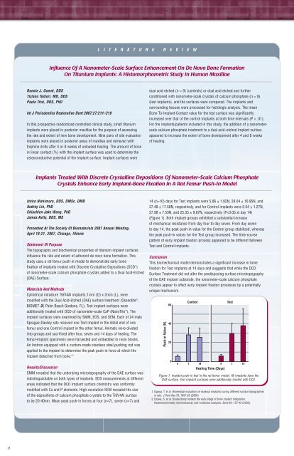
L I T E R A T U R E R E V I E WInfluence Of A Nanometer-Scale Surface Enhancement On De Novo Bone FormationOn Titanium Implants: A Histomorphometric Study In Human MaxillaeRonnie J. Goené, DDSTiziano Testori, MD, DDSPaolo Trisi, DDS, PhDInt J Periodontics Restorative Dent <strong>2007</strong>;27:211–219In this prospective randomized controlled clinical study, small titaniumimplants were placed in posterior maxillae for the purpose of assessingthe rate and extent of new bone development. Nine pairs of site evaluationimplants were placed in posterior areas of maxillae and retrieved withtrephine drills after 4 or 8 weeks of unloaded healing. The amount of bonein linear contact (%) with the implant surface was used to determine theosteoconductive potential of the implant surface. Implant surfaces weredual acid etched (n = 9) (controls) or dual acid etched and furtherconditioned with nanometer-scale crystals of calcium phosphate (n = 9)(test implants), and the surfaces were compared. The implants andsurrounding tissues were processed for histologic analysis. The meanBone-To-Implant-Contact value for the test surface was significantlyincreased over that of the control implants at both time intervals (P < .01).For the implants/patients included in this study, the addition of a nanometerscalecalcium phosphate treatment to a dual acid–etched implant surfaceappeared to increase the extent of bone development after 4 and 8 weeksof healing.Implants Treated With Discrete Crystalline Depositions Of Nanometer-Scale Calcium PhosphateCrystals Enhance Early Implant-Bone Fixation In A Rat Femur Push-In ModelIchiro Nishimura, DDS, DMSc, DMDAudrey Lin, PhDChiachien Jake Wang, PhDJames Kelly, DDS, MSPresented At The Society Of Biomaterials <strong>2007</strong> Annual Meeting,April 18-21, <strong>2007</strong>, Chicago, IllinoisStatement Of PurposeThe topography and biochemical properties of titanium implant surfacesinfluence the rate and extent of adherent de novo bone formation. Thisstudy uses a rat femur push-in model to demonstrate early bonefixation of implants treated with Discrete Crystalline Depositions (DCD )of nanometer-scale calcium phosphate crystals added to a Dual Acid-Etched(DAE) Surface.Materials And MethodsCylindrical miniature Ti6V4Al implants,1mm (D) x 2mm (L), weremodified with the Dual Acid-Etched (DAE) surface treatment (Osseotite ® ,BIOMET <strong>3i</strong>, Palm Beach Gardens, FL). Test implant surfaces wereadditionally treated with DCD of nanometer-scale CaP (NanoTite ). Theimplant surfaces were examined by SMM, EDS, and SEM. Each of 24 maleSprague-Dawley rats received one Test implant in the distal end of onefemur and one Control implant in the other femur. Animals were dividedinto groups and sacrificed after four, seven and 14 days of healing. Thefemur-implant specimens were harvested and embedded in resin blocks.An Instron equipped with a custom-made stainless steel pushing rod wasapplied to the implant to determine the peak push-in force at which theimplant detached from bone. 1,214 (n=10) days for Test implants were 5.86 ± 1.82N, 29.04 ± 10.95N, and37.48 ± 17.58N, respectively, and for Control implants were 5.54 ± 1.27N,27.98 ± 7.53N, and 25.35 ± 9.87N, respectively (P
G L O B A L E D U C A T I O N A N D E V E N T S C O N T I N U E DEuropeAdvanced Course On SurgicalTechniques And AestheticImplantologyOctober 5-6, <strong>2007</strong>Munich, GermanySpeakers: Dr. Wolfgang Bolz, Prof. Dr. MarkusHürzeler, Prof. Dr. Hannes Wachtel, Dr. Otto ZuhrThe course offers live surgeries, hands-on-trainingand scientific presentations to be able tounderstand and eventually practice the moderntechniques of oral surgery for implants andaesthetic dentistry. For additional informationand registration, call Ms. Barbara De Wildemanat +34-93-445-81-00 or emaileducation.eu@<strong>3i</strong>mplant.com.Expert Forum At NYUDiscussion leaders and topics include Dr. MichaelSonick, Soft Tissue Management For AnteriorAesthetics; Dr. Stephen Wallace, Complex SinusLift Surgery; Dr. Dennis Tarnow, ComprehensiveTreatment Planning For Ultimate Aesthetics AndFunction With Implants.For questions or more information, please callMs. Barbara De Wildeman at +34-93-445-8100or email education.eu@<strong>3i</strong>mplant.com.Find Out About The NanoTite Implant In The U.K.November 16-17, <strong>2007</strong>Birmingham, U.K.Speaker: Dr. Alan MeltzerDecember 14, <strong>2007</strong>Manchester, U.K.Speaker: Dr. Tiziano TestoriThe 7th BIOMET <strong>3i</strong> IbericSymposiumJanuary 11-12, 2008Madrid, SpainPacific RimAustralian OsseointegrationSociety (AOS) 6th BiennialConferenceOctober 17-20, <strong>2007</strong>Grand Hyatt, Melbourne, AustraliaKeynote speakers include Dr. Dennis Tarnow,sponsored by BIOMET <strong>3i</strong>, who will present twotopics pertaining to Aesthetics and OsseointegratedImplants: Buccal Site Preservation AndDevelopment and The Interdental PapillaPreservation And Formation. For more information,please go to http://www.aos07.com.au/.Take the opportunity to enjoy New York Cityby attending the NYU Expert Forum In OralImplantology to be held November 6-8, <strong>2007</strong>.This special three-day program for advancedimplant surgery is intended to generate creativeand comprehensive thinking on treatment planningalternatives for the most difficult of dental implantprocedures. The program has been developed inconjunction with Dr. Dennis Tarnow to provide abroad presentation of ideas and solutions.For information and registration, call Ms. OlgaBlanco at +34-93-470-59-50 or emailoblanco@<strong>3i</strong>mplant.com.The 5th French CongressMarch 13-15, 2008Paris, FranceFor more information, please call Ms. SylviePonthieux at +33-01-41-05-43-48 or emailsponthieux@<strong>3i</strong>mplant.com.Australian And New ZealandAssociation Of Oral AndMaxillofacial SurgeonsCongress (ANZAOMS)October 24-26, <strong>2007</strong>Hyatt Hotel, Perth, Western AustraliaThe conference features a distinguished list ofexperts in surgical education including Dr. MichaelBlock. For more information, please go tohttp://www.conferenceaction.com.au/ANZAOMS/.O N L I N E A C C E S SOur Universe ExpandsCustomers in Australia, Canada, Ireland, New Zealand and the U.K.can now access the BIOMET <strong>3i</strong> Product “Smart” Catalog.The “smart” pages are organized by implant systems, site preparation,regenerative technologies, restorative technologies and patienteducation. Designed for convenient selection, each page has linksthat guide you to related products.The BIOMET <strong>3i</strong> Product Catalog is available onlineat www.biomet<strong>3i</strong>.com.Please Note: Not all products are available outside the U.S.ARCHITECH PSR, CAM StructSURE, Certain, Encode, EP, OSSEOTITE, PREVAIL, PreFormance andQuickSeat are registered trademarks and BIOMET <strong>3i</strong>, Bone Bonding, DCD, NanoTite, Navigator,OsseoGuard, QuickBridge and Robocast are trademarks of BIOMET <strong>3i</strong>, Inc. BIOMET is a registeredtrademark and BIOMET <strong>3i</strong> and design are trademarks of BIOMET.©<strong>2007</strong> BIOMET <strong>3i</strong>, Inc. All rights reserved.REV A 08/0712


