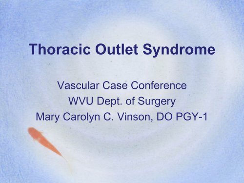
Thoracic Outlet Syndrome - WVU School of Medicine
Thoracic Outlet Syndrome - WVU School of Medicine
Thoracic Outlet Syndrome - WVU School of Medicine

You also want an ePaper? Increase the reach of your titles
YUMPU automatically turns print PDFs into web optimized ePapers that Google loves.
<strong>Thoracic</strong> <strong>Outlet</strong> <strong>Syndrome</strong>Vascular Case Conference<strong>WVU</strong> Dept. <strong>of</strong> SurgeryMary Carolyn C. Vinson, DO PGY-1
Definition• <strong>Thoracic</strong> outlet syndrome is a disease<strong>of</strong> extrinsic compression <strong>of</strong> theneurovascular structures thoracic outlet
Anatomy <strong>of</strong> <strong>Thoracic</strong> <strong>Outlet</strong>QuickTime and aTIFF (LZW) decompressorare needed to see this picture.
More Anatomyhttp://intraspec.ca/images/brachialplexus.jpg
Pathophysiology• Brachial plexus trunk & subclavian vesselsare subject to compression or irritation• Three narrow passageways @ base <strong>of</strong> necktoward the axilla & proximal arm.– Interscalene Triangle– Costoclavicular Triangle– Subcoracoid Space• Repetitive trauma to especially– Lower trunk– C8-T1 spinal nerves
Interscalene Triangle• Triangle borders– Anteriorly: anterior scalene muscle– Posteriorly: middle scalene muscle– Inferiorly: medial surface <strong>of</strong> the first rib• Area small at rest & becomes even smallerwith certain movements• Fibrous bands, cervical ribs, and anomalousmuscles, may further constrict this triangle
Costoclavicular Triangle• Borders– Anteriorly by middle 3rd <strong>of</strong> clavicle– Posteromedially by 1st rib– Posterolaterally by upper border <strong>of</strong> scapula
Subcoracoid Space• Is beneath the coracoid process justdeep to the pectoralis minor tendonQuickTime and aTIFF (LZW) decompressorare needed to see this picture.QuickTime and aTIFF (LZW) decompressorare needed to see this picture.
• Anatomic FactorsEtiology– Interscalene compression– Costoclavicular compression– Subcoracoid compression
Congenital• Cervical rib• Rudimentary first rib• Scalene muscle abnormalities• Fibrous bands• Bifid clavicle• First rib exostosis• Enlarged C7 transverse process• Omohyoid muscle abnormalities• Anomalous transverse cervical artery• Postfixed brachial plexus• Flat clavicle
Traumatic Factors• Fractured clavicle• Humeral head dislocation• Upper thorax crush injury• Sudden effort <strong>of</strong> shoulder girdlemuscles• C-spine injuries/cervical spondylosis
Clinical Presentation• Depends on which anatomic structure iscompressed in the area <strong>of</strong> the thoracic outlet– Axillary-subclavian artery–Vein• Paget-Schroetter syndrome, or effort thrombosis– Neurogenic• brachial plexus, or sympathetic nerves• Clinical syndrome results from any mixture oran isolated compression <strong>of</strong> structures
Neurologic Presentation• More common• Strenuous physical exercise precipitates• Pain & paresthesias 95% <strong>of</strong> patients– Neck, shoulder, arm & hand– Positional: arm abduction & neck hyperextension• True motor weakness w/ atrophy– Usually Ulnar nerve distribution :• hypothenar/interosseus muscles in 10%– medial arm & hand– 4th & 5th fingers• Sensory fibers on outside <strong>of</strong> nerve bundles1st affected
Arterial Presentation• Signs:– Distal embolization– Post-stenotic dilation or aneurysm <strong>of</strong> subclavian a.– True arterial occlusion• Symptoms:– Pain usually diffuse & assoc. w/ coldness,weakness, easy fatigability <strong>of</strong> hand & arm• Unilateral Raynaud's phenomenon– 7.5% patients– precipitated by hyperabduction or carrying heavyobjects
Venous Presentation• Venous obstruction less common– Effort thrombosis– Paget-Schroetter syndrome• Signs & Symptoms– Edema– Discolored– Aches
Differential Diagnosis• Neurologic,vascular, pulmonary,cardiac, and esophageal disorders.• More common Differential Diagnosisinclude– herniated cervical disk– cervical spondylosis– peripheral neuropathies
Clinical Diagnosis• Positive findings for all tests:– ⇓ or loss <strong>of</strong> the radial pulse– reproduction <strong>of</strong> symptoms• Adson/Scalene test:– Deep Breath, fully extends neck, and turns headto the side• Costoclavicular test:– shoulders drawn inferiorly and posteriorly• Hyperabduction test:– arm is hyperabducted to 180 degrees
Imaging• CXR & C-spine films:– detect cervical ribs & degenerative changes• Cervical CT performed if:– osteophytic changes & intervertebral spacenarrowing present• Angiography indicated for:– Pulsating paraclavicular mass– Absent radial pulse– Paraclavicular bruit
AngiogramShows compression <strong>of</strong> subclavian artery at two levels: proximally between clavicle and cervical rib (longarrow) and distally by subclavius muscle (short arrow).
Venogram:R subclavian vein
VenogramComplete occlusion <strong>of</strong> Left subclavian vein (arrow) where it crosses the first rib
Treatment• Physical therapy is initial treatment• Many patients get relief from non-operativetherapy– esp. for neurogenic TOS• Simple changes in posture may result inopening the thoracic outlet– PT= Strengthen muscles supporting improvedposture
Surgical Treatment for TOS• Reserved for patients w/ symptoms persistingafter aggressive physical therapy• Equals about 5% <strong>of</strong> PTs w/ TOS requiresurgery• “There are multiple compressive forces, thefirst rib is the common denominator, andextirpation <strong>of</strong> this structure is the “gold”standard for therapy.”• Urschel et al. 2003
Surgical Outcomes• > 2200 patients showed excellent orgood results after operation in over 90%<strong>of</strong> cases• Urschel et. al 1997• Symptoms recur in approx 10%• Less than 2% require re-operation
Surgical Pictures1st thoracic rib removed to decompressneurovascular structures <strong>of</strong> TOS
Recurrent <strong>Thoracic</strong> <strong>Outlet</strong><strong>Syndrome</strong>• Approx 1-2% <strong>of</strong> PTs have persistent orworsening symptoms after operation– Most have recurrence within 3 months• History, physical examination, andnerve conduction studies shouldpreformed
Types <strong>of</strong> Recurrence• Pseudorecurrence– Cervical rib or the second rib was resected instead<strong>of</strong> the first rib– First rib was resected instead <strong>of</strong> the causativecervical rib• True recurrence– First rib was incompletely resected– Excessive scar development around the brachialplexus
Re-operation for FailedInitial Operation on TOS• 80% <strong>of</strong> patients after re-operation =improvement in symptoms• 7% required a second re-operation
Summary• TOS mimics many other processes• Compression is the causative agent• 1st rib is <strong>of</strong>ten the culprit• History ⇒ PE ⇒ UNVC• XR ⇒ CT ⇒ Angio/Venogram• Physical therapy ⇒ Surgery• Note: DVT and Arterial Occlusions are treatedwith Anticoagulation/Thrombectomy
References• Thomas S. Maxey, MD, T. Brett Reece, MD, Peter I. Ellman, MD, Curtis G. Tribble, MD,Nancy Harthun, MD, Irving L. Kron, MD, John A. Kern, MD. Safety and efficacy <strong>of</strong> thesupraclavicular approach to thoracic outlet decompression.Ann Thorac Surg 2003;76:396-400• Harold C. Urschel, Jr, MD,, Amit N. Patel, MD. Surgery Remains the Most EffectiveTreatment for Paget-Schroetter <strong>Syndrome</strong>: 50 Years' Experience. Ann Thorac Surg2008;86:254-260.• Urschel HC Jr and Razzuk MA. Upper plexus thoracic outlet syndrome: optimal therapy. .Annals <strong>of</strong> <strong>Thoracic</strong> Surgery 1997 63(4):935-9.• Harold C. Urschel Jr and Amit Patel. <strong>Thoracic</strong> outlet syndromes. Current Treatment Optionsin Cardiovascular <strong>Medicine</strong>. Vol 5, No 2. April 2003. 1092-8464• Urschel HC Jr, Razzuk MA.The failed operation for thoracic outlet <strong>Syndrome</strong>: the difficulty <strong>of</strong>diagnosis and management.Ann Thorac Surg. 1986 Nov;42(5):523-8• http://brighamrad.harvard.edu/Cases/bwh/hcache/170/full.html• http://www.ajronline.org/cgi/content-nw/full/183/1/113/FIG9• http://www.ctsnet.org/doc/7628