Myositis ossificans traumatica of the deltoid ligament in a 34 year old ...
Myositis ossificans traumatica of the deltoid ligament in a 34 year old ...
Myositis ossificans traumatica of the deltoid ligament in a 34 year old ...

Create successful ePaper yourself
Turn your PDF publications into a flip-book with our unique Google optimized e-Paper software.
0008-3194/2010/229–242/$2.00/©JCCA 2010<strong>Myositis</strong> ossifi cans <strong>traumatica</strong> <strong>of</strong> <strong>the</strong> <strong>deltoid</strong><strong>ligament</strong> <strong>in</strong> a <strong>34</strong> <strong>year</strong> <strong>old</strong> recreational ice hockeyplayer with a 15 <strong>year</strong> post-trauma follow-up: acase report and review <strong>of</strong> <strong>the</strong> literatureDr. Brad Muir, HBSc(K<strong>in</strong>), DC, FCCSS(C)*<strong>Myositis</strong> <strong>ossificans</strong> <strong>traumatica</strong> is a relatively common<strong>in</strong>jury associated with sports especially those <strong>in</strong>volv<strong>in</strong>gcontact. It cont<strong>in</strong>ues to frustrate both athlete and healthpractitioner alike due to its cont<strong>in</strong>ued lack <strong>of</strong> treatmentoptions and a lengthy natural history. This case studychronicles <strong>the</strong> observation <strong>of</strong> a <strong>34</strong> <strong>year</strong> <strong>old</strong> recreationalice hockey player who presented 7 <strong>year</strong>s post-trauma,was diagnosed with myositis <strong>ossificans</strong> <strong>traumatica</strong>and was followed up on 8 <strong>year</strong>s later (15 <strong>year</strong>s posttrauma).This case report is suspected to be <strong>the</strong> fi rstpublished case study <strong>of</strong> its k<strong>in</strong>d. The literature reviewoutl<strong>in</strong>es <strong>the</strong> various types <strong>of</strong> myositis <strong>ossificans</strong>, its<strong>in</strong>cidence, pathogenesis, differential diagnoses <strong>in</strong>clud<strong>in</strong>gosteosarcoma, and <strong>the</strong> various methods/modalitiesreported <strong>in</strong> its treatment.(JCCA 2010; 54(4):229–242)key words: myositis <strong>ossificans</strong> <strong>traumatica</strong>,heterotopic ossification, post-traumatic myositis<strong>ossificans</strong>, ice hockey, <strong>deltoid</strong> <strong>ligament</strong>La myosite ossifiante traumatique est une blessurerelativement courante liée à la pratique des sports,particulièrement les sports de contact. Elle cont<strong>in</strong>uede frustrer les athlètes et les pr<strong>of</strong>essionnels de la santéen raison de l’absence d’options de traitement et dufait qu’elle existe depuis longtemps. Cette étude de casraconte l’observation d’un joueur de hockey sur glacede ligue récréative de <strong>34</strong> ans qui reçut un diagnosticde myosite ossifiante traumatique après 7 ans, etqu’on exam<strong>in</strong>a ensuite 8 ans plus tard (15 ans après letraumatisme), Cette étude de cas est probablement lapremière en son genre. Une analyse de la documentationà ce sujet souligne les divers types de myosite ossifiante,sa fréquence, sa pathogénie, les différents diagnostics,notamment l’ostéosarcome, et les diverses méthodes/modalités signalées dans son traitement.(JCCA 2010; 54(4):229–242)mots clés : myosite ossifiante traumatique,ossification hétéropique, myosite ossifiante posttraumatique,hockey sur glace, <strong>ligament</strong> deltoïdeIntroduction<strong>Myositis</strong> <strong>ossificans</strong> <strong>traumatica</strong> (MOT) is <strong>of</strong>ten def<strong>in</strong>ed asheterotopic, non-neoplastic proliferation <strong>of</strong> bone <strong>in</strong> anarea previously exposed to trauma and hematoma. 1,2The most common areas that are affected by MOTare <strong>the</strong> quadriceps femoris, brachialis anticus, and <strong>the</strong>adductor muscles <strong>of</strong> <strong>the</strong> thigh 3 although it may occuranywhere. It can happen at any age, but occurs most frequently<strong>in</strong> adolescents and young athletes, with over half<strong>of</strong> <strong>the</strong> cases occurr<strong>in</strong>g <strong>in</strong> <strong>the</strong> third decade. 1,3 MOT is consideredrare <strong>in</strong> children under 10 <strong>year</strong>s <strong>of</strong> age 1 and malesare more <strong>of</strong>ten affected than females. 3The <strong>in</strong>cidence <strong>of</strong> MOT follow<strong>in</strong>g quadriceps contusionsis reported at vary<strong>in</strong>g percentages depend<strong>in</strong>g on <strong>the</strong>severity <strong>of</strong> <strong>the</strong> <strong>in</strong>jury. Mild contusions have shown an <strong>in</strong>cidence<strong>of</strong> MOT <strong>of</strong> 0–9%, moderate to severe 17–72%and recurrent contusion 100%. 4,5 Contusions <strong>of</strong> <strong>the</strong> thighare generally graded accord<strong>in</strong>g to <strong>the</strong> subsequent loss <strong>of</strong>range <strong>of</strong> motion at <strong>the</strong> knee jo<strong>in</strong>t. Contusions are gradedas mild (active ROM greater than 90º), moderate (45–90º)* Assistant Pr<strong>of</strong>essor, Canadian Memorial Chiropractic College, 177 Carnwith Drive East, Brookl<strong>in</strong>, ON L1M 2J5.Tel: (416) 482-2546 ext. 123 (CMCC), (905) 428-9370 (practice)© JCCA 2010J Can Chiropr Assoc 2010; 54(4) 229
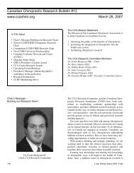
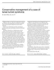
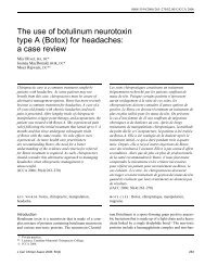
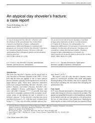
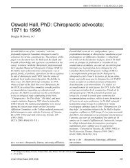
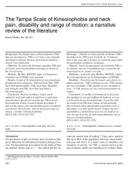
<strong>Myositis</strong> ossifi cans <strong>traumatica</strong>Figure 1 Photograph <strong>of</strong> patient’s bilateral ankles.The arrow <strong>in</strong>dicates <strong>the</strong> area <strong>of</strong> myositis <strong>ossificans</strong><strong>traumatica</strong> <strong>of</strong> <strong>the</strong> left medial ankle.Figure 2 Photograph <strong>of</strong> <strong>the</strong> patient’s left medial ankle.The arrow <strong>in</strong>dicates <strong>the</strong> area <strong>of</strong> myositis <strong>ossificans</strong><strong>traumatica</strong>.and severe (less than 45º). 5 Arr<strong>in</strong>gton 6 grades thigh contusionsas: mild – near normal ROM, local tenderness,no gait abnormality; moderate – swollen tender muscle,75% ROM, antalgic gait; and severe – marked tenderness/swell<strong>in</strong>g, 50% ROM with a severe limp. Muscle contusion<strong>in</strong>juries are second only to stra<strong>in</strong> <strong>in</strong>juries as a majorcause <strong>of</strong> morbidity <strong>in</strong> <strong>the</strong> modern athlete. 2This paper presents a case study <strong>of</strong> a <strong>34</strong> <strong>year</strong> <strong>old</strong> recreationalice hockey player who was diagnosed with MOT <strong>of</strong><strong>the</strong> <strong>deltoid</strong> <strong>ligament</strong> <strong>of</strong> <strong>the</strong> ankle 7 <strong>year</strong>s post-trauma andfollowed up on 8 <strong>year</strong>s later (15 <strong>year</strong>s post-trauma).Case ReportHistoryA <strong>34</strong> <strong>year</strong> <strong>old</strong> recreational hockey player presented withan unusual bony lump just anterior and <strong>in</strong>ferior to hisleft medial malleolus (see Figure 1 and 2). He reportedthat <strong>the</strong> lump had started follow<strong>in</strong>g block<strong>in</strong>g a shot <strong>in</strong> ahockey game 7 <strong>year</strong>s previously. He was able to f<strong>in</strong>ish<strong>the</strong> game and had partial weightbear<strong>in</strong>g follow<strong>in</strong>g <strong>the</strong>game. The foot became <strong>in</strong>creas<strong>in</strong>gly swollen and wastreated with rest and ice. No radiographs were taken at<strong>the</strong> time <strong>of</strong> <strong>the</strong> <strong>in</strong>itial <strong>in</strong>jury. He reported that <strong>the</strong> lumphad <strong>in</strong>creased <strong>in</strong> size slowly over <strong>the</strong> next few <strong>year</strong>s. Two<strong>year</strong>s after <strong>the</strong> <strong>in</strong>jury, he had returned to play competitivehockey and didn’t recall any limitation although <strong>the</strong>lump had not resolved. Four <strong>year</strong>s post <strong>in</strong>jury, he beganto notice <strong>the</strong> lump was larger and was becom<strong>in</strong>g <strong>in</strong>creas<strong>in</strong>glyirritated from <strong>the</strong> direct pressure <strong>of</strong> tighten<strong>in</strong>g hisskate laces. He was also gett<strong>in</strong>g sharp pa<strong>in</strong> <strong>in</strong> his footwith pivot<strong>in</strong>g while skat<strong>in</strong>g and noticed decreased range<strong>of</strong> motion especially <strong>in</strong> dorsiflexion. Seven <strong>year</strong>s post<strong>in</strong>jury,he began to notice <strong>in</strong>creased pa<strong>in</strong> when turn<strong>in</strong>garound pylons while demonstrat<strong>in</strong>g drills as a hockeycoach. Stopp<strong>in</strong>g, and backward skat<strong>in</strong>g had also becomepa<strong>in</strong>ful and pivot<strong>in</strong>g was still difficult due to pa<strong>in</strong>. Healso noted <strong>in</strong>creas<strong>in</strong>g difficulty go<strong>in</strong>g up stairs and fel<strong>the</strong> had to lift his leg higher to get his foot flat on <strong>the</strong> nextstair. At this po<strong>in</strong>t, he presented to <strong>the</strong> cl<strong>in</strong>ic for exam<strong>in</strong>ationand possible imag<strong>in</strong>g.There was no history <strong>of</strong> recent <strong>in</strong>fection, sudden unexpla<strong>in</strong>edweight loss or night pa<strong>in</strong>. Systems review revealeda history <strong>of</strong> seasonal bronchitis but was o<strong>the</strong>rwiseunremarkable. Previous medical history revealed a previousright shoulder surgery for recurrent subluxation butno o<strong>the</strong>r major traumas or motor vehicle accidents. Hedid not report any previous <strong>in</strong>jury to <strong>the</strong> left foot or ankle.Family history did not reveal anyth<strong>in</strong>g relevant. He is ano<strong>the</strong>rwise healthy <strong>in</strong>dividual who exercises regularly and230 J Can Chiropr Assoc 2010; 54(4)
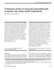
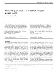
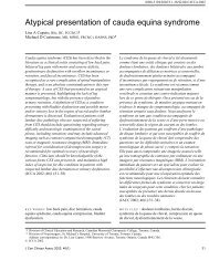
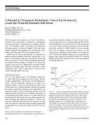
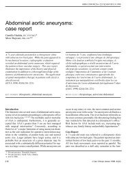
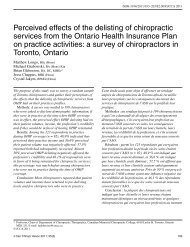
B MuirFigure 3 Medial sagittal CT image <strong>of</strong> <strong>the</strong> left ankle 7<strong>year</strong>s post <strong>in</strong>jury show<strong>in</strong>g an irregular ossifi c densityantero<strong>in</strong>ferior to <strong>the</strong> medial malleolus <strong>in</strong>dicat<strong>in</strong>gmyositis <strong>ossificans</strong> <strong>traumatica</strong> <strong>of</strong> <strong>the</strong> <strong>deltoid</strong> <strong>ligament</strong>.The white arrow <strong>in</strong>dicates <strong>the</strong> area <strong>of</strong> ossification.does not smoke. This patient is a factory worker and apart-time hockey coach.He did not report any numbness or t<strong>in</strong>gl<strong>in</strong>g <strong>in</strong> his footor ankle.Physical exam<strong>in</strong>ationUpon exam<strong>in</strong>ation, <strong>the</strong> patient was able to ambulate normally.A visible prom<strong>in</strong>ence was noted anterior and <strong>in</strong>feriorto <strong>the</strong> medial malleolus <strong>of</strong> his left foot. Active andpassive range <strong>of</strong> motion revealed a decrease <strong>of</strong> 40% <strong>in</strong>dorsiflexion, <strong>in</strong>version and eversion due to restriction notpa<strong>in</strong>. Plantar flexion was decreased 10–20% due to s<strong>of</strong>ttissue restrictions. (The patient felt he could go fur<strong>the</strong>rbut was tight and no bony end feel was noted.) Jo<strong>in</strong>t play<strong>of</strong> <strong>the</strong> midtarsal jo<strong>in</strong>ts was with<strong>in</strong> normal limits. O<strong>the</strong>rranges <strong>of</strong> motion were full and pa<strong>in</strong> free. Strength wasslightly decreased compared to <strong>the</strong> right side <strong>in</strong> all rangesbut <strong>the</strong>re was no pa<strong>in</strong>. Palpation <strong>of</strong> <strong>the</strong> bony mass revealedsome po<strong>in</strong>t tenderness but <strong>the</strong> foot and ankle wereo<strong>the</strong>rwise pa<strong>in</strong> free. Radiographs and a CT were orderedfor this patient at this po<strong>in</strong>t <strong>in</strong> <strong>the</strong> exam<strong>in</strong>ation (see Figure3–6) .DiagnosisThe patient was subsequently diagnosed with myositis <strong>ossificans</strong><strong>traumatica</strong> <strong>of</strong> <strong>the</strong> left <strong>deltoid</strong> <strong>ligament</strong> <strong>of</strong> <strong>the</strong> ankle.Figure 4 Axial CT image <strong>of</strong> <strong>the</strong> left ankle 7 <strong>year</strong>s post<strong>in</strong>jury show<strong>in</strong>g an irregular ossifi c density medial to <strong>the</strong>medial malleolus. The white arrow <strong>in</strong>dicates <strong>the</strong> area <strong>of</strong>ossifi cation.Plan <strong>of</strong> managementThe <strong>in</strong>itial plan <strong>of</strong> management for this patient was to ruleout any <strong>in</strong>sidious diagnosis, <strong>in</strong>clud<strong>in</strong>g osteosarcoma, with<strong>the</strong> imag<strong>in</strong>g. The diagnosis <strong>of</strong> osteosarcoma was unlikelydue to <strong>the</strong> slow, progressive nature <strong>of</strong> <strong>the</strong> <strong>in</strong>jury and itslack <strong>of</strong> pa<strong>in</strong> but it was felt imag<strong>in</strong>g was prudent <strong>in</strong> thiscase. Follow<strong>in</strong>g <strong>the</strong> diagnosis, <strong>the</strong> patient was <strong>in</strong>formed<strong>of</strong> <strong>the</strong> natural history <strong>of</strong> MOT and that <strong>the</strong>re were fewproven treatment options available at that time. Due to<strong>the</strong> relatively m<strong>in</strong>or irritation experienced dur<strong>in</strong>g hockeyand activities <strong>of</strong> daily liv<strong>in</strong>g <strong>the</strong> patient was not <strong>in</strong>terested<strong>in</strong> surgery and decided to treat <strong>the</strong> area symptomaticallywith ice and rest as needed. A “wait and see” approachwas taken with <strong>in</strong>structions that if <strong>the</strong> lump was <strong>in</strong>creas<strong>in</strong>g<strong>in</strong> size and/or pa<strong>in</strong> that <strong>the</strong> patient was to return forfur<strong>the</strong>r exam<strong>in</strong>ation.15 Year Follow-upThe patient, now 42 <strong>year</strong>s <strong>old</strong>, was contacted to follow-upon his status. The patient reported that he felt <strong>the</strong>re waslittle change <strong>in</strong> <strong>the</strong> size <strong>of</strong> <strong>the</strong> lesion on his left ankle. Hestill had similar restrictions with regard to his on-ice activities<strong>in</strong>clud<strong>in</strong>g quick pivot<strong>in</strong>g and demonstrat<strong>in</strong>g drillsand his activities <strong>of</strong> daily liv<strong>in</strong>g <strong>in</strong>clud<strong>in</strong>g go<strong>in</strong>g up stairs.He felt <strong>the</strong>se activities were difficult due to his decreasedJ Can Chiropr Assoc 2010; 54(4) 231
<strong>Myositis</strong> ossifi cans <strong>traumatica</strong>Figure 5 Coronal CT image <strong>of</strong> <strong>the</strong> left ankle 7<strong>year</strong>s post <strong>in</strong>jury show<strong>in</strong>g an irregular ossifi c densityantero<strong>in</strong>ferior to <strong>the</strong> medial malleolus. The white arrow<strong>in</strong>dicates <strong>the</strong> area <strong>of</strong> ossifi cation.ability to dorsiflex his ankle. The patient reported that hehad learned to avoid certa<strong>in</strong> aggravat<strong>in</strong>g activities because<strong>of</strong> <strong>the</strong> length <strong>of</strong> time <strong>of</strong> his <strong>in</strong>jury. No follow-up physicalexam was performed. Radiographic images were obta<strong>in</strong>edat follow-up. The follow-up radiographs show that littlechange has occurred <strong>in</strong> <strong>the</strong> size <strong>of</strong> <strong>the</strong> MOT as visualized(see Figure 7 and 8).DiscussionTypes <strong>of</strong> <strong>Myositis</strong> Ossifi cansHeterotopic bone formation <strong>in</strong> s<strong>of</strong>t tissue has historicallybeen classified as myositis <strong>ossificans</strong>. Many authors nowconsider <strong>the</strong> term myositis <strong>ossificans</strong> to be a misnomerargu<strong>in</strong>g that <strong>the</strong>re is no <strong>in</strong>flammatory process and <strong>the</strong> conditionmay or may not <strong>in</strong>volve bone or muscle tissue. 7,8<strong>Myositis</strong> <strong>ossificans</strong> consists <strong>of</strong> three ma<strong>in</strong> types – congenital,idiopathic, and traumatic (see chart 1). <strong>Myositis</strong><strong>ossificans</strong> <strong>traumatica</strong> (MOT) is also known <strong>in</strong> <strong>the</strong> literatureby specific names given to MOT <strong>in</strong> specific areas <strong>of</strong><strong>the</strong> body (see chart 2). 3,7,9,10 Arr<strong>in</strong>gton 6 classifies MOT<strong>in</strong>to three types as does Mestan and Bassano (see chart3). 10The pathogenesis <strong>of</strong> this <strong>in</strong>jury rema<strong>in</strong>s unclear at thistime. Some <strong>of</strong> <strong>the</strong> common <strong>the</strong>ories <strong>in</strong>clude: 10Figure 6 Radiograph left ankle AP view 7 <strong>year</strong>s post<strong>in</strong>jury show<strong>in</strong>g an irregular ossifi c density <strong>in</strong>ferior to <strong>the</strong>medial malleolus. The white arrow <strong>in</strong>dicates <strong>the</strong> area <strong>of</strong>ossifi cation.1. transformation <strong>of</strong> muscle hematoma to bone;2. hematoma calcification;3. <strong>in</strong>tramuscular bone formation from detached periostealflaps;4. osteoblast proliferation from periosteal rupture;5. metaplasia <strong>of</strong> <strong>in</strong>tramuscular connective tissue cells;6. <strong>in</strong>dividual predisposition.It is believed that blunt trauma to <strong>the</strong> extremity creates acompression wave travell<strong>in</strong>g through s<strong>of</strong>t-tissue crush<strong>in</strong>g<strong>the</strong> deepest muscle aga<strong>in</strong>st <strong>the</strong> bone. The force is transmittedthrough <strong>the</strong> fluid compartment <strong>of</strong> all <strong>of</strong> <strong>the</strong> layers<strong>of</strong> muscles but <strong>the</strong> damage usually occurs <strong>in</strong> <strong>the</strong> layer thatis next to <strong>the</strong> bone. 5232 J Can Chiropr Assoc 2010; 54(4)
B MuirFigure 7 Radiograph left ankle Oblique view 15 <strong>year</strong>spost <strong>in</strong>jury show<strong>in</strong>g an irregular ossifi c density <strong>in</strong>feriorto <strong>the</strong> medial malleolus. The black arrow <strong>in</strong>dicates <strong>the</strong>area <strong>of</strong> ossifi cation.Wang 7 suggests that <strong>the</strong>re are several steps follow<strong>in</strong>g<strong>the</strong> <strong>in</strong>jury to <strong>the</strong> tissue that leads to <strong>the</strong> development <strong>of</strong>MOT. There is cellular damage caus<strong>in</strong>g necrotic debristhat is subsequently removed through <strong>the</strong> <strong>in</strong>vasion <strong>of</strong> histiocytes.Fibroblasts from <strong>the</strong> endomysium <strong>the</strong>n assail <strong>the</strong>Figure 8 Radiograph left ankle AP view 15 <strong>year</strong>s post<strong>in</strong>jury show<strong>in</strong>g an irregular ossifi c density <strong>in</strong>ferior to <strong>the</strong>medial malleolus. The white arrow <strong>in</strong>dicates <strong>the</strong> area <strong>of</strong>ossifi cation.<strong>in</strong>jured cells and mesenchymal cells beg<strong>in</strong> to proliferate.The fibroblasts and mesenchymal cells produce osteoidand chondroid tissue that lays down <strong>the</strong> groundworkfor <strong>the</strong> formation <strong>of</strong> bone with<strong>in</strong> <strong>the</strong> damaged tissue. Thiscan occur as early as 4 to 5 days after <strong>the</strong> <strong>in</strong>jury has occurred.A particularly important aspect <strong>of</strong> MOT occurs at thisJ Can Chiropr Assoc 2010; 54(4) 233
<strong>Myositis</strong> ossifi cans <strong>traumatica</strong>Chart 1 Types <strong>of</strong> <strong>Myositis</strong> Ossifi cans1. Congenital 24• three <strong>in</strong>herited diseases that are characterized by heterotopic bone formation 24• <strong>the</strong>se are: fibrousdysplasia <strong>ossificans</strong> progressive (FOP), progressive osseous heteroplasia (POH), andAlbright’s hereditary osteodystrophy (AHO)• although rare, <strong>the</strong>y should be <strong>in</strong>cluded <strong>in</strong> children present<strong>in</strong>g with myositis <strong>ossificans</strong> <strong>of</strong> an idiopathicnature.Fibrousdysplasia <strong>ossificans</strong>progressiva (FOP):• also called “stone mandisease” and myositis<strong>ossificans</strong> progressive• first described <strong>in</strong> <strong>the</strong> late 17 thcentury• characterized by <strong>the</strong>formation <strong>of</strong> bone <strong>in</strong> tendonsand muscles.• manifests itself <strong>in</strong> childhoodand is slowly progressiveresult<strong>in</strong>g <strong>in</strong> <strong>the</strong> formation<strong>of</strong> a “second skeleton” <strong>in</strong>patients.• very uncommon occurr<strong>in</strong>g <strong>in</strong>approximately 1 <strong>in</strong> 2 million.2. IdiopathicProgressive osseousheteroplasia (POH):• first described by Kaplan <strong>in</strong>1994• orig<strong>in</strong>ally thought to beatypical FOP• <strong>in</strong> contrast to FOP,ossification beg<strong>in</strong>s <strong>in</strong> <strong>the</strong>dermis and spreads to <strong>the</strong>subcutaneous tissue andfascia with no skeletalabnormalities.• POH is very rare and beg<strong>in</strong>s<strong>in</strong> childhood, similar to FOP.Albright’s hereditaryosteodystrophy (AHO):• manifests with a certa<strong>in</strong>phenotypical presentationalong with subcutaneouscalcifications about <strong>the</strong> limbjo<strong>in</strong>ts• phenotypical features<strong>in</strong>cludes: short stature, around facies, hypertelorism,and shortness <strong>of</strong> <strong>the</strong> 4 th and5 th digits.• mental retardation is alsocommon <strong>in</strong> <strong>the</strong>se patients.• controversy regard<strong>in</strong>g <strong>the</strong> idiopathic nature <strong>of</strong> myositis <strong>ossificans</strong> and its nam<strong>in</strong>g.• some authors use <strong>the</strong> term myositis <strong>ossificans</strong> circumscripta (MOC) to describe <strong>the</strong> heterotopicossification <strong>of</strong> s<strong>of</strong>t tissue <strong>of</strong> an idiopathic nature 25• o<strong>the</strong>r authors use MOC as synonymous with myositis <strong>ossificans</strong> <strong>traumatica</strong>8, 16• has been reported follow<strong>in</strong>g burns, tetanus, and polio and neurogenic <strong>in</strong>juries <strong>in</strong>clud<strong>in</strong>g sp<strong>in</strong>al cord<strong>in</strong>juries, closed head <strong>in</strong>juries, central nervous system <strong>in</strong>fections, and stroke 1,93. Traumatic• difficult to adequately research MOT due to its many synonyms.• <strong>the</strong>se <strong>in</strong>clude post-traumatic myositis <strong>ossificans</strong>, myositis <strong>ossificans</strong> circumscripta, pseudomalignan<strong>the</strong>terotopic ossification, pseudomalignant myositis <strong>ossificans</strong>, pseudomalignant osseous tumor <strong>of</strong> s<strong>of</strong>ttissue, extraosseous localized non-neoplastic bone or cartilage formation 3• some radiologists consider <strong>the</strong> traumatic ossification <strong>of</strong> <strong>ligament</strong>s specifically to be called dystrophiccalcification.• dystrophic calcification is def<strong>in</strong>ed as “calcium salt deposits <strong>in</strong> <strong>the</strong> presence <strong>of</strong> normal calcium/phosphorus metabolism <strong>in</strong>volv<strong>in</strong>g tissues that do not physiologically calcify” 26 which may notadequately describe our case2<strong>34</strong> J Can Chiropr Assoc 2010; 54(4)
B Muir“Rider’s Bone”, Prussian’s Disease or“Saddle Tumor”Chart 2 Specifi c Areas <strong>of</strong> MOT with Specifi c Names 3,7,10,27,28• found <strong>in</strong> <strong>the</strong> hip adductors <strong>in</strong> horseback riders and jockeysfrom <strong>the</strong> pressure <strong>of</strong> <strong>the</strong> saddle aga<strong>in</strong>st upper/<strong>in</strong>ner thigh.• described by Geschickter and Maseritz <strong>in</strong> 1938 27 asossification <strong>in</strong> <strong>the</strong> thighs <strong>of</strong> cavalry men.Author’s Note: <strong>the</strong> terms Prussian’s Disease and saddletumor were attributed to Ackerman 11 by Danchik et al. 3 butno mention <strong>of</strong> <strong>the</strong>se terms were found when exam<strong>in</strong><strong>in</strong>g <strong>the</strong>Ackerman paper. A subsequent literature search <strong>of</strong> <strong>the</strong>se termsdid not reveal any articles on PubMed.Pelligr<strong>in</strong>i-Stieda Disease • found <strong>in</strong> <strong>the</strong> medial collateral <strong>ligament</strong> <strong>of</strong> <strong>the</strong> knee.• first described by Pellegr<strong>in</strong>i <strong>in</strong> 1905. Kohler reporteda similar case <strong>in</strong> 1905 as well. Stieda, unaware <strong>of</strong> <strong>the</strong>previous works <strong>of</strong> Pellegr<strong>in</strong>i or Kohler reported a similarcase <strong>in</strong> 1907. 28• most commonly found <strong>in</strong> football players.“Rider’s Bone” • found near <strong>the</strong> attachment <strong>of</strong> <strong>the</strong> hamstr<strong>in</strong>gs at <strong>the</strong> ischialtuberosity.• Danchik et al. 3 suggests it is secondary to hypervascularityfollow<strong>in</strong>g an avulsion <strong>of</strong> <strong>the</strong> hamstr<strong>in</strong>gs• has also been called “Rider’s Bone” due to <strong>the</strong> proclivity <strong>of</strong>this <strong>in</strong>jury <strong>in</strong> horseback riders 3• Common <strong>in</strong> spr<strong>in</strong>ters, hurdlers and cheerleadersAuthor’s Note: <strong>in</strong> check<strong>in</strong>g Danchik’s reference <strong>of</strong> Ackerman 11no mention <strong>of</strong> “Rider’s Bone” was found suggest<strong>in</strong>gthat <strong>the</strong> “Rider’s Bone” mentioned may be similar to <strong>the</strong>aforementioned with “Prussian’s Disease” and <strong>the</strong> ischialtuberosity <strong>in</strong>jury as described is misnamed.“Rifle Shoulder” • found <strong>in</strong> <strong>the</strong> pectoralis major or anterior <strong>deltoid</strong> muscledue to <strong>the</strong> repeated kickback <strong>of</strong> a rifle or due to carry<strong>in</strong>g<strong>the</strong> rifle on <strong>the</strong> shoulder.• most common <strong>in</strong> s<strong>old</strong>iers particularly <strong>the</strong> <strong>in</strong>fantry.• described by Geschickter and Maseritz <strong>in</strong> 1938 27 as <strong>the</strong>“rifle shoulders” <strong>of</strong> <strong>in</strong>fantry men.“Fencer’s Bone” • found <strong>in</strong> <strong>the</strong> brachialis anticus due to <strong>the</strong> overextension <strong>of</strong><strong>the</strong> forearm 7• most common <strong>in</strong> fencers and baseball pitchers.“Dancer’s Bone” • found <strong>in</strong> <strong>the</strong> soleus muscle 7Author’s Note: a subsequent literature search <strong>of</strong> <strong>the</strong> terms“Fencer’s Bone” and “Dancer’s Bone” did not reveal anyarticles on PubMed.J Can Chiropr Assoc 2010; 54(4) 235
<strong>Myositis</strong> ossifi cans <strong>traumatica</strong>Chart 3 Classifi cation <strong>of</strong> MOTArr<strong>in</strong>gton Classification 6 Mestan and Bassano Classification 101. stalk <strong>the</strong>re is a stalk attachmentto <strong>the</strong> underly<strong>in</strong>g bone2. periosteal <strong>the</strong>re is total cont<strong>in</strong>uitybetween <strong>the</strong> underly<strong>in</strong>gbone and <strong>the</strong> heterotopicbone1. parosteal <strong>the</strong> ossification is found near oraga<strong>in</strong>st bone2. periosteal <strong>the</strong> periosteum is torn awayfrom <strong>the</strong> underly<strong>in</strong>g cortex dueto <strong>the</strong> trauma. The result<strong>in</strong>g<strong>in</strong>jury may have a sunburstappearance.Note: 60% <strong>of</strong> MOT’s have aperiosteal reaction secondary to<strong>the</strong> trauma. 123. broad-based is a large base <strong>of</strong>attachment to <strong>the</strong>underly<strong>in</strong>g muscle3. extraosseus <strong>the</strong> heterotopic bone is found<strong>in</strong> <strong>the</strong> substance <strong>of</strong> <strong>the</strong> <strong>in</strong>volvedmusclestage and is critical to <strong>the</strong> differentiation <strong>of</strong> MOT andosteosarcoma. The previously mentioned steps beg<strong>in</strong> <strong>in</strong><strong>the</strong> least damaged area mov<strong>in</strong>g toward <strong>the</strong> area <strong>of</strong> mostdamage. As a result, lamellar new bone is laid down <strong>in</strong>a centripetal pattern that is referred to as “<strong>the</strong> zonal phenomenon”(see Figure 9). 7,11Zonal phenomenon seems to be first mentioned byAckerman <strong>in</strong> 1958. 11 He states, “I have been impressed bywhat I have designated as zone phenomena (<strong>in</strong>ner, middleand outer zones).” He fur<strong>the</strong>r states, “… <strong>the</strong>y are welldocumented <strong>in</strong> examples <strong>of</strong> classic myositis <strong>ossificans</strong>,but no one has mentioned it, probably because <strong>the</strong> processis <strong>of</strong>ten recognized by typical histories and locations.” 11Beg<strong>in</strong>n<strong>in</strong>g <strong>in</strong> <strong>the</strong> second week to second month, boneis formed surround<strong>in</strong>g <strong>the</strong> area and <strong>the</strong>n moves <strong>in</strong>wardtoward <strong>the</strong> site <strong>of</strong> <strong>in</strong>jury giv<strong>in</strong>g layers <strong>of</strong> vary<strong>in</strong>g tissueactivity. Mov<strong>in</strong>g <strong>in</strong>ward, <strong>the</strong> outside layer is bone, <strong>the</strong>middle layer is an area <strong>of</strong> extensive osteoblastic and fibroblasticactivity, and <strong>the</strong> central layer is an area <strong>of</strong> highlycellular tissue with a lot <strong>of</strong> cell differentiation and necroticcells. 7,8 Osteosarcoma does not show this zonal phenomenon.The calcification seen with osteosarcoma beg<strong>in</strong>s <strong>in</strong><strong>the</strong> central areas <strong>of</strong> <strong>the</strong> tumour. Unfortunately, if a biopsycentre <strong>of</strong> impact siteFirst zone <strong>of</strong> bone formation.Figure 9 The “Zonal Phenomenon” <strong>of</strong> MOT.Direction <strong>of</strong> new boneformation <strong>in</strong> MOT. Inosteosarcoma, <strong>the</strong>calcification beg<strong>in</strong>scentrally and movesoutward from <strong>the</strong> lesion.<strong>of</strong> <strong>the</strong> area is done and <strong>the</strong> majority <strong>of</strong> <strong>the</strong> cells come from<strong>the</strong> middle and central layers, an <strong>in</strong>correct diagnosis <strong>of</strong>osteosarcoma can been made and improper treatment hasfollowed, <strong>in</strong>clud<strong>in</strong>g amputation. 3,12Typical Patient PresentationA typical patient presentation <strong>in</strong>cludes a history <strong>of</strong> traumato <strong>the</strong> affected area with <strong>in</strong>creased difficulty <strong>in</strong> mov<strong>in</strong>g236 J Can Chiropr Assoc 2010; 54(4)
B MuirChart 4 <strong>Myositis</strong> Ossifi cans Traumatica and OsteosarcomaA comparison <strong>of</strong> <strong>the</strong> two conditions <strong>in</strong>cludes <strong>the</strong> follow<strong>in</strong>g: 23<strong>Myositis</strong> Ossificans TraumaticaOsteosarcoma• no history <strong>of</strong> trauma (although trauma can• history <strong>of</strong> traumabut can be elevated 2• found <strong>in</strong> <strong>the</strong> diaphysisoccur <strong>in</strong> up to 40% <strong>of</strong> cases) 17• cortex usually <strong>in</strong>tact even with a periosteal • found <strong>in</strong> <strong>the</strong> metaphysisreaction• cortex <strong>of</strong>ten violated• pa<strong>in</strong> and mass decrease with time• pa<strong>in</strong> and mass <strong>in</strong>crease with time• activity-related pa<strong>in</strong>• night pa<strong>in</strong> alkal<strong>in</strong>e phosphatase levels• alkal<strong>in</strong>e phosphatase levels <strong>of</strong>ten normal elevatedand us<strong>in</strong>g <strong>the</strong> affected limb. In football, an oppos<strong>in</strong>g player’shelmet to <strong>the</strong> thigh and <strong>in</strong> hockey and soccer, an oppos<strong>in</strong>gplayers knee to <strong>the</strong> thigh are common mechanisms<strong>of</strong> <strong>in</strong>jury. 12 Trauma (direct and repetitive) is reported <strong>in</strong>60–70% <strong>of</strong> <strong>the</strong> cases. 3 As previously mentioned <strong>the</strong> onsetseems to be related to <strong>the</strong> severity <strong>of</strong> <strong>the</strong> <strong>in</strong>jury.In our case, <strong>the</strong> athlete reported a severe, yet commoncause <strong>of</strong> trauma (block<strong>in</strong>g a shot). However, he did notfeel it was severe enough to get a radiograph or seek treatmentat <strong>the</strong> time <strong>of</strong> <strong>the</strong> <strong>in</strong>jury.A cl<strong>in</strong>ician should <strong>in</strong>itially suspect <strong>the</strong> early stages <strong>of</strong>MOT if <strong>the</strong> pa<strong>in</strong>, swell<strong>in</strong>g and tenderness <strong>of</strong> <strong>the</strong> affectedarea has not responded to conservative managementwith<strong>in</strong> <strong>the</strong> first 4–5 days. 12 This suspicion should <strong>in</strong>creasefur<strong>the</strong>r if <strong>the</strong> athlete reports that <strong>the</strong> symptoms have notimproved significantly or have worsened with<strong>in</strong> 10–14days <strong>of</strong> <strong>the</strong> <strong>in</strong>jury. 12A physical exam<strong>in</strong>ation <strong>of</strong> an athlete suspected <strong>of</strong> hav<strong>in</strong>gMOT beg<strong>in</strong>s with observation. If <strong>the</strong> <strong>in</strong>jury is to <strong>the</strong>lower limb, particularly <strong>the</strong> thigh, an antalgic gait withlimited weight bear<strong>in</strong>g <strong>of</strong> <strong>the</strong> affected limb will <strong>of</strong>ten benoted as <strong>the</strong> athlete enters <strong>the</strong> treat<strong>in</strong>g room. The athletewill <strong>of</strong>ten have difficulty gett<strong>in</strong>g <strong>in</strong>to <strong>the</strong> seated positiondue to an <strong>in</strong>ability to flex <strong>the</strong> adjacent jo<strong>in</strong>ts. A significantsuperficial hematoma is <strong>of</strong>ten noted with palpable tenseness<strong>of</strong> <strong>the</strong> affected area. However, a deep <strong>in</strong>tramuscularhematoma may not produce visual signs <strong>of</strong> <strong>in</strong>jury butshould be suspected <strong>in</strong> cases <strong>of</strong> a severe blow to <strong>the</strong> limb.Active, passive and resisted range <strong>of</strong> motion will benotably decreased <strong>in</strong> <strong>the</strong> affected limb with more severe<strong>in</strong>juries. In our case, <strong>the</strong> athlete’s ankle range <strong>of</strong> motionwas restricted but he was still able to function <strong>in</strong> his activities<strong>of</strong> daily liv<strong>in</strong>g. While play<strong>in</strong>g hockey, he reportedthat he was still able to play but certa<strong>in</strong> movements whileskat<strong>in</strong>g were difficult. Booth reports that muscle atrophymay be noted with persistent local tenderness and loss <strong>of</strong>range <strong>of</strong> motion. 12 There was no muscle atrophy noted <strong>in</strong>our case.Beauchesne reported a case <strong>of</strong> MOT <strong>of</strong> <strong>the</strong> piriformis <strong>in</strong>a patient that fell down <strong>the</strong> stairs. 13 The patient presentedwith radicular symptoms <strong>of</strong> <strong>the</strong> lower limb <strong>in</strong>clud<strong>in</strong>g pares<strong>the</strong>siasand decreased sensation on <strong>the</strong> lateral border <strong>of</strong><strong>the</strong> left foot as well as weakness <strong>in</strong> <strong>the</strong> left ankle. Our patientdid not present with any lack or change <strong>in</strong> sensation.Hyder reported a case <strong>of</strong> MOT <strong>of</strong> <strong>the</strong> tibialis anterior<strong>in</strong> a 31 <strong>year</strong> <strong>old</strong> man 14 <strong>year</strong>s post <strong>in</strong>jury. 14 The man presentedwith extreme limitation <strong>of</strong> ankle movements withsome pa<strong>in</strong> <strong>in</strong> <strong>the</strong> leg, especially with walk<strong>in</strong>g.In ano<strong>the</strong>r case report by Mestan and Bassano, a 54<strong>year</strong> <strong>old</strong> man presented with acute pa<strong>in</strong> localized to <strong>the</strong>middle aspect <strong>of</strong> his left lower leg after a log rolled ontoit. 10 The patient was subsequently diagnosed with a fracture<strong>of</strong> a previous heterotopic bone formation secondaryto a karate <strong>in</strong>jury 32 <strong>year</strong>s previously.Booth identifies three important criteria to considerwhen diagnos<strong>in</strong>g MOT. 12 These <strong>in</strong>clude:1. a history <strong>of</strong> significant local <strong>in</strong>jury;2. cl<strong>in</strong>ical and radiological evidence <strong>of</strong> ossification with<strong>in</strong>2 months <strong>of</strong> <strong>the</strong> <strong>in</strong>itial <strong>in</strong>jury;J Can Chiropr Assoc 2010; 54(4) 237
<strong>Myositis</strong> ossifi cans <strong>traumatica</strong>3. <strong>the</strong> location <strong>of</strong> <strong>the</strong> lesion <strong>in</strong> proximal limb areas morecommonly associated with MOT <strong>in</strong>clud<strong>in</strong>g <strong>the</strong> brachialisanticus and quadriceps femoris.O<strong>the</strong>r reasonable differential diagnoses <strong>in</strong>clude: 8 extraosseousosteosarcoma, synovial osteosarcoma, osteochondroma,posttraumatic periostitis, osteomyelitis, andtumoral calc<strong>in</strong>osis. There have been cases <strong>of</strong> osteosarcomathat have developed from traumatic myositis <strong>ossificans</strong>but it is very rare. 1 Aboulafia reported a case <strong>of</strong>osteosarcoma aris<strong>in</strong>g from heterotopic ossification thatoccurred as a result <strong>of</strong> an electrical burn 10 <strong>year</strong>s previous.15 (Chart 3 provides a differential diagnoses betweenMOT and osteosarcoma).There are several risk factors that have been identified<strong>in</strong> those athletes that have developed MOT. These are described<strong>in</strong> Table 1.TreatmentNatural HistoryMOT is generally considered to be a self-limit<strong>in</strong>g conditionand can have spontaneous resolution. 1,3,16 Hamidareports that without treatment <strong>the</strong> radiological and cl<strong>in</strong>icalf<strong>in</strong>d<strong>in</strong>gs will have stabilized and/or resolved with<strong>in</strong> 1.5 to3 <strong>year</strong>s follow<strong>in</strong>g <strong>the</strong> onset <strong>of</strong> symptoms. 16 Danchik notesthat with<strong>in</strong> 4 to 6 months, <strong>the</strong> lesion has stabilized. 3 Fullreabsorption can take place especially <strong>in</strong> lesions occurr<strong>in</strong>gwith<strong>in</strong> <strong>the</strong> muscle belly. 3 Lesions located near an orig<strong>in</strong>or <strong>in</strong>sertion <strong>of</strong> a muscle are less likely to reabsorb andmay result <strong>in</strong> functional impairment. 3 Parikh reports thatfull reabsorption is more likely to occur <strong>in</strong> smaller upperextremity lesions compared to a larger lesion located<strong>in</strong> <strong>the</strong> lower extremity. 1 Le Roux noted that smaller upperextremity lesions were more likely to reabsorb exceptfor those lesions occurr<strong>in</strong>g at <strong>the</strong> elbow. 8 There are severalmedical management options that are used for MOT.These are listed <strong>in</strong> Table 2.PreventionThere is little to be done once <strong>the</strong> process <strong>of</strong> MOT hasbeen <strong>in</strong>itiated. 10,12 However, as a sport medic<strong>in</strong>e cl<strong>in</strong>icianthat may be responsible for <strong>the</strong> diagnosis and treatment<strong>of</strong> MOT <strong>in</strong> athletes, <strong>the</strong>re are several areas <strong>in</strong> which <strong>the</strong>proper management <strong>of</strong> <strong>the</strong> risk factors may decrease <strong>the</strong>possibility <strong>of</strong> its onset. These <strong>in</strong>clude:1. early consideration <strong>of</strong> MOT as a differential diagnosisand <strong>the</strong> proper management <strong>of</strong> <strong>the</strong> hematoma (seePost Injury Protocol);2. protection <strong>of</strong> <strong>the</strong> athlete from recurrent <strong>in</strong>jury to <strong>the</strong>area by restriction <strong>of</strong> return to play and <strong>the</strong> addition<strong>of</strong> extra padd<strong>in</strong>g when <strong>the</strong> athlete does return. Someauthors recommend extra protection 3 to 6 months post<strong>in</strong>jury; 173. education <strong>of</strong> <strong>the</strong> athletes on <strong>the</strong> causes and consequences<strong>of</strong> MOT and <strong>the</strong> importance <strong>of</strong> early treatment<strong>in</strong> its possible prevention. This should occurafter an <strong>in</strong>jury and also prior to <strong>the</strong> season before seriouscontusions are likely to occur.Fracture <strong>of</strong> <strong>the</strong> myositis <strong>ossificans</strong> is rare but has been reported.10 Care should be taken to protect exist<strong>in</strong>g cases <strong>of</strong>MOT from fracture <strong>in</strong> those athletes that have returned toplay and are at an <strong>in</strong>creased risk <strong>of</strong> significant <strong>in</strong>jury to <strong>the</strong>area.Post Injury ProtocolRyan <strong>in</strong>vestigated post <strong>in</strong>jury protocols and <strong>the</strong> <strong>in</strong>cidence<strong>of</strong> myositis <strong>in</strong> 117 contusions at West Po<strong>in</strong>t. 5 In a previousstudy done by Jackson and Feag<strong>in</strong> at West Po<strong>in</strong>t <strong>in</strong> 1973,early knee extension was emphasized. 4 In <strong>the</strong> 1991 study,early return <strong>of</strong> knee flexion was emphasized <strong>in</strong> an effort toreduce <strong>the</strong> disability and <strong>in</strong>cidence <strong>of</strong> myositis <strong>ossificans</strong>.Of <strong>the</strong> 117 contusions, 71 were mild, 38 were moderateand 8 were classified as severe. Their protocol consisted<strong>of</strong> 3 phases: Phase I – <strong>the</strong> limitation <strong>of</strong> hemorrhage withRICE. Weightbear<strong>in</strong>g was to tolerance with crutches if necessary.The hip and knee were immobilized <strong>in</strong> flexion topa<strong>in</strong> tolerance as opposed to extension. The patient wasadvanced to <strong>the</strong> next phase when <strong>the</strong>y were pa<strong>in</strong>-free atrest and <strong>the</strong>ir thigh girth had stabilized; Phase II – <strong>the</strong> restoration<strong>of</strong> motion with emphasis on knee flexion and cont<strong>in</strong>uedRICE as necessary. The patient was advanced to <strong>the</strong>next phase when <strong>the</strong>ir pa<strong>in</strong>-free active knee range <strong>of</strong> motionwas greater than 120˚ and <strong>the</strong>ir thigh girth was equalbilaterally; Phase III – functional rehabilitation emphasiz<strong>in</strong>g<strong>the</strong> return <strong>of</strong> strength and endurance with always pa<strong>in</strong>free cycl<strong>in</strong>g, swimm<strong>in</strong>g, walk<strong>in</strong>g and jogg<strong>in</strong>g. The patientwas advanced to full activities if <strong>the</strong>y had full active range<strong>of</strong> motion, full squat, and all o<strong>the</strong>r activities were pa<strong>in</strong> free.They were to wear extra thigh protection for 3 to 6 monthsfor all contact sports. It is <strong>in</strong>terest<strong>in</strong>g to note that mild and238 J Can Chiropr Assoc 2010; 54(4)
B MuirTable 1 Risk Factors for develop<strong>in</strong>g MOT 3,4,51. Severity <strong>of</strong> <strong>the</strong> <strong>in</strong>jury. Although MOT has occurred follow<strong>in</strong>g relatively m<strong>in</strong>or trauma oreven repetitive microtrauma, <strong>the</strong> more severe <strong>the</strong> <strong>in</strong>itial <strong>in</strong>jury, <strong>the</strong> greater <strong>the</strong> likelihood<strong>of</strong> develop<strong>in</strong>g MOT.2. Localized tenderness and swell<strong>in</strong>g.3. Knee flexion 72 hours.6. Improper management <strong>of</strong> <strong>the</strong> muscle contusion. 3These <strong>in</strong>clude:i) massage, heat, ultrasound, whirlpool or specific analgesic l<strong>in</strong>iments used <strong>in</strong> <strong>the</strong> <strong>in</strong>itialstages <strong>of</strong> <strong>the</strong> <strong>in</strong>jury recovery is applied to <strong>the</strong> area <strong>of</strong> hematoma;ii) s<strong>of</strong>t tissue manipulation, passive exercises and active stretch<strong>in</strong>g <strong>of</strong> <strong>the</strong> contused area isperformed too soon <strong>in</strong> <strong>the</strong> rehabilitation <strong>of</strong> <strong>the</strong> <strong>in</strong>jury;iii) <strong>the</strong> athlete returns to competition too soon;iv) <strong>in</strong>creased bleed<strong>in</strong>g <strong>in</strong> <strong>the</strong> tissue secondary to hemophilia or anti-coagulant <strong>the</strong>rapy.Author’s Note:Danchik did not provide references for this list <strong>of</strong> improper management strategies and is<strong>the</strong>refore thought to be evidence based on <strong>the</strong> expert op<strong>in</strong>ion <strong>of</strong> <strong>the</strong> authors. 3moderate quadriceps contusions were treated daily and severecontusions were treated twice daily. 5Extracorporeal Shock WaveThe results <strong>of</strong> this study showed that average disabilitytime was reduced for moderate (19 versus 56 days)and severe (21 versus 72 days) contusions but <strong>in</strong>creasedfor mild contusions (13 versus 7 days). The <strong>in</strong>cidence <strong>of</strong>MOT occurr<strong>in</strong>g was 11 <strong>of</strong> 117 (9%) follow<strong>in</strong>g contusionsoverall (mild contusion 3 <strong>of</strong> 71 (4%), moderate 7 <strong>of</strong> 38(18%) and severe 1 <strong>of</strong> 8 (13%)). Compared to <strong>the</strong> 1973study, <strong>the</strong>re was a reduction <strong>in</strong> overall <strong>in</strong>cidence from20% to 9% and <strong>in</strong> severe contusions 72% to 13%. 4,5In a prospective study done by Buseli et al <strong>in</strong> 2010, threesessions <strong>of</strong> extracorporeal shock wave <strong>the</strong>rapy was performedover a 6 week period occurr<strong>in</strong>g once every 2weeks. 18 The shockwave was adm<strong>in</strong>istered at 100 impulsesper cm 2 <strong>of</strong> ossification at a medium power sett<strong>in</strong>g<strong>of</strong> 0.13 to 0.23 mJ/mm 2 <strong>in</strong> an effort to keep pa<strong>in</strong> at a tolerablelevel. The shockwave treatments were started anaverage <strong>of</strong> 4 +/– 2.09 months after <strong>the</strong> trauma and treatmenteffectiveness evaluations were made at 1,2,3,6 and12 months after <strong>the</strong> last treatment.J Can Chiropr Assoc 2010; 54(4) 239
<strong>Myositis</strong> ossifi cans <strong>traumatica</strong>AspirationBooth 12 reported on astudy done by Molloyand McGuirk <strong>in</strong> 1976.Alendronate (Fosamax)Case report. 16 A 22<strong>year</strong> <strong>old</strong> police <strong>of</strong>ficerand judoist presentedfollow<strong>in</strong>g a period <strong>of</strong><strong>in</strong>tense tra<strong>in</strong><strong>in</strong>g.Table 2 Medical Management <strong>of</strong> MOT• early aspiration <strong>of</strong> <strong>the</strong> hematoma with subsequent <strong>in</strong>jection <strong>of</strong>steroids, lysosomal enzymes, and xyloca<strong>in</strong>e.• able to return <strong>the</strong> majority <strong>of</strong> patients to full activity• <strong>in</strong>tervention did not prevent <strong>the</strong> onset <strong>of</strong> MOT or <strong>the</strong> reabsorption<strong>of</strong> mature lesions.• efficacy <strong>in</strong> reduc<strong>in</strong>g <strong>the</strong> <strong>in</strong>cidence <strong>of</strong> MOT is questionable.• started on Alendronate six months post presentation• six months later <strong>the</strong> symptoms had resolved although a smallerossified mass was still present <strong>in</strong> <strong>the</strong> affected tissue.• larger trial necessary to rule out <strong>the</strong> effects <strong>of</strong> natural history <strong>of</strong>MOTNSAIDs 17,23 • Indomethac<strong>in</strong> (25 mg, 3 X/day for 6 weeks) suggested forprevention <strong>of</strong> heterotopic ossification post surgery.• Indomethac<strong>in</strong> and naproxen sodium are effective if used for 2 to 6weeks but not effective with shorter-term usage.• NSAID <strong>the</strong>rapy is especially endorsed follow<strong>in</strong>g hip replacementsurgery.• common side effects <strong>in</strong>clude <strong>the</strong> spectrum <strong>of</strong> gastro<strong>in</strong>test<strong>in</strong>aldistress to bleed<strong>in</strong>g ulcers and death, make <strong>the</strong>ir usage a lessviable option.Acetic acidiontophoresisCase report. 2916 <strong>year</strong> <strong>old</strong> malethat developed MOTfollow<strong>in</strong>g a div<strong>in</strong>gaccident.• 2% acetic acid solution was used with iontophoresis <strong>in</strong> an attemptto decrease <strong>the</strong> size and progression <strong>of</strong> <strong>the</strong> calcium formation.• treatment <strong>in</strong>itiated 4 weeks post <strong>in</strong>jury• consisted <strong>of</strong> iontophoresis, ultrasound and passive pa<strong>in</strong> free ROM3X/wk for 3 weeks.• after 3 week period, a follow-up radiograph showed a 98.9%decrease <strong>in</strong> <strong>the</strong> size <strong>of</strong> <strong>the</strong> mass• athlete rega<strong>in</strong>ed full ROM and full, pa<strong>in</strong> free ADL’s.• a randomized control trial needed to rule out natural history.Author’s Note: Iontophoresis <strong>in</strong>volves <strong>the</strong> movement <strong>of</strong> an electricallycharged substance (<strong>in</strong> this case acetic acid) through <strong>the</strong> sk<strong>in</strong> us<strong>in</strong>g lowelectrical current. This is not to be confused with phonophoresis that<strong>in</strong>volves <strong>the</strong> use <strong>of</strong> ultrasound.Surgery 2,17 • surgery may be necessary if <strong>the</strong> lesion has not resorbed andfunctional disability exists.• universally accepted that if surgery is performed, it is only after<strong>the</strong> lesion has reached osseous maturity as <strong>in</strong>dicated on bone scanor radiograph. This usually occurs 6 to 12 months post-<strong>in</strong>jury.• lesion is likely to reoccur if excised prior to osseous maturity andeven <strong>in</strong> up to 67% <strong>of</strong> cases after maturity.240 J Can Chiropr Assoc 2010; 54(4)
B MuirThere was a complete cl<strong>in</strong>ical and functional resolution<strong>in</strong> 21 <strong>of</strong> <strong>the</strong> 24 athletes. Return to previous sport activity<strong>in</strong> <strong>the</strong>se athletes occurred with<strong>in</strong> <strong>the</strong> 12 months <strong>of</strong> follow-up.Interest<strong>in</strong>gly, <strong>the</strong>re were statistically significantimprovements <strong>in</strong> range <strong>of</strong> motion and pa<strong>in</strong> (visual analogueand Fischer algometer) but no significant difference<strong>in</strong> <strong>the</strong> size <strong>of</strong> <strong>the</strong> lesion with radiographic follow-up.In our case, <strong>the</strong> athlete did not seek any assessment <strong>of</strong><strong>the</strong> <strong>in</strong>jury until 7 <strong>year</strong>s post-trauma. Prior to that, he didnot feel that his activities were restricted enough to seekassessment and/or treatment but now felt assessment waswarranted.ConclusionsThe report<strong>in</strong>g <strong>of</strong> myositis <strong>ossificans</strong> <strong>traumatica</strong> (MOT) <strong>in</strong><strong>the</strong> medical literature is, <strong>in</strong> general, a series <strong>of</strong> case reportsthat most commonly <strong>in</strong>volve athletes but can rangefrom <strong>in</strong>juries as a result <strong>of</strong> college fraternity haz<strong>in</strong>g 19 torepetitive physical assault and battery found at autopsy 20to subcutaneous <strong>in</strong>jections 21 to fall<strong>in</strong>g down <strong>the</strong> stairs on<strong>the</strong> buttocks. 13 Previous MOT <strong>of</strong> <strong>the</strong> ankle <strong>in</strong> athletes hasbeen reported as heterotopic ossification <strong>in</strong>volv<strong>in</strong>g <strong>the</strong><strong>in</strong>terosseous membrane follow<strong>in</strong>g a syndesmotic spra<strong>in</strong><strong>in</strong> football players. 22 To our knowledge, <strong>the</strong> present casestudy is <strong>the</strong> first report <strong>of</strong> MOT <strong>in</strong>volv<strong>in</strong>g <strong>the</strong> <strong>deltoid</strong> <strong>ligament</strong><strong>of</strong> <strong>the</strong> ankle follow<strong>in</strong>g blunt trauma from a hockeypuck <strong>in</strong> a recreational ice hockey player.MOT must be carefully exam<strong>in</strong>ed and must be properlydiagnosed due to its similarity <strong>in</strong> presentation to osteosarcoma.10,17,23 An <strong>in</strong>appropriate diagnosis can lead to devastat<strong>in</strong>gresults for <strong>the</strong> patient.Until recently, <strong>the</strong>re have been very few new approachesto <strong>the</strong> treatment <strong>of</strong> MOT o<strong>the</strong>r than prevention 10,12 anda proper post <strong>in</strong>jury protocol 5 . Buselli et al have shownextracorporeal shock wave to have some real potential as aviable treatment option for athletes suffer<strong>in</strong>g from MOT. 18More studies are needed <strong>in</strong> an effort to determ<strong>in</strong>e <strong>the</strong> mosteffective dose/response ratio for <strong>the</strong> use <strong>of</strong> shock wave <strong>in</strong>its treatment <strong>of</strong> MOT.References1 Parikh J, Hyare H, Saifudd<strong>in</strong> A. The imag<strong>in</strong>g features <strong>of</strong>post-traumatic myositis <strong>ossificans</strong>, with emphasis on MRI.Cl<strong>in</strong> Radiol. 2002; 57(12):1058–66.2 Be<strong>in</strong>er JM, Jokl P. Muscle contusion <strong>in</strong>jury and myositis<strong>ossificans</strong> <strong>traumatica</strong>. Cl<strong>in</strong> Orthop. 2002; 403S:S110–9.3 Danchik JJ, Yochum TR, Aspegren DD. <strong>Myositis</strong><strong>ossificans</strong> <strong>traumatica</strong>. J Manipulative Physiol Ther. 1993;16(9):605–14.4 Jackson DW, Feag<strong>in</strong> JA. Quadriceps contusions <strong>in</strong> youngathletes. J Bone Jo<strong>in</strong>t Surg [Am]. 1973; 55-A:95–105.5 Ryan JB, Wheeler JH, Hopk<strong>in</strong>son WJ, Arciero RA,Kolakowski KR. Quadriceps contusions. West Po<strong>in</strong>tupdate. Am J Sports Med. 1991 May-Jun;19(3):299–304.6 Arr<strong>in</strong>gton ED, Miller MD. Skeletal muscle <strong>in</strong>juries.Orthop Cl<strong>in</strong> North Am. 1995; 26(3):411–22.7 Wang SY, Lomasney LM, Demos TC, Hopk<strong>in</strong>son WJ.Radiologic case study. Traumatic myositis <strong>ossificans</strong>.Orthopedics. 1999; 22(10):1000, 991–5.8 Le Roux DA. Chondrosarcoma and myositis <strong>ossificans</strong>. JManipulative Physiol Ther. 1998; 21(9):640–648.9 Patton, C., Tew, M. Periarticular heterotopic ossificationafter multiple knee <strong>ligament</strong> reconstructions: a report <strong>of</strong>three cases. Am J Sports Med. 2000; 28(3):398–401.10 Mestan MA, Bassano JM. Fractured heterotopic bone <strong>in</strong>myositis <strong>ossificans</strong> <strong>traumatica</strong>. J Manipulative PhysiolTher. 2001; 24(4):296–9.11 Ackerman LV. Extra-osseous localized non-neoplasticbone and cartilage formation (so-called myositis<strong>ossificans</strong>): cl<strong>in</strong>ical and pathological confusion withmalignant neoplasms. J Bone Jo<strong>in</strong>t Surg Am. 1958;40:279–298.12 Booth DW, Westers BM. The management <strong>of</strong> athletes withmyositis <strong>ossificans</strong> <strong>traumatica</strong>. Can J Sport Sci. 1989;14(1):10–6.13 Beauchesne RP, Schutzer SF. <strong>Myositis</strong> <strong>ossificans</strong> <strong>of</strong><strong>the</strong> piriformis muscle: an unusual cause <strong>of</strong> piriformissyndrome. A case report. J Bone Jo<strong>in</strong>t Surg Am. 1997;79(6):906–10.14 Hyder N, Shaw DL, Bollen SR. <strong>Myositis</strong> <strong>ossificans</strong>:calcification <strong>of</strong> <strong>the</strong> entire tibialis anterior after ischaemic<strong>in</strong>jury (compartment syndrome). J Bone Jo<strong>in</strong>t Surg [Br].1996; 78-B:319–320.15 Aboulafia AJ, Brooks F, Piratzky J, Weiss S. Osteosarcomaaris<strong>in</strong>g from heterotopic ossification after an electricalburn: a case report. J Bone Jo<strong>in</strong>t Surg [Am]. 1999; 81-A(4):564–570.16 Ben Hamida KS, Hajri R, Kedadi H, Bouhaouala H,Salah MH, Mestiri A, Zakraoui L, Doughi MH. <strong>Myositis</strong><strong>ossificans</strong> circumscripta <strong>of</strong> <strong>the</strong> knee improved byalendronate. Jo<strong>in</strong>t Bone Sp<strong>in</strong>e. 2004; 71(2):144–146.17 Larson CM, Almek<strong>in</strong>ders LC, Karas SG, Garrett WE.Evaluat<strong>in</strong>g and manag<strong>in</strong>g muscle contusions and myositis<strong>ossificans</strong>. The Physician and Sportsmedic<strong>in</strong>e. 2002;30(2):41–48.18 Buselli P, Coco V, Notarnicola A, Mess<strong>in</strong>a S, Sagg<strong>in</strong>iR, Tafuri S, Moretti L, Moretti B. Shock waves <strong>in</strong> <strong>the</strong>treatment <strong>of</strong> post-traumatic myositis <strong>ossificans</strong>. Ultrasound<strong>in</strong> Med & Biol. 2010; 36(3):397–409.19 Sodl JF, Bassora R, Guffman GR, Keenan M. CaseJ Can Chiropr Assoc 2010; 54(4) 241
<strong>Myositis</strong> ossifi cans <strong>traumatica</strong>report: traumatic myositis <strong>ossificans</strong> as a result <strong>of</strong> collegefraternity haz<strong>in</strong>g. Cl<strong>in</strong> Orthop Relat Res. 2008; 466:225–230.20 Takahashi S, Kanetake J, Kanawaku Y, Funayama M.Adult autopsy case with marked myositis <strong>ossificans</strong>:association with repetitive physical assault and battery.Legal Medic<strong>in</strong>e. 2008; 10:274–276.21 Gunduz B, Erhan B, Demir Y. Subcutaneous <strong>in</strong>jections as arisk factor <strong>of</strong> myositis <strong>ossificans</strong> <strong>traumatica</strong> <strong>in</strong> sp<strong>in</strong>al cord<strong>in</strong>jury. Int J Rehab Res. 2007; 30:87–90.22 Taylor DC, Englehardt DL, Bassett FH. Syndesmosisspra<strong>in</strong>s <strong>of</strong> <strong>the</strong> ankle: <strong>the</strong> <strong>in</strong>fluence <strong>of</strong> heterotopicossification. Am J Sports Med. 1992; 20(2):146–150.23 Kaed<strong>in</strong>g CC, Sanko WA, Fischer RA. <strong>Myositis</strong>Ossificans. M<strong>in</strong>imiz<strong>in</strong>g Downtime. The Physician andSportsmedic<strong>in</strong>e. 1995; 23(2):77–82.24 Job-Deslandre C. Inherited ossify<strong>in</strong>g diseases. Jo<strong>in</strong>t BoneSp<strong>in</strong>e. 2004; 71(2):98–101.25 Tsuno MM, Shu GJ. <strong>Myositis</strong> <strong>ossificans</strong>. J ManipulativePhysiol Ther. 1990; 13(6):<strong>34</strong>0–2.26 Sencimen M, Gulses A, Ogretir O, Gunham O, OzkaynakO, Okcu KM. Dystrophic calcifications aris<strong>in</strong>g <strong>in</strong> <strong>the</strong>masseter muscle: a case report. Qu<strong>in</strong>tessence Int. 2010;Apr; 41(4):295–7.27 Geschickter CF, Maseritz IH. <strong>Myositis</strong> Ossificans. J BoneJo<strong>in</strong>t Surg [Am]. 1938; 20:661–674.28 Kulowski J. Pellegr<strong>in</strong>i-Stieda’s disease: a report <strong>of</strong>one case surgically treated. J Am Med Assoc. 1933;100(13):1014–1017.29 Wieder DL. Treatment <strong>of</strong> traumatic myositis <strong>ossificans</strong>with acetic acid iontophoresis. Phys Ther. 1992;72(2):133–7.242 J Can Chiropr Assoc 2010; 54(4)