Conservative management of a case of tarsal tunnel syndrome
Conservative management of a case of tarsal tunnel syndrome
Conservative management of a case of tarsal tunnel syndrome

You also want an ePaper? Increase the reach of your titles
YUMPU automatically turns print PDFs into web optimized ePapers that Google loves.
<strong>Conservative</strong> <strong>management</strong> <strong>of</strong> a <strong>case</strong> <strong>of</strong> <strong>tarsal</strong> <strong>tunnel</strong> <strong>syndrome</strong>on the left foot only. A separation between the 2nd and3rd digits was noted on the right foot. The patient reportedthat this was due to an injury that she had suffered 2–3years prior in which she reports that she tore the ligamentbetween the meta<strong>tarsal</strong>s. Deep palpation and pressure inthis location did not reveal any pain. Morton’s test (graspthe foot around the transverse meta<strong>tarsal</strong> arch and squeezethe heads <strong>of</strong> the meta<strong>tarsal</strong>s together 5 to rule out Morton’sneuroma), and Tinel’s sign (tap over the posterior tibialnerve inferior and posterior to the medial malleolus 5 )were negative bilaterally. No hallux rigidus was notedbilaterally. Lower back evaluation was conducted to ensurethe symptoms were not referred and was found to beunremarkable, including the following orthopaedic tests(bilaterally where applicable): active and passive straightleg raise, Patrick’s test (passive flexion, abduction, andexternal rotation <strong>of</strong> the hip placing the lateral malleolus<strong>of</strong> the involved leg over the knee <strong>of</strong> the uninvolved leg 5 ),sacral thrust test (posterior to anterior pressure on the sacrumwhile the patient is prone), Hibb’s test (while the patientis prone bend the knee passively to 90 degrees andinternally rotate the hip by moving the lower leg awayfrom the midline), and the thigh thrust test (passively flexingthe hip and knee to 90 degrees while putting an anteriorto posterior pressure through the sacroiliac joint).Upon weight bearing, the height <strong>of</strong> the plantar longitudinalarches diminished bilaterally but noticeably more onthe right side. Gait analysis revealed toeing out bilaterallyand over-pronation <strong>of</strong> the forefoot bilaterally. Rearfootvalgus deformity was noted bilaterally on standing. Loss<strong>of</strong> the transverse <strong>tarsal</strong> arch was noted bilaterally with increasedseverity on the left. No calluses were noted onthe plantar surface <strong>of</strong> the feet. A diagnosis <strong>of</strong> <strong>tarsal</strong> <strong>tunnel</strong><strong>syndrome</strong> was made based on physical findings includingover pronation <strong>of</strong> the rear foot, loss <strong>of</strong> transverse archheight with standing as well as reported symptoms andlocation <strong>of</strong> pain.As the patient reported some relief with the makeshiftmeta<strong>tarsal</strong> pad that she had placed in her shoe, she expresseddiscomfort and dissatisfaction with her currentcustom orthotics and was cast in non weight-bearing subtalarneutral for a new pair <strong>of</strong> custom fit orthotics. Thenew orthotics were made with a meta<strong>tarsal</strong> pad built into the top cover bilaterally and were dispensed to her 10days after the initial visit. The patient was given the followinginstructions on the use <strong>of</strong> her orthotics: wear theorthotics for one hour the first day, two hours the secondday after which time doubling the length <strong>of</strong> time the orthoticsare worn each day up to eight hours. She was toldto discontinue use if she found the orthotics uncomfortableand contact the practitioner. She was then dischargedfrom care as she stated that she was leaving for vacationout <strong>of</strong> the country within the week and did not desirefurther treatment. The patient was given instructions toreturn for further care and to call the clinic if her paindid not diminish or if she had any further questions. Afollow up call was placed approximately 1 month afterdispensing the orthotics with no response. A further followup call was placed 10 weeks after the initial visit atwhich time the patient reported very little change in hercondition and a VRS <strong>of</strong> 8/10 in intensity. She reportedthat she had been wearing her orthotics as instructed. Atreatment plan using fascial stripping techniques includingcross friction massage and instrument assisted fascialstripping to the lateral heel over the <strong>tarsal</strong> <strong>tunnel</strong>, and overthe plantar and dorsal surfaces <strong>of</strong> the forefoot, and highvelocity low amplitude (HVLA) toggle board adjustments<strong>of</strong> the talonavicular joint and mobilizations <strong>of</strong> the cuboidas well were initiated based on motion palpation. Treatmentwas given twice per week for two weeks, followedby a break in treatment <strong>of</strong> two weeks due to the patientleaving on holiday. Upon her return, she reported that hersymptoms had decreased to 2/10 in intensity and that shedid not have any pain while flying, or while walking inthe aforementioned sandals. Treatment was resumed at afrequency <strong>of</strong> once per week for 2 weeks at which timethe patient reported that the pain had decreased to 0/10 inintensity with intermittent recurrences <strong>of</strong> a low level sensationover the dorsal and plantar surfaces <strong>of</strong> the forefootthat she could no longer classify as burning. Treatmentwas decreased to once every 2 weeks for the following 6weeks during which time the patient reported that she didnot have any recurrences <strong>of</strong> pain. The patient was subsequentlydischarged from active care and advised to returnas needed for supportive care.DiscussionFoot pain is a common complaint in a chiropracticpractice. Common causes <strong>of</strong> foot pain include arthriticchanges, plantar fasciitis, stress fractures, and Morton’sneuroma, while less common causes may include painfulaccessory bones, complex regional pain <strong>syndrome</strong>102 J Can Chiropr Assoc 2010; 54(2)
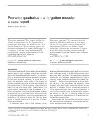
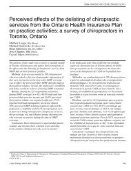
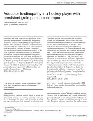
K HudesFigures A and BATibiaPosteriortibial arteryBTendon <strong>of</strong> flexordigitorum longusTendon <strong>of</strong>tibialis posteriorTalusTibial nerveTarsal <strong>tunnel</strong>Tendon <strong>of</strong> flexorhallucis longusFlexor retinaculumPulse <strong>of</strong> post-tibial arterymidway between heeland medial malleolusCalcaneusDrake: Gray’s Anatomy for Students, 2nd Edition.Copyright © 2009 by Churchill Livingstone, an imprint <strong>of</strong> Elsevier, Inc. All rights reserved.(CRPS), Baxter’s nerve entrapment, and <strong>tarsal</strong> <strong>tunnel</strong><strong>syndrome</strong>. 6 Even more rare causes include Freiberg’s disease(avascular necrosis <strong>of</strong> the meta<strong>tarsal</strong> head) and <strong>tarsal</strong>coalition, 5 as well as Sever’s Disease (calcaneal apophysitis).With the presentation <strong>of</strong> foot pain <strong>of</strong> 6 months duration,all <strong>of</strong> the aforementioned conditions should be on alist <strong>of</strong> differential diagnoses.Tarsal <strong>tunnel</strong> <strong>syndrome</strong> is associated with complaints<strong>of</strong> tingling and/or numbness around the medial ankle andon the plantar surface <strong>of</strong> the foot extending towards thetoes, 7,8,9 caused by stretching or compression <strong>of</strong> the posteriortibial nerve in the <strong>tarsal</strong> <strong>tunnel</strong>. The <strong>tarsal</strong> <strong>tunnel</strong> isbordered by the calcaneus, medial malleolus, talus, andflexor retinaculum, and houses the posterior tibial nerve,artery and vein, the tibialis posterior and the tendons andmuscles <strong>of</strong> the flexor digitorum longus and flexor hallucislongus. 3,7 It is illustrated in figures A and B (reprintedwith permission). 10The pain is <strong>of</strong>ten worse with activity, certain shoes thatthe patient may find aggravating or standing, and can berelieved by rest (with permission). 3,8 Symptoms can occursuddenly as a result <strong>of</strong> direct trauma or related to an inversionsprain <strong>of</strong> the ankle but may also be a result <strong>of</strong> overuseas in the <strong>case</strong> <strong>of</strong> excessive standing, walking, or exercise.Symptoms are <strong>of</strong>ten diffuse and poorly localized. Thephysical findings may vary, 3 and in 43% <strong>of</strong> <strong>case</strong>s the painis worse at night. 11 The patient may report significant orrelatively minor trauma to the foot. 3 The symptoms can bemisinterpreted as plantar fasciitis or even radicular painfrom the lumbar spine. 5 Proximal radiation <strong>of</strong> tinglingand numbness is seen in approximately one third <strong>of</strong> <strong>case</strong>sand is called the Valleix phenomenon. 3As the <strong>tarsal</strong> <strong>tunnel</strong> has limited space available, anycompromise to the space in the <strong>tunnel</strong> can put pressureon the structures within, and may cause symptoms. It isimportant to recall that this may include systemic diseaseswhich cause edema such as arthritis and diabetes. Directinjuries and hyperpronation, may compromise the struc-J Can Chiropr Assoc 2010; 54(2) 103
<strong>Conservative</strong> <strong>management</strong> <strong>of</strong> a <strong>case</strong> <strong>of</strong> <strong>tarsal</strong> <strong>tunnel</strong> <strong>syndrome</strong>tures within the <strong>tunnel</strong> by physically decreasing its crosssectional area, highlighting the need to record valgus orvarus deformities <strong>of</strong> the foot. 9 Plain film radiography,bone scan or CT is useful for identifying causes <strong>of</strong> <strong>tarsal</strong><strong>tunnel</strong> <strong>syndrome</strong> such as fractures or osteophytes, whereasMRI is more appropriate for other causes <strong>of</strong> <strong>tarsal</strong> <strong>tunnel</strong><strong>syndrome</strong> including: varicosities, trauma, fibrosis, accessorymuscles, ganglion cysts, lipoma, and nerve sheathtumours. 6 Two point discrimination on the plantar surface<strong>of</strong> the foot is the first sign <strong>of</strong> sensory loss, which mayprogress to pinprick hypoesthesia. 3 Sensory testing shouldtherefore be repeated periodically throughout the course<strong>of</strong> treatment in order to appropriately monitor the condition.Percussion <strong>of</strong> the posterior tibial nerve (Tinel’s sign)may cause parethesias along the course <strong>of</strong> the nerve, 3,8,9with one study reporting that Tinel’s sign is positive inonly 67% <strong>of</strong> <strong>case</strong>s. 11 The Dorsiflexion-eversion test for<strong>tarsal</strong> <strong>tunnel</strong> <strong>syndrome</strong> (dorsiflexion and eversion <strong>of</strong> thefoot with extension <strong>of</strong> the metatarsophalangeal (MTP)joints) may also be positive and create pain in the heel orreproduce the patient’s pain. 12 This test has been shown toincrease the tension on the structures <strong>of</strong> the <strong>tarsal</strong> <strong>tunnel</strong>,though it is not specific enough to differentiate between<strong>tarsal</strong> <strong>tunnel</strong> <strong>syndrome</strong> and plantar fasciitis. 12Severe presentations <strong>of</strong> <strong>tarsal</strong> <strong>tunnel</strong> <strong>syndrome</strong> mayexhibit weakness <strong>of</strong> intrinsic foot muscles. 11 Weakenedplantar muscles may cause the patient to have difficultyspreading their toes. 13 Atrophy may develop in the intrinsicand plantar muscles if the condition runs unchecked. 13Detection <strong>of</strong> minor weaknesses in the intrinsic foot musculatureis difficult in the clinical setting and referral for anerve conduction study should be made if compromise tothe motor nerves is suspected. 10 Signs <strong>of</strong> muscle atrophymay warrant a surgical consult.Tarsal <strong>tunnel</strong> <strong>syndrome</strong> that is not complicated bymuscle atrophy may be managed conservatively. Treatmentmay include: reassurance, custom orthotics, taping,bracing, stretching, strengthening, icing, s<strong>of</strong>t tissuemanipulation, chiropractic adjustments, massage, fascialstripping, non steroidal anti-inflammatory medication,corticosteroid injection, analgesic medication, or opioidmedication. 13,14,15,16 If conservative intervention fails torelieve symptoms, surgical approaches may be explored,such as microsurgical decompression <strong>of</strong> the tibial nervewith splitting <strong>of</strong> the flexor retinaculum. 13,15As with other chronic conditions, beneficial effects <strong>of</strong>manipulation and s<strong>of</strong>t tissue treatment such as massageand stretching done with the intent to relieve pain and restorenormal my<strong>of</strong>ascial movement have been noted forplantar fasciitis and other foot disorders. 16 Manipulationand mobilization <strong>of</strong> hypomobile foot joints has been recommendedin treatment <strong>of</strong> foot disorders such as plantarfasciitis. 17 Other foot conditions such as Morton’s neuromamay also derive short term relief after manipulationand mobilization <strong>of</strong> the foot. 18 Manual therapy such asGraston technique, an instrument assisted s<strong>of</strong>t tissue mobilization,Active Release Therapy and other s<strong>of</strong>t tissuemobilization techniques administered with the clinicianshands have been used to treat a wide variety <strong>of</strong> conditionsincluding, but not limited to, relieving the signs andsymptoms <strong>of</strong> sprains, strains, muscular adhesions and entrapment<strong>syndrome</strong>s such as carpal <strong>tunnel</strong> <strong>syndrome</strong>. 19,20One study outlined the efficiency <strong>of</strong> both instrument assistedtechniques as well as s<strong>of</strong>t tissue mobilization donewith the clinician’s hands noting that while the clinicalimprovements were not different between the therapygroups, improvement in both groups was maintained ona 3 month follow up. 19Orthotic therapy using semi-rigid orthotics cast in nonweight-bearing subtalar neutral has also been widely usedto treat a variety <strong>of</strong> chronic foot conditions and are foundto be beneficial in the <strong>management</strong> <strong>of</strong> plantar fasciitis. 17 Arandomized controlled trial <strong>of</strong> chiropractic manipulationand Achilles stretching versus orthotics found that bothtreatments appeared successful when used individuallyfor treatment <strong>of</strong> plantar fasciitis. 21 In a clinical setting,a practitioner will <strong>of</strong>ten use several <strong>of</strong> the tools availablesimultaneously to shorten the course <strong>of</strong> a complaint.While the symptom presentation and history in this<strong>case</strong> ruled out CPRS, and a negative Morton’s test decreasedthe likelihood <strong>of</strong> Morton’s neuroma, the remaining7 conditions remained on the differential. Tarsal <strong>tunnel</strong><strong>syndrome</strong> may be under-diagnosed as it can be difficultto diagnose due to the ease <strong>of</strong> confusing symptoms withplantar fasciitis and other foot conditions. Additionally, itwas found while researching this topic that two clinicaltests for plantar heel pain, the dorsiflexion-eversion testfor <strong>tarsal</strong> <strong>tunnel</strong> <strong>syndrome</strong> and the Windlass test for plantarfasciitis (“passive extension <strong>of</strong> the first MTP joint orall MTP joints with the ankle in neutral {90 degrees}” 12 ),“seem unable to differentiate between conditions that leadto plantar heel pain.” 12 Although the dorsiflexion-ever-104 J Can Chiropr Assoc 2010; 54(2)
K Hudession test may not be specific to <strong>tarsal</strong> <strong>tunnel</strong> <strong>syndrome</strong>,if performed it may have provided further support for thediagnosis. Tinel’s test, which has been traditionally usedto determine if an entrapment neuropathy is present, ispositive in 67% <strong>of</strong> <strong>case</strong>s <strong>of</strong> <strong>tarsal</strong> <strong>tunnel</strong> <strong>syndrome</strong>, butwas not present in this <strong>case</strong>. If the patient did not respondto conservative <strong>management</strong>, radiographs and/or a bonescan could have been performed to help to rule out rheumatologiccauses as well as stress fracture and the rare possibility<strong>of</strong> infiltration by tumour. Unless there are signs<strong>of</strong> muscle atrophy or motor involvement, a conservativeapproach to treating <strong>tarsal</strong> <strong>tunnel</strong> <strong>syndrome</strong> should be attemptedbefore referral for a nerve conduction study andprior to considering a surgical referral.Previous studies have reported successful <strong>management</strong><strong>of</strong> <strong>tarsal</strong> <strong>tunnel</strong> <strong>syndrome</strong> with: custom orthotics, 3,14,15,16taping, bracing, stretching, icing, s<strong>of</strong>t tissue manipulation,chiropractic adjustments, massage, fascial stripping, nonsteroidal anti-inflammatory medication, corticosteroid injection,analgesic medication, or opioid medication. 13,14,15,16The patient in this <strong>case</strong> was prescribed a pair <strong>of</strong> custom orthoticsbut reported little change in her condition after wearingthe orthotic devices for 10 weeks. A course <strong>of</strong> fascialstripping techniques to the lateral heel over the <strong>tarsal</strong> <strong>tunnel</strong>,and over the plantar and dorsal surfaces <strong>of</strong> the forefoot, andHVLA toggle board adjustments <strong>of</strong> the talonavicular jointand mobilizations <strong>of</strong> the cuboid were initiated as restrictions<strong>of</strong> joint motion were noted when evaluated for joint play. Assignificant improvement was reported by the patient after 4treatments; the same treatment was continued throughout thecourse <strong>of</strong> therapy.There are several factors that may have influenced thefavourable outcome <strong>of</strong> this <strong>case</strong>. HVLA adjustments wereused to attempt to re-establish normal motion <strong>of</strong> the cuboidand the talonavicular joint. Although orthotics alonedid not ease the symptoms, orthotics were used to attemptto correct faulty biomechanics and to address dysfunctionalfoot mechanics, which play a role in influencingchanges along the kinetic chain. S<strong>of</strong>t tissue techniquesincluding fascial stripping were used to attempt to breakdown scar tissue that may have accumulated in the area. 22With the onset <strong>of</strong> manual therapy, the patient seemed tohave a rapid reduction <strong>of</strong> subjective symptoms, but it isimportant to note other factors that may have produced afavourable outcome in this <strong>case</strong> such as the use <strong>of</strong> orthoticsprior to manual intervention.Further study is needed to identify other possible treatmentavenues such as specific rehabilitative exercises.Exercises that influence the strength and stability <strong>of</strong> theintrinsic musculature <strong>of</strong> the foot may prove a useful toolin the treatment <strong>of</strong> <strong>tarsal</strong> <strong>tunnel</strong> <strong>syndrome</strong>. This researchmight take the form <strong>of</strong> other <strong>case</strong> reports or a small scaleclinical trial to compare the effectiveness <strong>of</strong> treatmentwith and without specific exercise prescription.ConclusionAlthough favourable results were obtained, it is importantto remember that the nature <strong>of</strong> this investigation wasthat <strong>of</strong> a <strong>case</strong> study, and therefore treatment was appliedto only one patient. Limited as it may be, this <strong>case</strong> doesdemonstrate the conservative <strong>management</strong> using customorthotics, manipulation, and fascial stripping <strong>of</strong> one <strong>case</strong><strong>of</strong> <strong>tarsal</strong> <strong>tunnel</strong> <strong>syndrome</strong>. <strong>Conservative</strong> <strong>management</strong> <strong>of</strong><strong>tarsal</strong> <strong>tunnel</strong> <strong>syndrome</strong> should be explored prior to moreinvasive procedures such as injection <strong>of</strong> corticosteroids orsurgery.AcknowledgementsI would like to thank Drs. Richard Goldford, Kat Linaker,Clinton Eliason and Mr. Kenneth Clasper for their assistancewith editing and pro<strong>of</strong> reading.References1 Stovitz S, Coetzee J. Hyperpronation and foot pain. PhysSports Med. 2004; 32(8):19–26.2 Badlissi F, Dunn J, Link C, Keysor J, McKinlay J, FelsonD. Foot musculoskeletal disorders, pain and foot-relatedfunctional limitation in older persons. JAGS. 2005;53(6):1029–1033.3 Lau J, Daniels T. Tarsal Tunnel Syndrome: a review <strong>of</strong> theliterature. Foot Ankle Intern. 1999: 20(3):201–209.4 NBCE 2005 Job Analysis <strong>of</strong> Chiropractic http://ibce.org/publication/job-analysis.html5 Magee D. Orthopaedic Physical Measurement 5th Edition.Saunders Elsevier. 2008.6 Ahn J, El-Khoury G. Radiologic evaluation <strong>of</strong> chronic footpain. Am Fam Phys. 2007; 76(7):975–983.7 Sousa T. Differential Diagnosis for the Chiropractor.Apsen Publication 1997;350–355.8 Merck Manual <strong>of</strong> Diagnosis and Therapy. SeventeenthEdition; Merck Research Laboratories 1999, 487–488.9 Hammer W. Functional S<strong>of</strong>t Tissue Examination andTreatment by Manual Methods 2nd Edition. AspenPublication 1999; 322–324.10 Drake R, Vogl W, Mitchell A. Gray’s Anatomy for StudentsJ Can Chiropr Assoc 2010; 54(2) 105
<strong>Conservative</strong> <strong>management</strong> <strong>of</strong> a <strong>case</strong> <strong>of</strong> <strong>tarsal</strong> <strong>tunnel</strong> <strong>syndrome</strong>Second Edition. Churchill Livingstone 2010/2005, 612.Copyright Elsevier. (Reprinted with permission)11 Mondelli M, Morana P, Pauda L. An electrophysiologicalseverity scale in Tarsal Tunnel Syndrome. ACTA NeurolScand. 2004; 109:284–289.12 Alshami A, Babri A, Souvlis T, Coppieters M.Biomechanical evaluation <strong>of</strong> two clinical tests for plantarheel pain: the Dorsiflexion-Eversion test for Tarsal TunnelSyndrome and the Windlass test for plantar fasciitis. Foot& Ankle Intern. 2007; 28(4):499–505.13 Brockmann K, Schneider-Sickert F, Kolenda H, Aden I,Hanefeld F. Tarsal Tunnel Syndrome in a 7-year-old boy.Eur J Pediatr. 2004;163:46–47.14 Zhang J. Chiropractic adjustments and orthotics reducedsymptoms for standing workers. J Chiro Med. 2005;4(4):177–181.15 Franson J, Baravarian B. Tarsal Tunnel Syndrome: acompression neuropathy involving four distinct <strong>tunnel</strong>s.Clin Podiatr Med Surg. 2006; 23:597–609.16 Scheer P, Water L, Choate C, Huppin L. Is there pro<strong>of</strong> inthe evidence-based literature that custom orthoses work.Podiatry Management. 2007; 109:122.17 Brantigham J, Snyder R, Dishman R, Hubka M, Brown R,Brantingham C, Markham D. Plantar Fascitis. ChiropracticTechnique. 1992; (4–3):75–83.18 Govender N, Jretzmann H, Price J, Brantingham J, GlobeG. A single-blinded randomized placebo-controlledclinical trial <strong>of</strong> manipulation and mobilization in thetreatment <strong>of</strong> Morton’s neuroma. JACA. 2007; 8:18.19 Burke J, Buchberger D, Carey-Loghmani T, Dougherty P,Greco D, Dishman J. A pilot study comparing two manualtherapy interventions for carpal <strong>tunnel</strong> <strong>syndrome</strong>. J ManipPhysiol Ther. 2007; 30(1):50–61.20 Pajaczkowski J. Mimicking turf-toe: my<strong>of</strong>asopathy <strong>of</strong> thefirst dorsal interosseous muscle treated with ART. J CanChiropr Assoc. 2003; 47(1):28–32.21 Dimou E, Brantingham J, Wood T. A randomizedcontrolled trial (with blinded observer) <strong>of</strong> chiropracticmanipulation and achilles stretching vs. orthotics for thetreatment <strong>of</strong> plantar fasciitis. JACA. 2004; 41(9):32–42.22 Aspergen D et al. <strong>Conservative</strong> treatment <strong>of</strong> a femalecollegiate volleyball player with costochondritis. JMPT.2007; May:321–325.106 J Can Chiropr Assoc 2010; 54(2)