The new england journal of medicine - PULSION Medical Systems SE
The new england journal of medicine - PULSION Medical Systems SE
The new england journal of medicine - PULSION Medical Systems SE

Create successful ePaper yourself
Turn your PDF publications into a flip-book with our unique Google optimized e-Paper software.
<strong>The</strong> <strong>new</strong> <strong>england</strong><br />
<strong>journal</strong> <strong>of</strong> <strong>medicine</strong><br />
established in 1812 may 25, 2006 vol. 354 no. 21<br />
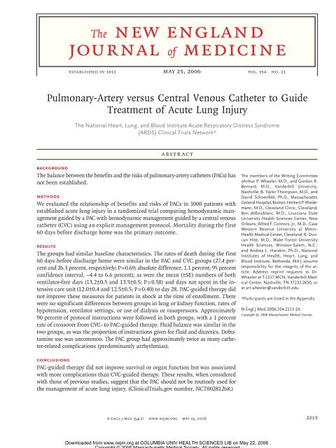
Pulmonary-Artery versus Central Venous Catheter to Guide<br />
Treatment <strong>of</strong> Acute Lung Injury<br />
<strong>The</strong> National Heart, Lung, and Blood Institute Acute Respiratory Distress Syndrome<br />
(ARDS) Clinical Trials Network*<br />
Abstract<br />
Background<br />
<strong>The</strong> balance between the benefits and the risks <strong>of</strong> pulmonary-artery catheters (PACs) has<br />
not been established.<br />
Methods<br />
We evaluated the relationship <strong>of</strong> benefits and risks <strong>of</strong> PACs in 1000 patients with<br />
established acute lung injury in a randomized trial comparing hemodynamic management<br />
guided by a PAC with hemodynamic management guided by a central venous<br />
catheter (CVC) using an explicit management protocol. Mortality during the first<br />
60 days before discharge home was the primary outcome.<br />
Results<br />
<strong>The</strong> groups had similar baseline characteristics. <strong>The</strong> rates <strong>of</strong> death during the first<br />
60 days before discharge home were similar in the PAC and CVC groups (27.4 percent<br />
and 26.3 percent, respectively; P = 0.69; absolute difference, 1.1 percent; 95 percent<br />
confidence interval, –4.4 to 6.6 percent), as were the mean (±<strong>SE</strong>) numbers <strong>of</strong> both<br />
ventilator-free days (13.2±0.5 and 13.5±0.5; P = 0.58) and days not spent in the intensive<br />
care unit (12.0±0.4 and 12.5±0.5; P = 0.40) to day 28. PAC-guided therapy did<br />
not improve these measures for patients in shock at the time <strong>of</strong> enrollment. <strong>The</strong>re<br />
were no significant differences between groups in lung or kidney function, rates <strong>of</strong><br />
hypotension, ventilator settings, or use <strong>of</strong> dialysis or vasopressors. Approximately<br />
90 percent <strong>of</strong> protocol instructions were followed in both groups, with a 1 percent<br />
rate <strong>of</strong> crossover from CVC- to PAC-guided therapy. Fluid balance was similar in the<br />
two groups, as was the proportion <strong>of</strong> instructions given for fluid and diuretics. Dobutamine<br />
use was uncommon. <strong>The</strong> PAC group had approximately twice as many catheter-related<br />
complications (predominantly arrhythmias).<br />
Conclusions<br />
PAC-guided therapy did not improve survival or organ function but was associated<br />
with more complications than CVC-guided therapy. <strong>The</strong>se results, when considered<br />
with those <strong>of</strong> previous studies, suggest that the PAC should not be routinely used for<br />
the management <strong>of</strong> acute lung injury. (ClinicalTrials.gov number, NCT00281268.)<br />
<strong>The</strong> members <strong>of</strong> the Writing Committee<br />
(Arthur P. Wheeler, M.D., and Gordon R.<br />
Bernard, M.D., Vanderbilt University,<br />
Nashville; B. Taylor Thompson, M.D., and<br />
David Schoenfeld, Ph.D., Massachusetts<br />
General Hospital, Boston; Herbert P. Wiedemann,<br />
M.D., Cleveland Clinic, Cleveland;<br />
Ben deBoisblanc, M.D., Louisiana State<br />
University Health Sciences Center, New<br />
Orleans; Alfred F. Connors, Jr., M.D., Case<br />
Western Reserve University at Metro-<br />
Health <strong>Medical</strong> Center, Cleveland; R. Duncan<br />
Hite, M.D., Wake Forest University<br />
Health Sciences, Winston-Salem, N.C.;<br />
and Andrea L. Harabin, Ph.D., National<br />
Institutes <strong>of</strong> Health, Heart, Lung, and<br />
Blood Institute, Bethesda, Md.) assume<br />
responsibility for the integrity <strong>of</strong> the article.<br />
Address reprint requests to Dr.<br />
Wheeler at T-1217 MCN, Vanderbilt <strong>Medical</strong><br />
Center, Nashville, TN 37232-2650, or<br />
at art.wheeler@vanderbilt.edu.<br />
*Participants are listed in the Appendix.<br />
N Engl J Med 2006;354:2213-24.<br />
Copyright © 2006 Massachusetts <strong>Medical</strong> Society.<br />
n engl j med 354;21 www.nejm.org may 25, 2006 2213<br />
Downloaded from www.nejm.org at COLUMBIA UNIV HEALTH SCIENCES LIB on May 22, 2006 .<br />
Copyright © 2006 Massachusetts <strong>Medical</strong> Society. All rights reserved.
2214<br />
<strong>The</strong> pulmonary-artery catheter (pac)<br />
provides unique hemodynamic data, including<br />
the cardiac index and pulmonaryartery–occlusion<br />
pressure. People who advocate<br />
the use <strong>of</strong> the PAC note that the clinician’s ability<br />
to predict intravascular pressure with the use <strong>of</strong><br />
this catheter is poor 1-3 ; central venous pressure, as<br />
obtained by means <strong>of</strong> the PAC, correlates imperfectly<br />
with pulmonary-artery–occlusion pressure<br />
4-6 ; and the insertion <strong>of</strong> a PAC <strong>of</strong>ten changes<br />
therapy. 6-8 Although many critically ill patients receive<br />
PACs, 9 no clear clinical benefit has been associated<br />
with their use. 10-12<br />
Practitioners <strong>of</strong>ten misinterpret the information<br />
obtained by means <strong>of</strong> a PAC or act incorrectly<br />
even when the data obtained with the use <strong>of</strong> this<br />
catheter are unambiguous, raising questions about<br />
the catheter’s value in usual practice. 13-18 A number<br />
<strong>of</strong> retrospective, prospective uncontrolled, and<br />
cohort studies 6,19-25 have raised questions about<br />
the safety <strong>of</strong> PACs, but because <strong>of</strong> their nonrandomized<br />
design, the results were not conclusive.<br />
Fears that the PAC could be harmful prompted<br />
calls for educational initiatives and even for a<br />
moratorium on its use until randomized trials<br />
were conducted. 26-29 <strong>The</strong> results <strong>of</strong> randomized<br />
studies also cast doubt on the value <strong>of</strong> the PAC, 30-35<br />
but even these were regarded as inconclusive because<br />
<strong>of</strong> the studies’ small size, population selection,<br />
lack <strong>of</strong> a comparison group randomly assigned<br />
to central venous catheter (CVC)–guided<br />
therapy, or most important, lack <strong>of</strong> an explicit<br />
management protocol. 36-40 To address these uncertainties,<br />
we conducted a randomized trial <strong>of</strong><br />
the management <strong>of</strong> acute lung injury using an<br />
explicit hemodynamic protocol guided by blood<br />
pressure, urinary output, and the results <strong>of</strong> a physical<br />
examination plus data obtained with either<br />
a PAC (i.e., cardiac index and pulmonary-artery–<br />
occlusion pressure) or a CVC (i.e., central venous<br />
pressure). Oxygen delivery and central or mixed<br />
venous oxygen saturation were not used in the<br />
management protocol.<br />
Methods<br />
Study Design<br />
<strong>The</strong> protocol for this multicenter factorial study,<br />
known as the Fluid and Catheter Treatment Trial<br />
(FACTT), can be found in the Supplementary Appendix,<br />
available with the full text <strong>of</strong> this article<br />
at www.nejm.org. Patients who had had acute lung<br />
<strong>The</strong> <strong>new</strong> <strong>england</strong> <strong>journal</strong> <strong>of</strong> <strong>medicine</strong><br />
n engl j med 354;21 www.nejm.org may 25, 2006<br />
injury for 48 hours or less were randomly assigned<br />
in permuted blocks <strong>of</strong> eight to receive a PAC or a<br />
CVC with the use <strong>of</strong> an automated system. Hemodynamic<br />
data obtained from the catheter were<br />
combined with clinical measures for use in a standardized<br />
management protocol. Patients were simultaneously<br />
randomly assigned to a strategy <strong>of</strong><br />
either liberal or conservative use <strong>of</strong> fluids guided<br />
by an explicit protocol (described in the Supplementary<br />
Appendix). Randomization was stratified<br />
according to hospital and the type <strong>of</strong> fluid<br />
therapy.<br />
Inclusion Criteria<br />
Eligible patients were receiving positive-pressure<br />
ventilation by tracheal tube and had a ratio <strong>of</strong> the<br />
partial pressure <strong>of</strong> arterial oxygen (PaO 2 ) to the<br />
fraction <strong>of</strong> inspired oxygen (FIO 2 ) below 300 (adjusted<br />
if the altitude exceeded 1000 m) and bilateral<br />
infiltrates on chest radiography consistent<br />
with the presence <strong>of</strong> pulmonary edema not due<br />
to left atrial hypertension. 41 If a potential participant<br />
did not have a CVC, the primary physician’s<br />
intent to insert one was required.<br />
Exclusion Criteria<br />
All reasons for exclusion are listed in Table 1 <strong>of</strong><br />
the Supplementary Appendix. Major exclusion criteria<br />
were the presence <strong>of</strong> a PAC after the onset <strong>of</strong><br />
acute lung injury; the presence <strong>of</strong> acute lung injury<br />
for more than 48 hours; an inability to obtain<br />
consent; the presence <strong>of</strong> chronic conditions<br />
that could independently influence survival, impair<br />
weaning, or compromise compliance with<br />
the protocol, such as dependence on dialysis or<br />
severe lung or neuromuscular disease; and irreversible<br />
conditions for which the estimated sixmonth<br />
mortality rate exceeded 50 percent, such<br />
as advanced cancer.<br />
Study Procedures<br />
Ventilation according to the Acute Respiratory<br />
Distress Syndrome (ARDS) Network protocol <strong>of</strong><br />
lower tidal volumes was begun within one hour<br />
after randomization and continued until day 28<br />
or until the patient was breathing without assistance.<br />
42 <strong>The</strong> assigned catheter was inserted within<br />
four hours after randomization. A CVC inserted<br />
before randomization could be used to<br />
determine intravascular pressure in the CVC group.<br />
Hemodynamic management as dictated by the<br />
protocol was started within the next 2 hours and<br />
Downloaded from www.nejm.org at COLUMBIA UNIV HEALTH SCIENCES LIB on May 22, 2006 .<br />
Copyright © 2006 Massachusetts <strong>Medical</strong> Society. All rights reserved.
pulmonary-artery or central venous catheter therapy for lung injury<br />
continued for seven days or until 12 hours after the<br />
patient was able to breathe without assistance. 42<br />
<strong>The</strong> PAC could be replaced by a CVC if hemodynamic<br />
stability (defined by the absence <strong>of</strong> the<br />
need for protocol-directed interventions for more<br />
than 24 hours) was achieved after day 3. We recorded<br />
complications from all central catheters<br />
present during the hemodynamic-management period<br />
and for three days after their removal. For<br />
the purposes <strong>of</strong> tracking complications, each introducer,<br />
PAC, and CVC was considered a separate<br />
catheter. We monitored compliance with protocol<br />
instructions twice each day: once during a<br />
morning reference period and again at a randomly<br />
selected time. A 100 percent audit <strong>of</strong> all instructions<br />
conducted after the first 82 patients were<br />
enrolled showed rates <strong>of</strong> protocol compliance similar<br />
to those obtained during the random checks<br />
(data not shown).<br />
All study personnel underwent extensive training<br />
in the conduct <strong>of</strong> the protocol and the measurement<br />
<strong>of</strong> vascular pressure. <strong>The</strong>y subsequently<br />
explained the study procedures to clinicians in<br />
the intensive care unit (ICU). Vascular pressures<br />
were measured in supine patients at end expiration;<br />
end expiration was identified with the use <strong>of</strong><br />
an airway-pressure signal, but the vascular pressures<br />
used in the protocol were not adjusted for<br />
airway pressure. 43 Four main protocol variables<br />
were measured at least every four hours. Blood<br />
pressure and urinary output guided management<br />
in both groups. Pulmonary-artery–occlusion pressure<br />
and the cardiac index were included in the<br />
protocol in the PAC group, whereas central venous<br />
pressure and clinical assessment <strong>of</strong> circulatory<br />
effectiveness (i.e., skin temperature, appearance<br />
<strong>of</strong> the skin, and the rate <strong>of</strong> capillary refilling)<br />
were used in the CVC group. Lactate levels, the<br />
rate <strong>of</strong> oxygen delivery, and mixed venous and superior<br />
vena caval oxygen saturation were not used<br />
as protocol variables. Prompt reversal <strong>of</strong> hypotension,<br />
oliguria, and ineffective circulation was the<br />
overriding goal <strong>of</strong> the protocol. <strong>The</strong> treatment <strong>of</strong><br />
patients in shock (defined by a mean systemic arterial<br />
pressure <strong>of</strong> less than 60 mm Hg or the need<br />
for vasopressors) was left to the judgment <strong>of</strong> the<br />
primary physician, with the exception that weaning<br />
from vasopressors was conducted according<br />
to the protocol after the patient’s blood pressure<br />
had stabilized. Patients who were not in shock<br />
were prescribed fluids for oliguria and for ineffective<br />
circulation if central venous pressure or<br />
pulmonary-artery–occlusion pressure was below<br />
the target range. Clinicians were free to select isotonic<br />
crystalloid, albumin, or blood products, although<br />
the protocol dictated the volume <strong>of</strong> each<br />
agent administered. Patients with ineffective circulation<br />
who were not in shock were given dobutamine<br />
with or without furosemide if their<br />
central venous pressure or pulmonary-artery–<br />
occlusion pressure exceeded the target range. Patients<br />
without hypotension who had adequate circulation<br />
and an intravascular pressure above the<br />
target range received furosemide. Patients who<br />
had a mean arterial pressure <strong>of</strong> at least 60 mm Hg<br />
without the use <strong>of</strong> vasopressors, a urinary output<br />
<strong>of</strong> at least 0.5 ml per kilogram <strong>of</strong> body weight<br />
per hour, and in the CVC group, adequate circulation<br />
on the basis <strong>of</strong> a physical examination or<br />
in the PAC group, a cardiac index <strong>of</strong> at least 2.5<br />
liters per minute per square meter <strong>of</strong> body-surface<br />
area, received furosemide or fluids to return their<br />
intravascular pressure to the target range.<br />
<strong>The</strong> study was approved by a protocol-review<br />
committee <strong>of</strong> the National Institutes <strong>of</strong> Health,<br />
National Heart, Lung, and Blood Institute, and the<br />
institutional review board at each participating<br />
location. Written consent was obtained from participants<br />
or legally authorized surrogates. An independent<br />
data and safety monitoring board conducted<br />
interim analyses after 82 patients had been<br />
enrolled and after each enrollment <strong>of</strong> approximately<br />
200 patients. Sequential stopping rules<br />
for safety and efficacy used the method <strong>of</strong> O’Brien<br />
and Fleming.<br />
Statistical Analysis<br />
<strong>The</strong> study had a statistical power <strong>of</strong> 90 percent to<br />
detect a reduction by 10 percentage points in the<br />
primary end point, death before hospital discharge<br />
home during the first 60 days after randomization,<br />
with the planned enrollment <strong>of</strong> 1000 patients.<br />
We assumed patients who went home alive and<br />
without the use <strong>of</strong> a ventilator before day 60 were<br />
alive at 60 days. Data on patients who were receiving<br />
ventilation or in a hospital were censored<br />
on the last day <strong>of</strong> follow-up. <strong>The</strong> Kaplan–Meier<br />
method was used to estimate the mean (±<strong>SE</strong>)<br />
60-day mortality rate, at the time <strong>of</strong> the last death<br />
occurring before 60 days. Differences in mortality<br />
between the groups were assessed by a z test.<br />
<strong>The</strong> primary analysis was conducted according to<br />
the intention to treat and on the basis <strong>of</strong> treatmentgroup<br />
assignment. Differences in continuous vari-<br />
n engl j med 354;21 www.nejm.org may 25, 2006 2215<br />
Downloaded from www.nejm.org at COLUMBIA UNIV HEALTH SCIENCES LIB on May 22, 2006 .<br />
Copyright © 2006 Massachusetts <strong>Medical</strong> Society. All rights reserved.
2216<br />
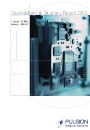
513 Assigned to PAC<br />
501 Received PAC<br />
12 Did not receive PAC<br />
0 Lost to follow-up<br />
513 Analyzed<br />
11,511 Patients screened<br />
1001 Underwent randomization<br />
ables were assessed by analysis <strong>of</strong> variance. Differences<br />
in categorical variables were assessed by<br />
the Mantel–Haenszel test. Differences between continuous<br />
variables over time were assessed by repeated-measures<br />
analysis <strong>of</strong> variance. All analyses<br />
were stratified according to the fluid-therapy<br />
assignment. For continuous variables, means ±<strong>SE</strong><br />
are reported. Two-sided P values <strong>of</strong> 0.05 were considered<br />
to indicate statistical significance. Analy-<br />
<strong>The</strong> <strong>new</strong> <strong>england</strong> <strong>journal</strong> <strong>of</strong> <strong>medicine</strong><br />
10,511 Excluded<br />
20.8% Had PAC<br />
15.9% Had physician decline<br />
13.8% Had chronic lung disease<br />
10.6% Had high risk <strong>of</strong> death<br />
within 6 mo<br />
9.3% Required dialysis<br />
8.4% Exceeded time window<br />
7.5% Had chronic liver disease<br />
6.4% Had acute MI<br />
5.8% Were unable to obtain<br />
consent<br />
4.3% Declined to give consent<br />
3.6% Were not committed<br />
to receiving full support<br />
2.9% Had neuromuscular<br />
disease<br />
488 Assigned to CVC<br />
480 Received CVC<br />
7 Crossed over to PAC<br />
1 Had unknown status<br />
1 Lost to follow-up<br />
(withdrew consent before study<br />
treatment was received)<br />
487 Analyzed<br />
1 Excluded from analysis<br />
Figure 1. Enrollment and Outcomes.<br />
Patients may have had more than one reason for exclusion. <strong>The</strong> exclusion<br />
criteria are listed in Table 1 <strong>of</strong> the Supplementary Appendix.<br />
n engl j med 354;21 www.nejm.org may 25, 2006<br />
sis was conducted with the use <strong>of</strong> SAS s<strong>of</strong>tware,<br />
version 8.2.<br />
Results<br />
Enrollment and Exclusions<br />
Screening for eligible patients was conducted at<br />
20 North American centers between June 8, 2000,<br />
and October 3, 2005. <strong>The</strong> trial was halted on July<br />
25, 2002, for a review by the Office <strong>of</strong> Human Research<br />
Protection and resumed unchanged except<br />
for the introduction <strong>of</strong> a modified consent form<br />
on July 23, 2003. 44-46 Figure 1 shows the most common<br />
reasons for exclusion for the 10,511 patients<br />
who were screened but not enrolled and the follow-up<br />
for the 513 patients who were randomly<br />
assigned to PAC-guided therapy and the 488 who<br />
were assigned to CVC-guided therapy. All exclusions<br />
are listed in Table 1 <strong>of</strong> the Supplementary<br />
Appendix.<br />
Baseline Characteristics<br />
<strong>The</strong> two groups were similar with respect to demographic<br />
characteristics, ICU location, cause <strong>of</strong><br />
lung injury, coexisting illnesses, and measures <strong>of</strong><br />
the severity <strong>of</strong> illness at baseline (Table 1). Approximately<br />
37 percent <strong>of</strong> patients in the PAC group<br />
and 32 percent <strong>of</strong> patients in the CVC group (P =<br />
0.06) met the criteria for shock, with 36 percent<br />
<strong>of</strong> patients in the PAC group receiving a vasopressor,<br />
as compared with 30 percent <strong>of</strong> patients in<br />
the CVC group (P = 0.05) (Table 1). Tidal volume,<br />
PaO 2 :FIO 2 , pH, plateau pressure, oxygenation index,<br />
lung injury score, and hemoglobin levels were<br />
similar in the two groups. Similar percentages <strong>of</strong><br />
each group were assigned to each fluid-therapy<br />
strategy (data not shown).<br />
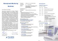
Main Outcomes<br />
<strong>The</strong> rate <strong>of</strong> death during the first 60 days after<br />
randomization was similar in the PAC group and<br />
the CVC group (27.4 percent and 26.3 percent,<br />
respectively; P = 0.69; absolute difference, 1.1 percent;<br />
95 percent confidence interval, –4.4 to 6.6<br />
percent), as were the number <strong>of</strong> ventilator-free<br />
days in the first 28 days (13.2±0.5 and 13.5±0.5,<br />
respectively; P = 0.58) (Fig. 2). CVC recipients had<br />
more ICU-free days during the first week <strong>of</strong> the<br />
study (0.88 day, vs. 0.66 day in the PAC group;<br />
P = 0.02); however, these differences were small<br />
and not significant at day 28 (12.5±0.5 vs. 12.0±0.4,<br />
P = 0.40). <strong>The</strong> number <strong>of</strong> days without various<br />
Downloaded from www.nejm.org at COLUMBIA UNIV HEALTH SCIENCES LIB on May 22, 2006 .<br />
Copyright © 2006 Massachusetts <strong>Medical</strong> Society. All rights reserved.
pulmonary-artery or central venous catheter therapy for lung injury<br />
Table 1. Baseline and Postrandomization Characteristics.*<br />
Characteristic<br />
PAC Group<br />
(N = 513)<br />
CVC Group<br />
(N = 487) P Value<br />
Age — yr 49.9±0.7 49.6±0.7 0.81<br />
Female sex — % 46 47 0.89<br />
Primary lung injury — % 0.81<br />
Pneumonia 48 46<br />
Severe sepsis 23 24<br />
Aspiration 15 15<br />
Trauma 8 7<br />
Other 7 8<br />
<strong>Medical</strong> ICU — % 66 66 0.91<br />
APACHE III score†<br />
Coexisting conditions — no./total no. (%)<br />
94.7±1.4 93.5±1.4 0.55<br />
Diabetes 89/500 (18) 84/467 (18) 0.94<br />
HIV infection or AIDS 30/500 (6) 41/467 (9) 0.10<br />
Cirrhosis 15/500 (3) 18/467 (4) 0.46<br />
Solid tumors 7/500 (1) 8/467 (2) 0.71<br />
Leukemia 14/500 (3) 8/467 (2) 0.27<br />
Lymphoma 7/500 (1) 6/467 (1) 0.88<br />
Immunosuppression<br />
Hemodynamic variables<br />
47/500 (9) 31/467 (7) 0.12<br />
Mean arterial pressure — mm Hg 77.5±0.7 76.8±0.6 0.41<br />
Met shock criteria — % 37 32 0.06<br />
Vasopressor use — %<br />
Respiratory variables<br />
36 30 0.05<br />
Tidal volume — ml/kg <strong>of</strong> PBW 7.4±0.1 7.4±0.1 0.88<br />
Plateau pressure — cm <strong>of</strong> water 26.2±0.4 26.2±0.4 0.93<br />
PEEP — cm <strong>of</strong> water 9.3±0.2 9.7±0.2 0.09<br />
pH 7.36±0.0 7.36±0.0 0.79<br />
PaO2:FIO2 158.9±3.3 151.3±3.1 0.10<br />
Bicarbonate — mmol/liter 22.3±0.2 22.3±0.2 0.93<br />
Oxygenation index‡ 12.8±0.4 13.3±0.5 0.48<br />
Lung injury score§<br />
Intervals — hr<br />
2.7±0.0 2.8±0.0 0.05<br />
From ICU admission to first instruction 44.4±1.8 40.8±2.5 0.23<br />
From qualification for acute lung injury to first instruction 25.2±0.7 23.0±0.6 0.02<br />
From randomization to first protocol instruction<br />
Prerandomization fluids — ml<br />
3.45±0.1 2.15±0.1
Proportion <strong>of</strong> Patients<br />
2218<br />
1.0<br />
0.9<br />
0.8<br />
0.7<br />
0.6<br />
0.5<br />
0.4<br />
0.3<br />
0.2<br />
0.1<br />
Alive, PAC group<br />
Unassisted breathing,<br />
CVC group<br />
0.0<br />
0 10 20 30<br />
Days<br />
40 50 60<br />
types <strong>of</strong> organ failure did not differ significantly<br />
between groups (Table 2 <strong>of</strong> the Supplementary<br />
Appendix). In the subgroup with shock at study<br />
entry, there were no significant differences between<br />
groups in the mortality rate or the number<br />
<strong>of</strong> organ-failure–free days (Table 3 <strong>of</strong> the Supplementary<br />
Appendix). <strong>The</strong>re was no interaction between<br />
the type <strong>of</strong> catheter and the type <strong>of</strong> fluid<br />
therapy assigned.<br />
Adverse Events<br />
Complications were uncommon and were reported<br />
at similar rates in each group: 0.08±0.01 per<br />
catheter inserted in the PAC group and 0.06±0.01<br />
per catheter inserted in the CVC group (P = 0.35).<br />
As compared with the CVC group, the PAC group<br />
had roughly 50 percent more catheters inserted<br />
(2.47±0.05 vs. 1.64±0.04, P
pulmonary-artery or central venous catheter therapy for lung injury<br />
Table 2. Catheter-Related Complications.<br />
Complication PAC Group CVC Group<br />
Sheath PAC CVC Total Sheath CVC Total<br />
number <strong>of</strong> patients<br />
Technical and mechanical<br />
complications<br />
Difficult placement 1 8 1 10 0 2 2<br />
Catheter malfunction 0 4 0 4 0 0 0<br />
Pneumothorax 3 2 1 6 0 6 6<br />
Air embolism 1 1 1 3 0 0 0<br />
Arterial puncture<br />
Arrhythmia<br />
1 0 2 3 0 0 0<br />
Atrial 3 15 0 18 0 0 0<br />
Ventricular 4 15 0 19 1 5 6<br />
Conduction defect<br />
Bleeding and clotting<br />
1 4 0 5 1 0 1<br />
Hemothorax 2 1 0 3 1 0 1<br />
Insertion-site bleeding 2 1 3 6 1 2 3<br />
Thromboembolism 0 0 0 0 1 0 1<br />
Local thrombosis<br />
Infection<br />
1 1 1 3 0 6 6<br />
Local 3 2 7 12 1 8 9<br />
Bloodstream* 1 3 1 5 0 3 3<br />
Other 0 2 1 3 0 3 3<br />
Total 23 59 18 100 6 35 41<br />
* Positive blood cultures were believed to be related to the presence <strong>of</strong> the catheter. Overall, 19 percent <strong>of</strong> patients in the<br />
PAC group and 18 percent <strong>of</strong> patients in the CVC group had one or more positive blood cultures (P = 0.43).<br />
Lung Function<br />
Ventilator settings and lung-function measures<br />
were similar in the two groups over time, with<br />
no significant differences in the respiratory rate,<br />
tidal volume, positive end-expiratory pressure,<br />
plateau pressure, PaO 2 :FIO 2 , pH, partial pressure<br />
<strong>of</strong> arterial carbon dioxide, oxygenation index, or<br />
lung injury score (Table 4 <strong>of</strong> the Supplementary<br />
Appendix).<br />
Metabolic and Renal Function<br />
While the hemodynamic management protocol<br />
was in use, there were no significant differences<br />
between groups in electrolyte, albumin, or hemoglobin<br />
levels (data not shown), although a higher<br />
percentage <strong>of</strong> patients in the PAC group than in<br />
the CVC group received erythrocyte transfusions<br />
(38 percent vs. 30 percent, P = 0.008). <strong>The</strong>re were<br />
no significant differences between groups in<br />
the percentage <strong>of</strong> patients treated with kidneyreplacement<br />
therapy (14 percent in the PAC group<br />
vs. 11 percent in the CVC group, P = 0.15).<br />
Discussion<br />
Because the PAC provides unique physiological<br />
information, it has been assumed that the use <strong>of</strong><br />
this catheter would improve survival and decrease<br />
the duration <strong>of</strong> assisted ventilation and the rate <strong>of</strong><br />
organ failure among patients with acute lung injury.<br />
Eroding this belief are observational and prospective<br />
trials indicating that such outcomes are<br />
not improved and may even be worsened by PAC<br />
use. 19-25 Since the initiation <strong>of</strong> this study, random-<br />
n engl j med 354;21 www.nejm.org may 25, 2006 2219<br />
Downloaded from www.nejm.org at COLUMBIA UNIV HEALTH SCIENCES LIB on May 22, 2006 .<br />
Copyright © 2006 Massachusetts <strong>Medical</strong> Society. All rights reserved.
2220<br />
A<br />
B<br />
Percentage <strong>of</strong> Baseline Measurement<br />
Percentage <strong>of</strong> Baseline Measurement<br />
11<br />
10<br />
9<br />
8<br />
7<br />
6<br />
5<br />
4<br />
3<br />
2<br />
1<br />
0<br />
12<br />
11<br />
10<br />
9<br />
8<br />
7<br />
6<br />
5<br />
4<br />
3<br />
2<br />
1<br />
0<br />
ized trials <strong>of</strong> patients undergoing high-risk surgery,<br />
31 patients with the acute respiratory distress<br />
syndrome and sepsis, 32 those with congestive heart<br />
failure, 34 and those with general critical illness 33,35<br />
have reported no benefit from PAC insertion. How-<br />
<strong>The</strong> <strong>new</strong> <strong>england</strong> <strong>journal</strong> <strong>of</strong> <strong>medicine</strong><br />
1 2 3 4 5 6 7 8 9 1011121314151617181920212223242526272829303132<br />
Pulmonary-Artery–Occlusion Pressure (mm Hg)<br />
1 2 3 4 5 6 7 8 9 1011121314151617181920212223242526272829303132<br />
Central Venous Pressure (mm Hg)<br />
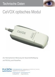
Figure 3. Distribution <strong>of</strong> Pulmonary-Artery–Occlusion Pressure (Panel A) and Central Venous Pressure (Panel B)<br />
before Receipt <strong>of</strong> the First Protocol-Mandated Instruction on Fluid Management.<br />
A total <strong>of</strong> 29 percent <strong>of</strong> patients had a pulmonary-artery–occlusion pressure that exceeded the traditionally accepted<br />
threshold <strong>of</strong> 18 mm Hg, though the majority <strong>of</strong> these pressures were 19 or 20 mm Hg, and 97 percent <strong>of</strong> patients with<br />
an initial pulmonary-artery–occlusion pressure <strong>of</strong> more than 18 mm Hg had a normal or elevated cardiac index.<br />
n engl j med 354;21 www.nejm.org may 25, 2006<br />
ever, these studies were limited by the inclusion <strong>of</strong><br />
relatively small numbers <strong>of</strong> patients and the lack<br />
<strong>of</strong> a strictly defined treatment protocol.<br />
Prevention or reversal <strong>of</strong> organ failure is a<br />
common justification to insert a PAC, but we<br />
Downloaded from www.nejm.org at COLUMBIA UNIV HEALTH SCIENCES LIB on May 22, 2006 .<br />
Copyright © 2006 Massachusetts <strong>Medical</strong> Society. All rights reserved.
A<br />
C<br />
E<br />
Mean Arterial Pressure (mm Hg)<br />
Net Fluid Balance (ml)<br />
pulmonary-artery or central venous catheter therapy for lung injury<br />
100<br />
90<br />
80<br />
70<br />
60<br />
0<br />
5000<br />
4000<br />
3000<br />
2000<br />
1000<br />
0<br />
PAC<br />
CVC<br />
0 1 2 3 4 5 6 7<br />
Day<br />
CVC<br />
PAC<br />
0 1 2 3 4 5 6 7<br />
Day<br />
Heart Rate (beats/min)<br />
110<br />
100<br />
were unable to identify any reduction in the incidence<br />
or the duration <strong>of</strong> any type <strong>of</strong> organ failure<br />
or the need for support (e.g., vasopressors, assisted<br />
ventilation, or kidney-replacement therapy) by<br />
using a PAC even in the subgroup <strong>of</strong> patients<br />
with shock at study entry. Likewise, PAC-guided<br />
therapy did not hasten discharge from the ICU; if<br />
90<br />
80<br />
70<br />
60<br />
0<br />
Cardiac index<br />
B<br />
D<br />
Vasopressor Use (%)<br />
PAOP and CVP (mm Hg)<br />
0 1 2 3 4 5 6 7<br />
Day<br />
40<br />
30<br />
20<br />
10<br />
0<br />
20<br />
15<br />
10<br />
5<br />
0<br />
anything, CVC use was associated with more ICUfree<br />
time during the first seven days. However,<br />
the small differences seen could be artifactual.<br />
For example, patients with a CVC might be able<br />
to be transferred from the ICU sooner than patients<br />
with a PAC because CVCs are <strong>of</strong>ten allowed<br />
on regular medical–surgical floors.<br />
n engl j med 354;21 www.nejm.org may 25, 2006 2221<br />
PAC<br />
CVC<br />
0 1 2 3 4 5 6 7<br />
Heart rate,<br />
CVC<br />
Heart rate,<br />
PAC<br />
6.0<br />
5.5<br />
5.0<br />
4.5<br />
4.0<br />
3.5<br />
3.0<br />
0<br />
Cardiac Index (liters/min/m 2 )<br />
Day<br />
0 1 2 3 4 5 6 7<br />
Day<br />
PAOP<br />
CVP, PAC<br />
CVP, CVC<br />
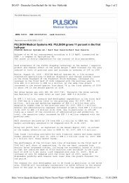
Figure 4. Mean Arterial Pressure (Panel A), Vasopressor Use (Panel B), Net Fluid Balance (Panel C), Pulmonary-Artery–Occlusion Pressure<br />
(PAOP) and Central Venous Pressure (CVP) (Panel D), and Heart Rate and Cardiac Index (Panel E) over Time.<br />
Mean (±<strong>SE</strong>) values are shown for mean arterial pressure obtained closest to 8 a.m. on specified study days in Panel A. Panel C shows<br />
the mean (±<strong>SE</strong>) net cumulative fluid balance as the sum <strong>of</strong> each day’s fluid balance. Day 0 is the day <strong>of</strong> randomization. For each variable,<br />
there were no significant differences between groups in baseline (day 0) values or values obtained during the study.<br />
Downloaded from www.nejm.org at COLUMBIA UNIV HEALTH SCIENCES LIB on May 22, 2006 .<br />
Copyright © 2006 Massachusetts <strong>Medical</strong> Society. All rights reserved.
2222<br />
<strong>The</strong> initial pulmonary-artery–occlusion pressure<br />
was greater than the traditional upper boundary<br />
<strong>of</strong> 18 mm Hg for acute lung injury in 29 percent<br />
<strong>of</strong> the patients. Since the cardiac index was<br />
normal in the vast majority <strong>of</strong> these patients (98<br />
percent), cardiac failure is an unlikely explanation<br />
for the elevated pressure. On the basis <strong>of</strong> the results<br />
<strong>of</strong> protocols with a conservative approach to<br />
fluid administration and protocols with a liberal<br />
approach to fluid administration, as explained by<br />
Wiedemann et al. (available at www.nejm.org), 47<br />
identification <strong>of</strong> an initially elevated pulmonaryartery–occlusion<br />
pressure did not translate into<br />
improved clinical outcomes, perhaps because both<br />
protocols mandated that diuretic therapy be given<br />
to lower the pulmonary-artery–occlusion pressure<br />
into a target range. Although uncommon, when<br />
the cardiac index was below 2.5 liters per minute<br />
per square meter, the protocol provided instructions<br />
for the administration <strong>of</strong> dobutamine, an<br />
inotropic and afterload-reducing agent.<br />
Even though serious catheter-related complications<br />
were uncommon and there were no deaths<br />
related to insertion, more catheter-related complications<br />
occurred among patients given a PAC<br />
than among those given a CVC. <strong>The</strong>se were predominantly<br />
arrhythmias: roughly half were atrial<br />
and half ventricular. Conduction block was also<br />
reported with the use <strong>of</strong> PACs but not CVCs. This<br />
observation is qualitatively similar to the increase<br />
in cardiac complications observed by Polanczyk<br />
et al. among patients undergoing noncardiac surgery.<br />
24 Analysis <strong>of</strong> catheter-related complications<br />
is complex. Each catheter inserted in the PAC<br />
group appeared to carry a risk similar to that <strong>of</strong><br />
a catheter inserted in the CVC group; however,<br />
almost one and a half times as many catheters<br />
were inserted in patients in the PAC group, a<br />
finding partly explained by the insertion <strong>of</strong> an<br />
introducer through which the PAC is typically<br />
passed. Ascertainment bias in arrhythmia reporting<br />
may also have occurred. Since we prohibited<br />
patients from having a PAC before entry, all PACs<br />
and many introducers were inserted under close<br />
observation during the study. In contrast, many<br />
CVCs were inserted before randomization; thus,<br />
arrhythmias occurring during insertion may not<br />
have been documented.<br />
<strong>The</strong> strengths <strong>of</strong> this study include its size;<br />
randomized, multicenter design with concealed<br />
allocation; explicit methods; use <strong>of</strong> objective end<br />
points; and high rate <strong>of</strong> clinician compliance. Ex-<br />
<strong>The</strong> <strong>new</strong> <strong>england</strong> <strong>journal</strong> <strong>of</strong> <strong>medicine</strong><br />
n engl j med 354;21 www.nejm.org may 25, 2006<br />
tensive pretrial training <strong>of</strong> study personnel in the<br />
conduct <strong>of</strong> the protocol and intravascular-pressure<br />
measurement, centralized review <strong>of</strong> pressure<br />
tracings, and use <strong>of</strong> airway-pressure signals to<br />
facilitate identification <strong>of</strong> end expiration most<br />
likely increased levels <strong>of</strong> accuracy and precision. 43<br />
<strong>The</strong> use <strong>of</strong> explicit protocols for hemodynamic<br />
and ventilator management, rather than usual<br />
care or general guidelines as in previous studies,<br />
makes it clear how patients were treated.<br />
Our study had several weaknesses. One common<br />
to all such trials is the inability to mask<br />
catheter assignment; however, the selection <strong>of</strong><br />
objective end points including death and organfailure–free<br />
days reduces the impact <strong>of</strong> this shortcoming.<br />
<strong>The</strong> low rate <strong>of</strong> crossover to the other<br />
catheter group and the high rate <strong>of</strong> compliance<br />
with the protocol in both groups, as assessed by<br />
scheduled daily and additional random checks,<br />
suggest both that absence <strong>of</strong> blinding had little<br />
effect on the results and that clinical equipoise<br />
was maintained after randomization. Owing to<br />
the small number <strong>of</strong> patients and the lack <strong>of</strong><br />
stratification, we were unable to exclude potentially<br />
beneficial effects <strong>of</strong> a PAC in subgroups <strong>of</strong><br />
patients. Nevertheless, we saw no hint <strong>of</strong> improved<br />
outcomes in the PAC group, with the mortality<br />
rate nominally lower in the CVC group. Furthermore,<br />
because the majority <strong>of</strong> patients were enrolled<br />
in medical ICUs, the relevance <strong>of</strong> our results<br />
to other types <strong>of</strong> patients is unclear. In<br />
addition, among others, we excluded patients with<br />
congestive heart failure, patients with severe obstructive<br />
and restrictive lung disease, and those<br />
receiving dialysis, so our study does not provide<br />
information on the value <strong>of</strong> the PAC in those<br />
groups. Finally, it could be argued that despite<br />
extensive development by experts, iterative pilot<br />
testing, and high rates <strong>of</strong> compliance with the<br />
protocol, the hemodynamic-protocol rules used<br />
did not optimize the benefits <strong>of</strong> the PAC as compared<br />
with the CVC.<br />
When considered with the results <strong>of</strong> previous<br />
randomized trials, our results suggest that the<br />
PAC is not useful for routine hemodynamic management<br />
in patients with established acute lung<br />
injury and is associated with more complications<br />
than the CVC. Our results do not address the<br />
safety or benefits <strong>of</strong> the PAC as a diagnostic tool<br />
or in other conditions, such as early resuscitation<br />
from septic shock. Similarly, our data do not address<br />
the safety or efficacy <strong>of</strong> PACs when they are<br />
Downloaded from www.nejm.org at COLUMBIA UNIV HEALTH SCIENCES LIB on May 22, 2006 .<br />
Copyright © 2006 Massachusetts <strong>Medical</strong> Society. All rights reserved.
pulmonary-artery or central venous catheter therapy for lung injury<br />
used with other protocols, in patients who have<br />
had acute lung injury for more than 48 hours, or<br />
in those with concomitant diseases who were excluded<br />
from our study.<br />
References<br />
1. Eisenberg PR, Jaffe AS, Schuster DP. lung injury: an observational study. Am<br />
Clinical evaluation compared to pulmo- J Respir Crit Care Med 1999;160:69-76.<br />
nary artery catheterization in the hemo- 7. Mimoz O, Rauss A, Rekik N, Brundynamic<br />
assessment <strong>of</strong> critically ill pa- Buisson C, Lemaire F, Brochard L. Pulmotients.<br />
Crit Care Med 1984;12:549-53. nary artery catheterization in critically ill<br />
2. Steingrub JS, Celoria G, Vickers-Lahti patients: a prospective analysis <strong>of</strong> outcome<br />
M, Teres D, Bria W. <strong>The</strong>rapeutic impact <strong>of</strong> changes associated with catheter-prompt-<br />
pulmonary artery catheterization in a meded changes in therapy. Crit Care Med 1994;<br />
ical/surgical ICU. Chest 1991;99:1451-5. 22:573-9.<br />
3. Ferguson ND, Meade MO, Hallett DC, 8. Connors AF Jr, McCaffree DR, Gray<br />
Stewart TE. High values <strong>of</strong> the pulmonary BA. Evaluation <strong>of</strong> right-heart catheteriza-<br />
artery wedge pressure in patients with acute tion in the critically ill patient without<br />
lung injury and acute respiratory distress acute myocardial infarction. N Engl J Med<br />
syndrome. Intensive Care Med 2002;28: 1983;308:263-7.<br />
1073-7.<br />
9. Chalfin DB. <strong>The</strong> pulmonary artery<br />
4. Civetta JM, Gabel JC. Flow directed- catheter: economic aspects. New Horiz<br />
pulmonary artery catheterization in sur- 1997;5:292-6.<br />
gical patients: indications and modifica- 10. Schultz RJ, Whitfield GF, LaMura JJ,<br />
tions <strong>of</strong> technic. Ann Surg 1972;176: Raciti A, Krishnamurthy S. <strong>The</strong> role <strong>of</strong><br />
753-6.<br />
physiologic monitoring in patients with<br />
5. Forrester JS, Diamond G, McHugh TJ, fractures <strong>of</strong> the hip. J Trauma 1985;25:309-<br />
Swan HJC. Filling pressures in the right 16.<br />
and left sides <strong>of</strong> the heart in acute myocar- 11. Shoemaker WC, Appel PL, Kram HB,<br />
dial infarction: a reappraisal <strong>of</strong> central- Waxman K, Lee TS. Prospective trial <strong>of</strong><br />
venous-pressure monitoring. N Engl J Med supranormal values <strong>of</strong> survivors as thera-<br />
1971;285:190-3.<br />
peutic goals in high-risk surgical patients.<br />
6. Marinelli WA, Weinert CR, Gross CR, Chest 1988;94:1176-86.<br />
et al. Right heart catheterization in acute 12. Shah MR, Hasselblad V, Stevenson<br />
Supported by contracts (NO1-HR-46054-64 and NO1-HR-16146-<br />
54) with the National Institutes <strong>of</strong> Health, National Heart, Lung,<br />
and Blood Institute.<br />
No potential conflict <strong>of</strong> interest relevant to this article was<br />
reported.<br />
appendix<br />
<strong>The</strong> following persons and institutions participated in the Fluid and Catheter Treatment trial: Writing Committee — A.P. Wheeler, H.P.<br />
Wiedemann, G.R. Bernard, B.T. Thompson, B. deBoisblanc, A.F. Connors, R.D. Hite, D.A. Schoenfeld, A.L. Harabin; Steering Committee<br />
chair — G.R. Bernard; Clinical Coordinating Center — D.A. Schoenfeld, B.T. Thompson, N. Ringwood, C. Oldmixon, F. Molay,<br />
A. Korpak, R. Morse, D. Hayden, M. Ancukiewicz, A. Minihan; Protocol-Review Committee — J.G.N. Garcia, R. Balk, S. Emerson, M.<br />
Shasby, W. Sibbald; Data and Safety Monitoring Board — R. Spragg, G. Corbie-Smith, J. Kelley, K. Leeper, A.S. Slutsky, B. Turnbull, C.<br />
Vreim; National Heart, Lung, and Blood Institute — A.L. Harabin, D. Gail, P. Lew, M. Waclawiw; Clinical Centers — University <strong>of</strong> Washington,<br />
Harborview — L. Hudson, K. Steinberg, M. Neff, R. Maier, K. Sims, C. Cooper, T. Berry-Bell, G. Carter, L. Andersson; University<br />
<strong>of</strong> Michigan — G.B. Toews, R.H. Bartlett, C. Watts, R. Hyzy, D. Arnoldi, R. Dechert, M. Purple; University <strong>of</strong> Maryland — H. Silverman, C.<br />
Shanholtz, A. Moore, L. Heinrich, W. Corral; Johns Hopkins University — R. Brower, D. Thompson, H. Fessler, S. Murray, A. Sculley;<br />
Cleveland Clinic Foundation — H.P. Wiedemann, A.C. Arroliga, J. Komara, T. Isabella, M. Ferrari; University Hospitals <strong>of</strong> Cleveland — J. Kern,<br />
R. Hejal, D. Haney; MetroHealth <strong>Medical</strong> Center — A.F. Connors; University <strong>of</strong> Colorado Health Sciences Center — E. Abraham, R. McIntyre, F.<br />
Piedalue; Denver Veterans Affairs <strong>Medical</strong> Center — C. Welch; Denver Health <strong>Medical</strong> Center — I. Douglas, R. Wolkin; St. Anthony Hospital — T.<br />
Bost, B. Sagel, A. Hawkes; Duke University — N. MacIntyre, J. Govert, W. Fulkerson, L. Mallatrat, L. Brown, S. Everett, E. VanDyne, N.<br />
Knudsen, M. Gentile; University <strong>of</strong> North Carolina — P. Rock, S. Carson, C. Schuler, L. Baker, V. Salo; Vanderbilt University — A.P. Wheeler,<br />
G.R. Bernard, T. Rice, B. Christman, S. Bozeman, T. Welch; University <strong>of</strong> Pennsylvania — P. Lanken, J. Christie, B. Fuchs, B Finkel, S.<br />
Kaplan, V. Gracias, C.W. Hanson, P. Reilly, M.B. Shapiro, R. Burke, E. O’Connor, D. Wolfe; Jefferson <strong>Medical</strong> College — J. Gottlieb, P. Park,<br />
D.M. Dillon, A. Girod, J. Furlong; LDS Hospital — A. Morris, C. Grissom, L. Weaver, J. Orme, T. Clemmer, R. Davis, J. Gleed, S. Pies, T.<br />
Graydon, S. Anderson, K. Bennion, P. Skinner; McKay–Dee Hospital — C. Lawton, J. d’Hulst, D. Hanselman; Utah Valley Regional <strong>Medical</strong><br />
Center — K. Sundar, T. Hill, K. Ludwig, D. Nielson; University <strong>of</strong> California, San Francisco, San Francisco — M.A. Matthay, M. Eisner, B. Daniel,<br />
O. Garcia; San Francisco General — J. Luce, R. Kallet; University <strong>of</strong> California, San Francisco, Fresno — M. Peterson, J. Lanford; Baylor College <strong>of</strong><br />
Medicine — K. Guntupalli, V. Bandi, C. Pope; Baystate <strong>Medical</strong> Center — J. Steingrub, M. Tidswell, L. Kozikowski; Louisiana State University<br />
— B. deBoisblanc, J. Hunt, C. Glynn, P. Lauto, G. Meyaski, C. Romaine; Louisiana State University–Earl K. Long — S. Brierre, C. LeBlanc,<br />
K. Reed; Alton–Ochsner Clinic Foundation — D. Taylor, C. Thompson; Tulane University <strong>Medical</strong> Center — F. Simeone, M. Johnston, M. Wright;<br />
University <strong>of</strong> Chicago — G. Schmidt, J. Hall, S. Hemmann, B. Gehlbach, A. Vinayak, W. Schweickert; Northwestern University — J. Dematte<br />
D’Amico, H. Donnelly; University <strong>of</strong> Texas Health Sciences Center — A. Anzueto, J. McCarthy, S. Kucera, J. Peters, T. Houlihan, R. Steward,<br />
D. Vines; University <strong>of</strong> Virginia — J. Truwit, M. Marshall, W. Matsumura, R. Brett; University <strong>of</strong> Pittsburgh — M. Donahoe, P. Linden, J.<br />
Puyana, L. Lucht, A. Verno; Wake Forest University — R.D. Hite, P. Morris, A. Howard, A. Nesser, S. Perez; Moses Cone Memorial Hospital<br />
— P. Wright, C. Carter-Cole, J. McLean; St. Paul’s Hospital–Vancouver — J. Russell, L. Lazowski, K. Foley; Vancouver General Hospital — D.<br />
Chittock, L. Grandolfo; Mayo Foundation — M. Murray.<br />
LW, et al. Impact <strong>of</strong> the pulmonary artery<br />
catheter in critically ill patients: meta-analysis<br />
<strong>of</strong> randomized clinical trials. JAMA<br />
2005;294:1664-70.<br />
13. Gnaegi A, Feihl F, Perret C. Intensive<br />
care physicians’ insufficient knowledge <strong>of</strong><br />
right-heart catheterization at the bedside:<br />
time to act? Crit Care Med 1997;25:213-<br />
20.<br />
14. Iberti TJ, Fischer EP, Leibowitz AB,<br />
Panacek EA, Silverstein JH, Albertson TE.<br />
A multicenter study <strong>of</strong> physicians’ knowledge<br />
<strong>of</strong> the pulmonary artery catheter.<br />
JAMA 1990;264:2928-32.<br />
15. Jacka MJ, Cohen MM, To T, Devitt JH,<br />
Byrick R. Pulmonary artery occlusion pressure<br />
estimation: how confident are anesthesiologists?<br />
Crit Care Med 2002;30:1197-<br />
203.<br />
16. Burns D, Burns D, Shively M. Critical<br />
care nurses’ knowledge <strong>of</strong> pulmonary artery<br />
catheters. Am J Crit Care 1996;5:49-<br />
54.<br />
17. Jain M, Canham M, Upadhyay D, Corbridge<br />
T. Variability in interventions with<br />
pulmonary artery catheter data. Intensive<br />
Care Med 2003;29:2059-62.<br />
18. Ontario Intensive Care Study Group.<br />
n engl j med 354;21 www.nejm.org may 25, 2006 2223<br />
Downloaded from www.nejm.org at COLUMBIA UNIV HEALTH SCIENCES LIB on May 22, 2006 .<br />
Copyright © 2006 Massachusetts <strong>Medical</strong> Society. All rights reserved.
2224<br />
pulmonary-artery or central venous catheter therapy for lung injury<br />
Evaluation <strong>of</strong> right heart catheterization<br />
in critically ill patients. Crit Care Med 1992;<br />
20:928-33.<br />
19. Afessa B, Spencer S, Khan W, LaGatta<br />
M, Bridges L, Freire AX. Association <strong>of</strong><br />
pulmonary artery catheter use with inhospital<br />
mortality. Crit Care Med 2001;29:<br />
1145-8.<br />
20. Gore JM, Goldberg RJ, Spodick DH,<br />
Alpert JS, Dalen JE. A community-wide assessment<br />
<strong>of</strong> the use <strong>of</strong> pulmonary artery<br />
catheters in patients with acute myocardial<br />
infarction. Chest 1987;92:721-7.<br />
21. Zion MM, Balkin J, Rosenmann D, et<br />
al. Use <strong>of</strong> pulmonary catheters in patients<br />
with acute myocardial infaction: analysis<br />
<strong>of</strong> experience in 5,841 patients in the<br />
SPRINT Registry. Chest 1990;98:1331-5.<br />
22. Sakr Y, Vincent JL, Reinhart K, et al.<br />
Use <strong>of</strong> the pulmonary artery catheter is<br />
not associated with worse outcome in the<br />
ICU. Chest 2005;128:2722-31.<br />
23. Cohen MG, Kelly RV, Kong DF, et al.<br />
Pulmonary artery catheterization in acute<br />
coronary syndromes: insights from the<br />
GUSTO IIb and GUSTO III trials. Am J Med<br />
2005;118:482-8.<br />
24. Polanczyk CA, Rohde LE, Goldman L,<br />
et al. Right heart catheterization and cardiac<br />
complications in patients undergoing<br />
noncardiac surgery: an observational<br />
study. JAMA 2001;286:309-14.<br />
25. Connors AF Jr, Sper<strong>of</strong>f T, Dawson NV,<br />
et al. <strong>The</strong> effectiveness <strong>of</strong> right heart catheterization<br />
in the initial care <strong>of</strong> critically<br />
ill patients. JAMA 1996;276:889-97.<br />
26. Bernard GR, Sopko G, Cerra F, et al.<br />
Pulmonary artery catheterization and clinical<br />
outcomes: National Heart, Lung, and<br />
Blood Institute and Food and Drug Administration<br />
Workshop report: consensus<br />
statement. JAMA 2000;283:2568-72.<br />
27. Robin ED. <strong>The</strong> cult <strong>of</strong> the Swan-Ganz<br />
catheter: overuse and abuse <strong>of</strong> pulmonary<br />
flow catheters. Ann Intern Med 1985;103:<br />
445-9.<br />
28. Dalen JE, Bone RC. Is it time to pull<br />
the pulmonary artery catheter? JAMA 1996;<br />
276:916-8.<br />
29. Dalen JE. <strong>The</strong> pulmonary artery catheter<br />
— friend, foe, or accomplice? JAMA<br />
2001;286:348-50.<br />
30. Guyatt G. A randomized control trial<br />
<strong>of</strong> right-heart catheterization in critically<br />
ill patients. J Intensive Care Med 1991;<br />
6:91-5.<br />
31. Sandham JD, Hull RD, Brant RF, et al.<br />
A randomized, controlled trial <strong>of</strong> the use<br />
<strong>of</strong> pulmonary-artery catheters in highrisk<br />
surgical patients. N Engl J Med 2003;<br />
348:5-14.<br />
32. Richard C, Warszawski J, Anguel N, et<br />
al. Early use <strong>of</strong> the pulmonary artery catheter<br />
and outcomes in patients with shock<br />
and acute respiratory distress syndrome:<br />
a randomized controlled trial. JAMA 2003;<br />
290:2713-20.<br />
33. Harvey S, Harrison DA, Singer M, et al.<br />
Assessment <strong>of</strong> the clinical effectiveness <strong>of</strong><br />
pulmonary artery catheters in management<br />
<strong>of</strong> patients in intensive care (PAC-<br />
Man): a randomised controlled trial. Lancet<br />
2005;366:472-7.<br />
34. Binanay C, Califf RM, Hasselblad V,<br />
et al. Evaluation study <strong>of</strong> congestive heart<br />
failure and pulmonary artery catheterization<br />
effectiveness: the ESCAPE trial. JAMA<br />
2005;294:1625-33.<br />
35. Rhodes A, Cusack RJ, Newman PJ,<br />
Grounds RM, Bennett ED. A randomized,<br />
controlled trial <strong>of</strong> the pulmonary artery<br />
catheter in critically ill patients. Intensive<br />
Care Med 2002;28:256-64.<br />
36. Morris AH. Developing and implementing<br />
computerized protocols for standardization<br />
<strong>of</strong> clinical decisions. Ann Intern<br />
Med 2000;132:373-83.<br />
37. Idem. Treatment algorithms and protocolized<br />
care. Curr Opin Crit Care 2003;<br />
9:236-40.<br />
38. Ivanov RI, Allen J, Sandham JD, Calvin<br />
JE. Pulmonary artery catheterization:<br />
CLINICAL TRIAL REGISTRATION<br />
<strong>The</strong> Journal encourages investigators to register their clinical trials<br />
in a public trials registry. <strong>The</strong> members <strong>of</strong> the International Committee<br />
<strong>of</strong> <strong>Medical</strong> Journal Editors plan to consider clinical trials for publication<br />
only if they have been registered (see N Engl J Med 2004;351:1250-1).<br />
<strong>The</strong> National Library <strong>of</strong> Medicine’s www.clinicaltrials.gov is a free registry,<br />
open to all investigators, that meets the committee’s requirements.<br />
n engl j med 354;21 www.nejm.org may 25, 2006<br />
a narrative and systematic critique <strong>of</strong> randomized<br />
controlled trials and recommendations<br />
for the future. New Horiz 1997;<br />
5:268-76.<br />
39. Walker MB, Waldmann CS. <strong>The</strong> use <strong>of</strong><br />
pulmonary artery catheters in intensive<br />
care: time for reappraisal? Clin Intensive<br />
Care 1994;5:15-9.<br />
40. Angus D, Black N. Wider lessons <strong>of</strong><br />
the pulmonary artery catheter trial. BMJ<br />
2001;322:446.<br />
41. Bernard GR, Artigas A, Brigham KL,<br />
et al. <strong>The</strong> American-European Consensus<br />
Conference on ARDS: definitions, mechanisms,<br />
relevant outcomes, and clinical trial<br />
coordination. Am J Respir Crit Care Med<br />
1994;149:818-24.<br />
42. <strong>The</strong> Acute Respiratory Distress Syndrome<br />
Network. Ventilation with lower<br />
tidal volumes as compared with traditional<br />
tidal volumes for acute lung injury and<br />
the acute respiratory distress syndrome.<br />
N Engl J Med 2000;342:1301-8.<br />
43. Rizvi K, Deboisblanc BP, Truwit JD, et<br />
al. Effect <strong>of</strong> airway pressure display on<br />
interobserver agreement in the assessment<br />
<strong>of</strong> vascular pressures in patients with<br />
acute lung injury and acute respiratory distress<br />
syndrome. Crit Care Med 2005;33:98-<br />
103.<br />
44. Drazen JM. Controlling research trials.<br />
N Engl J Med 2003;348:1377-80.<br />
45. Steinbrook R. How best to ventilate?<br />
Trial design and patient safety in studies<br />
<strong>of</strong> the acute respiratory distress syndrome.<br />
N Engl J Med 2003;348:1393-401.<br />
46. Steinbrook R. Trial design and patient<br />
safety — the debate continues. N Engl<br />
J Med 2003;349:629-30.<br />
47. National Heart, Lung, and Blood Institute<br />
ARDS Clinical Trials Network. Comparison<br />
<strong>of</strong> two fluid management strategies<br />
in acute lung injury. (Available at<br />
www.nejm.org.)<br />
Copyright © 2006 Massachusetts <strong>Medical</strong> Society.<br />
Downloaded from www.nejm.org at COLUMBIA UNIV HEALTH SCIENCES LIB on May 22, 2006 .<br />
Copyright © 2006 Massachusetts <strong>Medical</strong> Society. All rights reserved.