OCULAR LYMPHOMA - Ocular Immunology and Uveitis Foundation
OCULAR LYMPHOMA - Ocular Immunology and Uveitis Foundation
OCULAR LYMPHOMA - Ocular Immunology and Uveitis Foundation

You also want an ePaper? Increase the reach of your titles
YUMPU automatically turns print PDFs into web optimized ePapers that Google loves.
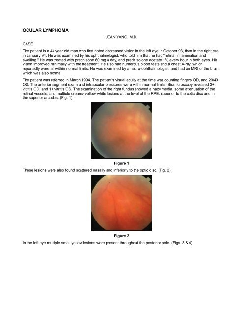
<strong>OCULAR</strong> <strong>LYMPHOMA</strong>CASEJEAN YANG, M.D.The patient is a 44 year old man who first noted decreased vision in the left eye in October 93, then in the right eyein January 94. He was examined by his ophthalmologist, who told him that he had "retinal inflammation <strong>and</strong>swelling." He was treated with prednisone 60 mg a day, <strong>and</strong> prednisolone acetate 1% every hour in both eyes. Hisvision improved minimally with the treatment. He also had numerous blood tests <strong>and</strong> a chest X-ray, whichreportedly were all within normal limits. He was examined by a neuro-ophthalmologist, <strong>and</strong> had an MRI of the brain,which was also normal.The patient was referred in March 1994. The patient’s visual acuity at the time was counting fingers OD, <strong>and</strong> 20/40OS. The anterior segment exam <strong>and</strong> intraocular pressures were within normal limits. Biomicroscopy revealed 3+vitritis OD, <strong>and</strong> 1+ vitritis OS. The examination of the right fundus showed a hazy media, some attenuation of theretinal vessels, <strong>and</strong> multiple creamy yellow-white lesions at the level of the RPE, superior to the optic disc <strong>and</strong> inthe superior arcades. (Fig. 1)Figure 1These lesions were also found scattered nasally <strong>and</strong> inferiorly to the optic disc. (Fig. 2)Figure 2In the left eye multiple small yellow lesions were present throughout the posterior pole. (Figs. 3 & 4)
Figure 3 ********************* Figure 4Fluorescein angiogram of the right fundus showed areas of hyperfluorescence in the central macula <strong>and</strong> superior tothe disc, which corresponded to some of the lesions noted on exam. (Fig. 5)Figure 5Late frames of the angiogram showed persistence of the hyperfluorescence.Fluorescein angiogram of the left fundus revealed multiple punctate hyperfluorescent lesions seen in the posteriorpole. (Fig. 6)These lesions remained stable throughout the angiogram.Figure 6The patient’s past medical history was significant for testicular cancer at age 2, which was treated in Pol<strong>and</strong> withorchiectomy <strong>and</strong> radiation therapy. Social history disclosed that he was a chemist <strong>and</strong> traveled to several foreigncountries. He grew up on a farm with cows, pigs <strong>and</strong> chickens. He enjoyed raw meat <strong>and</strong> deer hunting. In therecent past, he skinned his own deer without gloves. He also was a cat owner.The immunologic workup was significant for a white blood cell count of 15,700, <strong>and</strong> a markedly positive PPD,although he did receive BCG in childhood. Serologic tests were significant for a positive varicella IgG, with anegative IgM. Hepatitis A IgG was positive with a negative IgM, consistent with past infection. Other serologic tests
including herpes simplex, toxoplasmosis, toxocara, Lyme disease, FTA-abs, cat-scratch disease, tularemia,crytococcus, histoplasmosis, <strong>and</strong> cysticercosis were all negative.The patient was treated with IV acyclovir for presumed acute retinal necrosis. However his vision in the left eyedeteriorated to 20/100 despite the treatment. Multifocal choroiditis was suspected <strong>and</strong> the treatment was changedto Cyclosporin <strong>and</strong> Imuran, along with Rifampin <strong>and</strong> INH, given the markedly positive PPD. Diagnostic vitrectomywas delayed due to an acute viral conjunctivitis. Sulfadiazine <strong>and</strong> pyrimethamine were further added to thetreatment regimen by the Infectious Disease consultants for possible toxoplasmosis, despite the negative antibodytiters. The patient underwent a diagnostic vitrectomy of the right eye in April 1994. Cytologic study of the vitreousspecimen revealed markedly atypical lymphoid cells suspicious for lymphoma. (Fig. 7)Figure 7A complete neurologic workup including neurologic history <strong>and</strong> physical exam, lumbar puncture with cytology of theCSF, <strong>and</strong> MRI of the brain were all negative. In May, the patient’s vision further decreased to counting fingers ou.Radiation therapy was instituted. Vision improved to 20/500 OD, <strong>and</strong> 20/300 OS, <strong>and</strong> the deep retinal lesionsbegan to regress. (Fig. 8)Figure 8A year later the patient started experiencing memory loss. An MRI of the brain showed a large mass in the spleniumof corpus callosum. (Fig. 9)
Figure 9Brain biopsy of the lesion revealed CNS B-cell lymphoma. The patient was treated with chemotherapy <strong>and</strong> radiationtherapy.DISCUSSIONNon-Hodgkin’s lymphoma of the central nervous system (NHL-CNS) (previously called reticulum cell sarcoma),may also properly be referred to as intraocular large cell lymphoma. Reticulum cell sarcoma is a misnomer, sincethe tumor is neither a sarcoma nor comprised of reticulum cells. The incidence of Non-Hodgkin’s lymphoma of thecentral nervous system has slowly increased since 1960, <strong>and</strong> has trebled over the past 15 years. (1) This increasecannot be fully explained by AIDS or other causes of immunosuppression. The median age is between 50 <strong>and</strong> 60years. There is a slight male predominance. Three populations are at risk of developing NHL-CNS: patients withAIDS; transplant recipients; <strong>and</strong> patients with congenital immunodeficiencies, such as Wiskott-Aldrich syndrome<strong>and</strong> severe combined immunodeficiency.Patients with NHL-CNS can present with four distinct profiles: solitary or multiple discrete intracranial nodules,diffuse meningeal or periventricular lesions, subretinal infiltrates or vitritis, <strong>and</strong> localized spinal masses. (1) <strong>Ocular</strong>involvement may precede disease in other parts of the CNS. Both Whitcup <strong>and</strong> Freeman reported only half of theirpatients had CNS lesions on neuroradiologic studies by the time intraocular lymphoma was diagnosed. (2,3)Freeman also reported ocular symptoms preceded CNS symptoms in 82% of patients with CNS disease. The meantime between the onset of ocular symptoms <strong>and</strong> the onset of CNS symptoms was 29 months. (3)The most common presenting ocular symptoms are blurred vision <strong>and</strong> floaters. Pain <strong>and</strong> conjunctival hyperemiaare rare. Vision is often decreased. Biomicroscopic exam often shows mild anterior segment inflammation with cells<strong>and</strong> flare <strong>and</strong> keratic precipitates. Vitreous cells occurring in sheets are characteristic. Although the disease maybegin with one eye, bilateral involvement is common after several months. Fundus examination often showssubretinal yellow infiltrates. Many atypical fundus presentations have been reported, including hemorrhagic retinalvasculitis, <strong>and</strong> hemorrhagic retinal necrosis.(4,5) Multiple RPE detachments have been described by Gass as beingpathognomonic.(6) Exudative retinal detachments, thickening of the uveal tract (7) <strong>and</strong> papillitis have all beendescribed. Intraocular lymphoma should be suspected when chronic uveitis is poorly responsive to corticosteroids,<strong>and</strong> when there is a characteristic vitritis with deep retinal lesions. The vision is often better than expected based onthe clinical examination.All patients suspected of having intraocular lymphoma should undergo a neurologic workup. Neurologic symptomssuch as headache, focal weakness, sensory deficits, confusion, personality change, <strong>and</strong> difficulty with gait may bepresent. A history of recent seizure is also a strong indication of CNS involvement. The neurologic workup shouldinclude an MRI of the brain, <strong>and</strong> lumbar puncture with examination of the CSF. The cytology of CSF is diagnostic ofcourse if it is positive. Since lymphoma cells are fragile, it is important that samples be immediately transported tothe cytology laboratory for processing. At least 10 cc’s of CSF should be sent. A repeat lumbar puncture may benecessary for the diagnosis.If the CSF shows no malignant cells, a pars plana vitrectomy on the eye with the most severe vitritis or the worstvision is the next step. Char demonstrated loss of cellular detail when the vitreous specimen was obtained throughvitrectomy cutter, <strong>and</strong> therefore he advocates aspiration of 1 cc of vitreous, with or without cutting depending on theease of aspiration, prior to vitrectomy.(4) The vitreous specimen should be h<strong>and</strong>led with care <strong>and</strong> immediatelycarried to the cytology lab. Tissue culture medium enriched with 10% fetal calf serum can be added to the collection
chamber of the vitrectomy machine to improve cell viability. Multiple vitrectomies may be needed to make thediagnosis of intraocular lymphoma. Char <strong>and</strong> colleagues reported 3 of 14 patients required more than onevitrectomy.(8) Whitcup <strong>and</strong> colleagues reported 3 of 10 patients had one or more negative vitrectomies before thediagnosis was made.(2) Mish<strong>and</strong>ling of the vitreous specimens may partially account for the false negativediagnostic results. However, treatment with corticosteroids also may be responsible for the paucity of viablelymphoma cells in vitreous specimens. Steroids can be cytolytic to CNS lymphoma cells. The sensitivity of thetumor cells to corticosteroids is unique to CNS lymphoma <strong>and</strong> is not seen with any other CNS malignancies. Thissensitivity to steroids contributes to the difficulty in diagnosing this disease, since many patients with intraocularlymphoma are treated with corticosteroids at the time of vitrectomy for presumed chronic uveitis.Typical lymphoma cells are large <strong>and</strong> pleomorphic with scanty cytoplasm. The nuclei are round <strong>and</strong>hypersegmented with prominent nucleoli. Unlike systemic lymphomas that invade the choroid, most of CNSlymphoma cells are located between the RPE <strong>and</strong> Bruch’s membrane. (Fig. 13) Chorioretinal biopsies have beenused to diagnose intraocular lymphoma in some cases.(9) However, the risks of biopsy are significant, <strong>and</strong> thesensitivity is unclear. Immunohistochemical staining may be helpful by detecting monoclonal B cells with a kappa orlambda light chain, <strong>and</strong> can confirm the diagnosis.(10,11) However since tumor cells are mixed with inflammatorycells, immunologic analysis often reveals lymphocyte heterogeneity, making the study less reliable than cytology.Interleukin-10 is a growth <strong>and</strong> differentiation factor for B lymphocytes. Recently Chan <strong>and</strong> colleagues measured IL-10 levels in vitrectomy specimens from 3 patients with intraocular lymphoma <strong>and</strong> 5 patients with uveitis.(12) In theirreport, IL-10 was detected in the vitreous of all 3 patients with intraocualr lymphoma, but in none of the vitreousspecimens of the patients with uveitis. They also demonstrated the levels of IL-10 in the vitreous correlated withboth severity of vitritis <strong>and</strong> numbers of malignant cells by cytology. Given the difficulty of diagnosing intraocualrlymphoma accurately, IL-10 in the vitreous may provide a helpful diagnostic clue.Treatment consists of radiation therapy, <strong>and</strong> combined radiation therapy <strong>and</strong> chemotherapy if CNS disease ispresent.Go to Review QuestionsREFERENCES1. Hochberg FH, Miller DC. Primary central nervous system lymphoma. J Neurosurg 1988;68:835-853.2. Whitcup SM, deSmet MD, Rubin BI, et al. Intraocular lymphoma: Clinical <strong>and</strong> Histopathologic diagnosis.Ophthalmology 1993;100:1399-1406.3. Freeman LN, Schachat AP, Knox DL, et al. Clinical features, laboratory investigations, <strong>and</strong> survival in ocularreticulum cell sarcoma. Ophthalmology 1987;94:1631-9.4. de Smet MD, Nussenblatt RB, Davis JL, Palestine AG. Large cell lymphoma masquerading as a viral retinitis. IntOphthalmol 1990;14:413-17.5. Ridley ME, McDonald HR, Sternberg P Jr, et al. Retinal manifestations of ocular lymphoma (reticulum cellsarcoma). Ophthalmlogy 1992;99:1153-61.6. Gass JDM, Sever RJ, Grizzard WS, et al. Multifocal pigment epithelial detachments by reticulum cell sarcoma. Acharacteristic funduscopic picture. Retina 1984;4:135-143.7. Duker, JS, Shields, JA, Ross M. Intraocualr large cell lymphoma presenting as massive thickening of the uvealtract. Retina 1987;7:41-45.8. Char DH, Ljung B, Miller T, Phillips T. Primary intraocualr lymphoma (ocular reticulum cell sarcoma): Diagnosis<strong>and</strong> management. Ophthtalmology 1988;95:625-630.9. Kirmani MH, Thomas EL, Rao NA, Laborde RP. Intraocualr reticulum cell sarcoma: diagnosis by choroidalbiopsy. Br J Ophthalmol 1987;71:748-752.10. Char DH, Ljung B, Deschenes J, Miller TR. Intraocualr lymphoma: immunological <strong>and</strong> cytological analysis. Br JOphthalmol 1988;72:905-911.11. Ljung B, Char DH, Miller TR, Deschenes J. Intraocualr lymphoma: cytologic diagnosis <strong>and</strong> the role ofimmunologic markers. Acta Cytol 1987;32:840-847.12. Chan C, Whitcup SM, Solomon D, Nussenblatt, RB. Interleukin-10 in the vitreous of patients with primaryintraocualr lymphoma. Am J Ophthalmol 1995;120:671-673.