Ocular Sarcoidosis - Ocular Immunology and Uveitis Foundation
Ocular Sarcoidosis - Ocular Immunology and Uveitis Foundation
Ocular Sarcoidosis - Ocular Immunology and Uveitis Foundation

Create successful ePaper yourself
Turn your PDF publications into a flip-book with our unique Google optimized e-Paper software.
<strong>Ocular</strong> <strong>Sarcoidosis</strong><br />
by Panagiota Stavrou, F.R.C.S.<br />
<strong>Sarcoidosis</strong><br />
<strong>Sarcoidosis</strong> is a multisystem granulomatous disease which was first described by Jonathan Hutchinson in 1878. Its<br />
clinical manifestations <strong>and</strong> course can be variable in different ethnic groups. The organs affected more often are the<br />
lungs, skin <strong>and</strong> eyes.<br />
The frequency of ocular involvement ranges from 26% to 50%. The characteristics of ocular involvement are (1)<br />
when present, is seen generally early in the course of the disease (2) may co-exist with asymptomatic systemic<br />
disease <strong>and</strong> (3) can precede systemic involvement by several years. Most patients present between the ages of 20<br />
to 40 years; however, children <strong>and</strong> the elderly can be affected. Cases of familial sarcoidosis, including monozygotic<br />
twins, <strong>and</strong> husb<strong>and</strong>-wife pairs have also been reported.<br />
Diagnosis relies on demonstration of non caseating granuloma by tissue biopsy. In cases of suspected sarcoidosis<br />
where no affected tissue amenable to biopsy is identifiable, supportive evidence of the diagnosis can be obtained<br />
through non-invasive investigations including measurement of serum angiotensin converting enzyme (ACE) <strong>and</strong><br />
lysozyme, chest x-ray, chest computerized tomography (C-T), gallium scintillography, pulmonary function tests,<br />
bronchoalveolar lavage, <strong>and</strong> measurement of serum <strong>and</strong> urinary calcium.<br />
<strong>Ocular</strong> manifestations<br />
Anterior segment<br />
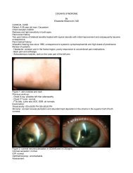
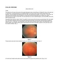
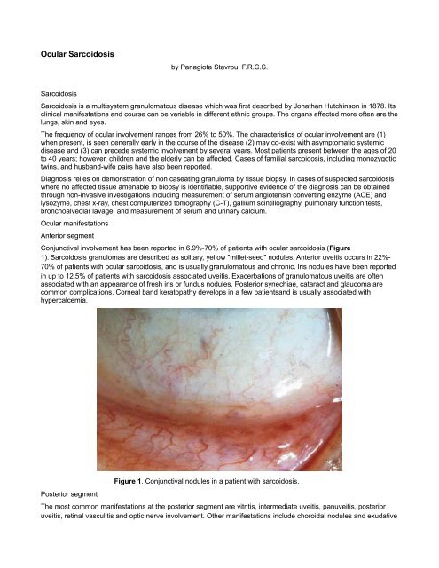
Conjunctival involvement has been reported in 6.9%-70% of patients with ocular sarcoidosis (Figure<br />
1). <strong>Sarcoidosis</strong> granulomas are described as solitary, yellow "millet-seed" nodules. Anterior uveitis occurs in 22%-<br />
70% of patients with ocular sarcoidosis, <strong>and</strong> is usually granulomatous <strong>and</strong> chronic. Iris nodules have been reported<br />
in up to 12.5% of patients with sarcoidosis associated uveitis. Exacerbations of granulomatous uveitis are often<br />
associated with an appearance of fresh iris or fundus nodules. Posterior synechiae, cataract <strong>and</strong> glaucoma are<br />
common complications. Corneal b<strong>and</strong> keratopathy develops in a few patients<strong>and</strong> is usually associated with<br />
hypercalcemia.<br />
Posterior segment<br />
Figure 1. Conjunctival nodules in a patient with sarcoidosis.<br />
The most common manifestations at the posterior segment are vitritis, intermediate uveitis, panuveitis, posterior<br />
uveitis, retinal vasculitis <strong>and</strong> optic nerve involvement. Other manifestations include choroidal nodules <strong>and</strong> exudative
etinal detachment. Clinical <strong>and</strong>/or angiographic cystoid macular edema (CME) has been reported in 19%-72% of<br />
patients. Overall, patients with chronic posterior uveitis <strong>and</strong> panuveitis have significantly more complications than<br />
do patients with anterior uveitis. "C<strong>and</strong>le wax drippings" <strong>and</strong> "punched-out" lesions can be seen in patients with<br />
uveitis secondary to sarcoidosis (Figure 2).<br />
Figure 2. Punched-out choroidoretinal lesions in a patient with sarcoidosis.<br />
Severe retinal vasculitis <strong>and</strong> ischemic retinopathy with neovascularization, requiring scatter photocoagulation has<br />
been described in some patients. Other less frequent complications include peripapillary <strong>and</strong> subfoveal choroidal<br />
neovascularization, posterior scleritis with annular ciliochoroidal detachment causing angle closure glaucoma, <strong>and</strong><br />
central retinal vein occlusion leading to a painful blind eye from neovascular glaucoma.<br />
Orbit<br />
Although sarcoid granulomas have been described in most areas inside the orbit, the lacrimal gl<strong>and</strong> appears to be<br />
the organ most commonly affected. The frequency of lacrimal gl<strong>and</strong> involvement varies from 7% to 69%. The<br />
nasolacrimal drainage system can also become involved in patients with sarcoidosis. The patients usually present<br />
with epiphora <strong>and</strong> nasal stuffiness. Extraocular muscle involvement can occur <strong>and</strong> presents with diplopia or painful<br />
external ophthalmoplegia.<br />
<strong>Sarcoidosis</strong> in childhood<br />
Early onset or pre-school sarcoidosis seen in children is relatively rare. The classic triad of symptoms consists of<br />
skin, eye <strong>and</strong> joint lesions; pulmonary involvement is rare, at least initially. It can be easily misdiagnosed as juvenile<br />
rheumatoid arthritis, as the latter also presents with symptoms from the joints <strong>and</strong> eyes.<br />
Extraocular involvement<br />
The lung is the most frequently affected organ in patients with sarcoidosis. Histologically the lesions are distributed<br />
primarily along the lymphatics around bronchi <strong>and</strong> blood vessels, although alveolar lesions are also seen. The<br />
relative frequency of granulomas in the bronchial submucosa accounts for the high diagnostic yield of<br />
bronchoscopic biopsies. Lymph nodes are involved in almost all cases, especially the hilar <strong>and</strong> mediastinal ones.<br />
The majority of patients are asymptomatic, while others may complain about cough or shortness of breath.<br />
The spleen is enlarged in only 18% of cases, although microscopical evidence of sarcoid granulomas in spleen is<br />
present in three-quarters of the cases. The liver is affected less often than is the spleen, but a finding of elevated<br />
liver enzymes in a sarcoidosis suspect may prompt percutaneous liver biopsy in the search for histopathologic
confirmation of the suspected diagnosis. Renal insufficiency has been reported in patients with histologically proven<br />
sarcoidosis of the kidneys <strong>and</strong> has been attributed to hypercalcemia <strong>and</strong> interstitial granulomatous nephritis. X-ray<br />
abnormalities of the bones can be identified in about one-fifth of patients. The radiologically visible lesions are<br />
usually seen in the phalangeal bones of the h<strong>and</strong>s <strong>and</strong> feet, creating small circumscribed areas of bone resorption<br />
within the marrow cavity.<br />
Skin lesions are found in 9%-37% of the patients. They may be specific, showing histologically non caseating<br />
granulomas, or non-specific, e.g. erythema nodosum. The specific skin lesions include lupus pernio, infiltrated<br />
plaques, maculopapular eruptions, subcutaneous nodules <strong>and</strong> infiltration of old scars (Figures 3 & 4).<br />
Figure 3 & 4. Skin manifestations in patients with sarcoidosis.<br />
Histology<br />
The hallmark of sarcoidosis is a non necrotizing granuloma. The center of the granuloma consists of histiocytes,<br />
epithelioid cells, <strong>and</strong> multinucleated giant cells which are surrounded by lymphocytes, plasma cells, <strong>and</strong> fibroblasts<br />
(Figure 5). Gross necrosis is not a feature of sarcoidosis, <strong>and</strong> suggests alternative diagnoses (e.g. tuberculosis,
fungal infection, vasculitis, etc). The epithelioid cells are transformed bone marrow monocytes <strong>and</strong> have marked<br />
secretory activity which includes over 40 different cytokines <strong>and</strong> other mediators. Among the enzymes <strong>and</strong> other<br />
chemicals secreted by granulomas are ACE, lysozyme, glucuroonidase, collagenase, <strong>and</strong> calcitriol.<br />
Kveim test<br />
Figure 5. Granulomas consisting of histiocytes, epithelioid cells, <strong>and</strong> multinucleated giant cells.<br />
In 1941, Kveim reported the use of a suspension derived from the spleen of a patient with sarcoidosis that, when<br />
injected intracutaneously into patients with biopsy proven sarcoidosis, yielded a cutaneous papule containing non<br />
caseating epithelioid granulomas. Four to six weeks following subcutaneous injection of the Kveim reagent, the<br />
typical lesion presents as a red or brownish raised papule ranging from a few millimeters up to 1.5 centimeters in<br />
diameter. Histopathologic analysis of the biopsied lesion reveals a granuloma composed of epithelioid cells,<br />
occasional Langhans cells <strong>and</strong> scattered lymphocytes at its center with a surrounding cuff of mononuclear cells,<br />
primarily lymphocytes.<br />
Cutaneous anergy<br />
In 1916, Boeck first described cutaneous anergy to tuberculin in patients with sarcoidosis. It was later on realized<br />
that this phenomenon was not limited to tuberculin alone, but that anergy to a variety of other skin tests-antigens<br />
such was also typical. In 1994, Kataria <strong>and</strong> Holter proposed a mechanism for the cutaneous anergy seen in<br />
sarcoidosis. At sites of granulomatous inflammation, there is a predominance of helper T lymphocytes, which<br />
proliferate <strong>and</strong> secrete large amounts of lymphokines, including interleukin (IL)-2, monocyte chemotactic factor<br />
(MCF) <strong>and</strong> migration inhibition factor (MIF). These lymphokines induce <strong>and</strong> amplify the immune response by<br />
enhancing T-lymphocyte proliferation as well as recruiting <strong>and</strong> retaining monocytes from the circulation. The<br />
lymphokines <strong>and</strong> monokines produced at sites of granulomatous inflammation have their highest concentration<br />
locally. Nevertheless, the protein molecules diffuse into blood, establishing a concentration gradient between the<br />
granulomatous inflammatory site <strong>and</strong> the remote site of the delayed type hypersensitivity (DTH) skin test. As a<br />
result, the traffic of T-helper lymphocytes <strong>and</strong> monocytes is preferentially directed towards site of granuloma<br />
formation. That leads to a preponderance of suppressor cells in the peripheral blood <strong>and</strong> competitively depletes the<br />
T-helper cells <strong>and</strong> monocytes available to sites of DTH.<br />
Etiology <strong>and</strong> pathogenesis<br />
The processes involved in the pathogenesis of sarcoidosis in the lungs include accumulation of CD 4<br />
+ lymphocytes<br />
at the affected site. The cytokines <strong>and</strong> factors secreted by these cells account for the influx of monocytes, alveolitis,
<strong>and</strong> non caseating granuloma formation in the lung <strong>and</strong> for the resulting progressive fibrosis, all characteristic<br />
features of pulmonary sarcoidosis. <strong>Sarcoidosis</strong> is characterized by "compartmentalization" of the T cells, such that<br />
the relative proportion of CD 4<br />
+ T cells in blood is reduced (e.g. CD 4<br />
/CD 8<br />
=0.8), while the reverse relation is<br />
observed in affected tissue (e.g. CD 4<br />
/CD 8<br />
=1.8 in lung). The CD 4<br />
+ cells in the involved organs are "activated" <strong>and</strong><br />
thus are releasing IL-2 <strong>and</strong> other mediators, while the CD 4<br />
+ cells in other sites, such as blood, are quiescent.<br />
In 1992, Holter et al demonstrated a Kveim-like granulomagenic activity in nonviable autologous BAL cells (NABC)<br />
recovered soon after symptomatic onset or relapse of sarcoidosis but not in patients with chronic stable<br />
sarcoidosis.The same group of investigators demonstrated a Kveim-like granolomagenic activity of peripheral blood<br />
monocytes, the progenitors of the alveolar macrophage. These findings suggest that the circulating monocyte is<br />
already primed with the granulomagenic factor before differentiation into alveolar macrophage. A monocyte source<br />
of the factor explains the multisystem distribution of granulomas in sarcoidosis.<br />
Chest radiology<br />
Sulavik et al suggested the following roentgen staging of sarcoidosis: 0= normal chest radiograph; 1= bilateral<br />
symmetrical hilar lymphadenopathy (BSHL) only; 2= BSHL with bilateral, symmetrical lung infiltration (Figure 6); 3=<br />
bilateral symmetrical lung infiltration only; 4a= BSHL with bilateral symmetrical lung infiltration indicative of<br />
pulmonary fibrosis (BSIF); 4b= BSIF only.<br />
Gallium ( 67 GA) scan<br />
Figure 6. Bilateral symmetrical hilar lymphadenopathy <strong>and</strong> lung infiltration.<br />
The combined abnormal bilateral symmetrical 67 GA uptake of the lacrimal <strong>and</strong> parotid gl<strong>and</strong>s (with or without<br />
subm<strong>and</strong>ibular gl<strong>and</strong> 67 GA uptake) is called the "p<strong>and</strong>a" pattern (Figure 7). The presence <strong>and</strong> pattern of 67 GA<br />
uptake in both (1) the parahilar <strong>and</strong> infrahilar bronchopulmonary lymph nodes <strong>and</strong> (2) the right paratracheal<br />
(azygous) mediastinal lymph nodes is called the "lambda" image after its resemblance with the Greek letter l.<br />
Pulmonary uptake of 67 GA is sensitive but not specific in the diagnosis of pulmonary sarcoidosis as it can occur in a<br />
wide variety of other inflammatory <strong>and</strong> neoplastic diseases.Abnormal 67 GA uptake in salivary <strong>and</strong> lacrimal gl<strong>and</strong>s<br />
may also occur in Sjögren's syndrome, tuberculosis <strong>and</strong> after radiation therapy.
Figure 7. Bilateral symmetrical gallium uptake by the lacrimal, parotid, <strong>and</strong> subm<strong>and</strong>ibular gl<strong>and</strong>s (P<strong>and</strong>a sign).<br />
Angiotensin converting enzyme (ACE)<br />
ACE is normally present in the vascular endothelium of many organs (lung, kidney, small intestine, uterus, prostate,<br />
thyroid, testes, adrenals) <strong>and</strong> in macrophages. The latter source accounts for elevated ACE levels in patients with<br />
sarcoidosis. The induction of ACE synthesis in epithelioid cells <strong>and</strong> macrophages is caused by a soluble ACEinducing<br />
factor (AIF). ACE is elevated in 60-90% of patients with active sarcoidosis. A normal serum ACE does not<br />
exclude the diagnosis, especially if the disease is in its early stages, localized to a small area (e.g. the eye), <strong>and</strong><br />
therefore with a small epithelioid cell "barden". False low values are also measured in patients taking ACE inhibitors<br />
or in patients with endothelial abnormalities, such as deep vein thrombosis, <strong>and</strong> in patients who have had<br />
chemotherapy or radiation. Treatment with systemic steroids or other immunosupressive agents can also affect<br />
ACE levels with values normalizing with adequate control of intraocular inflammation. Other disorders associated<br />
with elevated serum ACE include Gaucher's disease, leprosy, chronic pulmonary disease, rheumatoid arthritis,<br />
spondylitis, primary biliary cirrhosis, tuberculosis, histoplasmosis, histiocytic medullary fibrosis, hyperthyroidism <strong>and</strong><br />
diabetes mellitus.<br />
Pulmonary function tests<br />
Pulmonary function tests are useful in the initial diagnosis <strong>and</strong> follow up of patients with sarcoidosis. Their<br />
sensitivity in disease with <strong>and</strong> without roentgen evidence of parenchymal involvement has been reported as 70%<br />
<strong>and</strong> 40% respectively. The most common abnormalities seen early in the course of the disease are a post exercise<br />
increase in the alveolar-arterial oxygen gradient <strong>and</strong> diffusing capacity <strong>and</strong> a reduction in lung compliance.<br />
Bronchoalveolar lavage (BAL)- Transbronchial lung biopsy (TBLB)<br />
The earliest pathologic finding in patients with sarcoidosis is a mononuclear alveolitis composed of increased CD 4<br />
+<br />
lymphocytes (with an increased CD 4<br />
/CD 8<br />
ratio), monocyte-macrophages, <strong>and</strong> rare B lymphocytes. Tissue for<br />
biopsy from the bronchial mucosa or the adjacent lung is obtained through a fibreoptic bronchoscope. Non<br />
caseating granulomas have been reported in 54-88% of patients who underwent TBLB.The rate of positive findings<br />
by TBLB is higher in patients with radiologic evidence of pulmonary infiltration, <strong>and</strong> is approximately 60% among<br />
patients with hilar lymphadenopathy whose chest radiographs show normal lung parenchyme.<br />
Hypercalcemia<br />
Hypercalcemia has been reported in 10-15% of patients with sarcoidosis <strong>and</strong> is related to increased serum<br />
concentrations of 1,25-dihydroxy-vitamin D 3<br />
(calcitriol). Calcitriol is produced at sites of active disease by alveolar<br />
macrophages <strong>and</strong> possibly T lymphocytes. Elevated serum levels of calcitriol in hepercalcemic patients with<br />
sarcoidosis lead to increased absorption of calcium <strong>and</strong> phosphate from the gastrointestinal tract which leads to<br />
hypercalcemia <strong>and</strong> hypercalciuria.<br />
Lysozyme<br />
Lysozyme is an enzyme normally secreted by monocytes <strong>and</strong> polymorphonuclear leukocytes. Several reports have<br />
shown that the levels of serum lysozyme are raised in patients with sarcoidosis <strong>and</strong> may be related to disease<br />
activity. Raised lysozyme levels have been reported in parallel to raised serum ACE levels. It is thought that the<br />
epithelioid cell of the sarcoid granuloma is the source for both lysozyme <strong>and</strong> ACE.
Conjunctival biopsy<br />
The positive yield of conjunctival biopsy ranges from 14% to 40.4 %. The lower fornix is the most preferred site <strong>and</strong><br />
topical anaesthesia is sufficient. A higher positive yield has been reported in patients with follicles, those with ocular<br />
abnormality consistent with sarcoidosis, those with pulmonary infiltrates <strong>and</strong> those with histologically confirmed non<br />
ocular sarcoidosis. Furthermore, examination of multiple sections of each biopsy is also recommended as<br />
granulomas may be present in limited numbers which may be missed if the number of sections is not adequate.<br />
TREATMENT<br />
The treatment of ocular sarcoidosis depends on the type of involvement. Mild anterior uveitis is treated by topical<br />
steroids <strong>and</strong> cycloplegics. Systemic steroids are indicated in anterior uveitis not responding to topical steroids, <strong>and</strong><br />
in patients with posterior uveitis, neovascularization, or orbital disease with visual symptoms or optic nerve<br />
compromise. Patients who are refractory to steroids may respond to the addition of oral non steroidal antiinflammatory<br />
drugs. If inflammation persists, chemotherapy (azathioprine, cyclosporin, methotrexate) may be<br />
required. Secondary glaucoma not responding to medical treatment has been treated by trabeculectomy <strong>and</strong><br />
cryoablative therapy.Retinal neovascularization with evidence of ischemia on angiography responds well to<br />
panretinal photocoagulation.<br />
References<br />
Akova YA, Foster CS: Cataract surgery in patients with sarcoidosis-associated uveitis. Ophthalmology 101:473,<br />
1994.<br />
Crick RP, Hoyle C, Smellie H: The eyes in sarcoidosis. Br J Ophthalmol 45:461, 1961.<br />
Hunter DG, Foster CS: <strong>Ocular</strong> manifestations of sarcoidosis. In: Albert DM, Jakobiec FA, eds. Principles <strong>and</strong><br />
Practice of Ophthalmology. Philadelphia: Saunders, 1994.<br />
Jabs DA, Johns CJ: <strong>Ocular</strong> involvement in chronic sarcoidosis. Am J Ophthalmol 102:297, 1986.<br />
Karma A, Huhti E, Poukkula A: Course <strong>and</strong> outcome of ocular sarcoidosis. Am J Ophthalmol 106:467, 1988.<br />
Kataria YP, Holter JF: Cutaneous anergy in sarcoidosis. In James DG (ed): <strong>Sarcoidosis</strong> <strong>and</strong> Other Granulomatous<br />
Disorders (vol 73, Lung Biology in Health <strong>and</strong> Disease). New York, Markel Dekker, p. 181, 1994.<br />
Kataria YP, Holter JF: <strong>Immunology</strong> of sarcoidosis. Clin Chest Med 18:719, 1997.<br />
Holter JF, Park HK, Sjoerdsma KW, et al: Nonviable autologous bronchoalveolar lavage cell preparations induce<br />
intradermal epithelioid cell granulomas in sarcoidosis patients. Am Rev Respir Dis 145:864, 1992.<br />
Mañá J, Marcoval J, Graells J, et al: Cutaneous involvement in sarcoidosis. Relationship to systemic disease. Arch<br />
Dermatol 133:882, 1997.<br />
Power WJ, Neves RA, Rodriguez A, et al: The value of combined serum angiotensin-converting enzyme <strong>and</strong><br />
gallium scan in diagnosing ocular sarcoidosis. Ophthalmology 102:2007, 1995.<br />
Rothova A, Alberts C, Glasius E, et al: Risk factors for ocular sarcoidosis. Doc Ophthalmol 72:287, 1989.<br />
Rothova A, Suttorp-van Schulten MSA, Treffers WF, et al: Causes <strong>and</strong> frequency of blindness in patients with<br />
intraocular inflammatory disease. Br J Ophthalmol 80:332, 1996.<br />
Sulavik SB, Spencer RP, Weed DA, et al: Recognition of distinctive patterns of gallium-67 distribution in<br />
sarcoidosis. J Nucl Med 31:1909, 1990.<br />
Sulavik SB, Spencer RP, Palestro CJ, et al: Specificity <strong>and</strong> sensitivity of distinctive chest radiographic <strong>and</strong>/or 67Ga<br />
images in the noninvasive diagnosis of sarcoidosis. Chest 103:403, 1993.