Ocular Sarcoidosis - Ocular Immunology and Uveitis Foundation
Ocular Sarcoidosis - Ocular Immunology and Uveitis Foundation
Ocular Sarcoidosis - Ocular Immunology and Uveitis Foundation

Create successful ePaper yourself
Turn your PDF publications into a flip-book with our unique Google optimized e-Paper software.
fungal infection, vasculitis, etc). The epithelioid cells are transformed bone marrow monocytes <strong>and</strong> have marked<br />
secretory activity which includes over 40 different cytokines <strong>and</strong> other mediators. Among the enzymes <strong>and</strong> other<br />
chemicals secreted by granulomas are ACE, lysozyme, glucuroonidase, collagenase, <strong>and</strong> calcitriol.<br />
Kveim test<br />
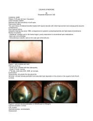
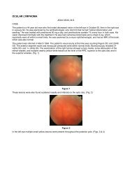
Figure 5. Granulomas consisting of histiocytes, epithelioid cells, <strong>and</strong> multinucleated giant cells.<br />
In 1941, Kveim reported the use of a suspension derived from the spleen of a patient with sarcoidosis that, when<br />
injected intracutaneously into patients with biopsy proven sarcoidosis, yielded a cutaneous papule containing non<br />
caseating epithelioid granulomas. Four to six weeks following subcutaneous injection of the Kveim reagent, the<br />
typical lesion presents as a red or brownish raised papule ranging from a few millimeters up to 1.5 centimeters in<br />
diameter. Histopathologic analysis of the biopsied lesion reveals a granuloma composed of epithelioid cells,<br />
occasional Langhans cells <strong>and</strong> scattered lymphocytes at its center with a surrounding cuff of mononuclear cells,<br />
primarily lymphocytes.<br />
Cutaneous anergy<br />
In 1916, Boeck first described cutaneous anergy to tuberculin in patients with sarcoidosis. It was later on realized<br />
that this phenomenon was not limited to tuberculin alone, but that anergy to a variety of other skin tests-antigens<br />
such was also typical. In 1994, Kataria <strong>and</strong> Holter proposed a mechanism for the cutaneous anergy seen in<br />
sarcoidosis. At sites of granulomatous inflammation, there is a predominance of helper T lymphocytes, which<br />
proliferate <strong>and</strong> secrete large amounts of lymphokines, including interleukin (IL)-2, monocyte chemotactic factor<br />
(MCF) <strong>and</strong> migration inhibition factor (MIF). These lymphokines induce <strong>and</strong> amplify the immune response by<br />
enhancing T-lymphocyte proliferation as well as recruiting <strong>and</strong> retaining monocytes from the circulation. The<br />
lymphokines <strong>and</strong> monokines produced at sites of granulomatous inflammation have their highest concentration<br />
locally. Nevertheless, the protein molecules diffuse into blood, establishing a concentration gradient between the<br />
granulomatous inflammatory site <strong>and</strong> the remote site of the delayed type hypersensitivity (DTH) skin test. As a<br />
result, the traffic of T-helper lymphocytes <strong>and</strong> monocytes is preferentially directed towards site of granuloma<br />
formation. That leads to a preponderance of suppressor cells in the peripheral blood <strong>and</strong> competitively depletes the<br />
T-helper cells <strong>and</strong> monocytes available to sites of DTH.<br />
Etiology <strong>and</strong> pathogenesis<br />
The processes involved in the pathogenesis of sarcoidosis in the lungs include accumulation of CD 4<br />
+ lymphocytes<br />
at the affected site. The cytokines <strong>and</strong> factors secreted by these cells account for the influx of monocytes, alveolitis,