Ocular Sarcoidosis - Ocular Immunology and Uveitis Foundation
Ocular Sarcoidosis - Ocular Immunology and Uveitis Foundation
Ocular Sarcoidosis - Ocular Immunology and Uveitis Foundation

You also want an ePaper? Increase the reach of your titles
YUMPU automatically turns print PDFs into web optimized ePapers that Google loves.
etinal detachment. Clinical <strong>and</strong>/or angiographic cystoid macular edema (CME) has been reported in 19%-72% of<br />
patients. Overall, patients with chronic posterior uveitis <strong>and</strong> panuveitis have significantly more complications than<br />
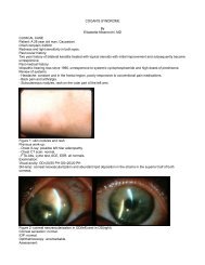
do patients with anterior uveitis. "C<strong>and</strong>le wax drippings" <strong>and</strong> "punched-out" lesions can be seen in patients with<br />
uveitis secondary to sarcoidosis (Figure 2).<br />
Figure 2. Punched-out choroidoretinal lesions in a patient with sarcoidosis.<br />
Severe retinal vasculitis <strong>and</strong> ischemic retinopathy with neovascularization, requiring scatter photocoagulation has<br />
been described in some patients. Other less frequent complications include peripapillary <strong>and</strong> subfoveal choroidal<br />
neovascularization, posterior scleritis with annular ciliochoroidal detachment causing angle closure glaucoma, <strong>and</strong><br />
central retinal vein occlusion leading to a painful blind eye from neovascular glaucoma.<br />
Orbit<br />
Although sarcoid granulomas have been described in most areas inside the orbit, the lacrimal gl<strong>and</strong> appears to be<br />
the organ most commonly affected. The frequency of lacrimal gl<strong>and</strong> involvement varies from 7% to 69%. The<br />
nasolacrimal drainage system can also become involved in patients with sarcoidosis. The patients usually present<br />
with epiphora <strong>and</strong> nasal stuffiness. Extraocular muscle involvement can occur <strong>and</strong> presents with diplopia or painful<br />
external ophthalmoplegia.<br />
<strong>Sarcoidosis</strong> in childhood<br />
Early onset or pre-school sarcoidosis seen in children is relatively rare. The classic triad of symptoms consists of<br />
skin, eye <strong>and</strong> joint lesions; pulmonary involvement is rare, at least initially. It can be easily misdiagnosed as juvenile<br />
rheumatoid arthritis, as the latter also presents with symptoms from the joints <strong>and</strong> eyes.<br />
Extraocular involvement<br />
The lung is the most frequently affected organ in patients with sarcoidosis. Histologically the lesions are distributed<br />
primarily along the lymphatics around bronchi <strong>and</strong> blood vessels, although alveolar lesions are also seen. The<br />
relative frequency of granulomas in the bronchial submucosa accounts for the high diagnostic yield of<br />
bronchoscopic biopsies. Lymph nodes are involved in almost all cases, especially the hilar <strong>and</strong> mediastinal ones.<br />
The majority of patients are asymptomatic, while others may complain about cough or shortness of breath.<br />
The spleen is enlarged in only 18% of cases, although microscopical evidence of sarcoid granulomas in spleen is<br />
present in three-quarters of the cases. The liver is affected less often than is the spleen, but a finding of elevated<br />
liver enzymes in a sarcoidosis suspect may prompt percutaneous liver biopsy in the search for histopathologic