measles.pdf (75.47 KB) - Ocular Immunology and Uveitis Foundation
measles.pdf (75.47 KB) - Ocular Immunology and Uveitis Foundation
measles.pdf (75.47 KB) - Ocular Immunology and Uveitis Foundation

Create successful ePaper yourself
Turn your PDF publications into a flip-book with our unique Google optimized e-Paper software.
Measlesby Erik Letko, M.D.DefintionThe Centers for Disease Control <strong>and</strong> Prevention defined "probable <strong>measles</strong>" as:1) generalized rash lasting 3 or more days, 2) temperature >101F, <strong>and</strong> 3) the presence of cough, coryza, orconjunctivitis.Measles can manifest as a congenital or acquired disease. Subacute sclerosing panencephalitis (SSPE) is a latecomplication of <strong>measles</strong> infection.Congenital <strong>measles</strong>In the pre-vaccine era, most people contracted <strong>measles</strong> in early childhood. For this reason, <strong>measles</strong> infection inpregnancy was unusual. Gershon et al estimated that four to six pregnancies per 100.000 were complicated by<strong>measles</strong> in the pre-vaccine era. In the post-vaccine era, a much larger proportion of <strong>measles</strong> cases has occurredamong adults of reproductive age. Measles virus can cross the placental barrier, causing spontaneous abortion orcongenital skeletal, cardiac or ocular anomalies.The congenital, ocular anomalies described in the literature include congenital cataract, dacryostenosis, <strong>and</strong> twocases of congenital retinopathy are reported as well (Figure 1). The visual acuities in both cases were normal.The diagnosis of congenital <strong>measles</strong> is made by the history of maternal <strong>measles</strong> <strong>and</strong> by the presence of congenitalanomalies. Congenital <strong>measles</strong> is a self limited disease that does not require treatment.Acquired <strong>measles</strong>EpidemiologyMeasles vaccine has been available in the United States since 1963. As a consequence, there is a markeddecrease of the disease with a shift in the ages of occurrence from children to adolescents <strong>and</strong> young adults.Vaccination in less developed countries is not widely available, <strong>and</strong> therefore the course of the disease is moresevere <strong>and</strong> often requires hospitalization. Dabis et al report on <strong>measles</strong> infection in a partially vaccinated populationin an African city. The mortality rate of hospitalized children was 16%.In spite of vaccine availability, not everyone in the United States receives it, mainly because of parental apathy,contraindications, <strong>and</strong> religious convictions. The failure of vaccination is present in 5 to 8 percent of population. Thispopulation remains susceptible to <strong>measles</strong>.Clinical courseClinical manifestations start with a prodromal phase of the disease 9 to 10 days after exposure, characterized byfever, malaise, lymphadenopathy, <strong>and</strong> mucosal lesions. The mucosal involvement consists of a catarrhal reaction ofthe conjunctiva <strong>and</strong> respiratory tract mucosa, as well as petechial lesions of the palate <strong>and</strong> pharynx. One to twodays before the onset of the rash, Koplik’s spots appear on the buccal mucosa. These are small, bluish-white dotssurrounded by a red areola typically localized on the buccal mucosa opposite the lower molars. The characteristicskin eruption starts as pink macules behind the ear, on the forehead, <strong>and</strong> on the neck.. The generallymphadenopathy <strong>and</strong> splenomegaly that arises early in the course of the acute disease may persist for severalweeks. The total duration of the rash is approximately 6 days.<strong>Ocular</strong> manifestationConjunctivitis, along with cough <strong>and</strong> coryza, is considered as a classical triad in <strong>measles</strong>. However, it is not alwayspresent. The conjunctivitis is usually mild, catarrhal, papillary <strong>and</strong> nonpurulent. Pseudomembranes may occur, <strong>and</strong>in debilitated patients, severe, true conjunctival membranes may develop. Koplik’s spots (on conjunctiva also calledHirschberg spots) are rarely present in the eye. Epithelial keratitis is even more common than conjunctivitis. It isusually mild <strong>and</strong> present in both eyes of all infected patients. The keratitis develops in the prodromal phase or at thetime of the onset of the skin rash <strong>and</strong> resolves without scarring several days afterwards. In some patients it maypersist as long as 4 months.Retinitis is, compared to keratitis <strong>and</strong> conjunctivitis, rarely present. It was described as macular edema,neuroretinitis, macular star formation, the presence of attenuated arterioles, <strong>and</strong> disc edema.
PATHOGENESISThe <strong>measles</strong> virus belongs to the genus Morbillivirus in the Paramyxoviridae family. It has an RNA core <strong>and</strong> ahelical capsid. The size of the virion is 100 to 200 nanometers in diameter. It is highly contagious. The first contactis at the mucous membrane of the respiratory tract. The conjunctiva also might act as a portal of entry for <strong>measles</strong>.If not inactivated by mucus or specific secretory IgA antibodies, the virus enters the ciliated columnar epithelium.During the primary viremia, two to six days after the infection, the virus is transported intracellularly inside theformed elements of the blood. A second viremia begins 10 days after the infection, with proliferation of the virusinside leukocytes. Neutralizing antibodies appear 14 days after infection, at the time of the appearance of the rash;rash is an expression of immunologic defense. In patients with severely impaired cell-mediated immunity, a<strong>measles</strong> infection may run its course without the appearance of rash. The <strong>measles</strong> antibodies cross the placenta<strong>and</strong> protect the infants against infection for the first six to twelve months of life.DiagnosisDiagnosis of <strong>measles</strong> is made by observing the regular sequence of the prodromal phase, the appearance ofKoplik’s spots <strong>and</strong> the generalized rash. The <strong>measles</strong> virus can be recovered from mucus membranes, urine, <strong>and</strong>blood for a few days before skin eruption <strong>and</strong> one or two days afterwards. The virus can be obtained from the urinefor an additional few days. Serum antibodies can be detected within one or two days after the onset of the rash.During the prodromal phase <strong>and</strong> early eruptive phase of <strong>measles</strong>, multi-nucleated giant cells with eosinophilicinclusions in both the nuclei <strong>and</strong> cytoplasm can be identified in sputum, nasal secretions, or urine. In electronmicroscopic studies, inclusion bodies are visible as granular masses, with the characteristic measurements of theRNA helix.TREATMENTUncomplicated <strong>measles</strong> infection is a self limiting disease. Supportive treatment is usually satisfactory enough. Thecourse of <strong>measles</strong> infection can be altered by treatment with gamma-globulin as soon as possible after exposure.After six days of exposure, the treatment with gamma globulin may not prevent or modify the disease.For the ocular manifestations, treatment is also symptomatic. No specific treatment for uveitis is described in theliterature.ComplicationsThe most serious complications of <strong>measles</strong> are those affecting central nervous system <strong>and</strong> heart. The incidence ofacute encephalitis in <strong>measles</strong> patients is 0.1%. It usually appears 6 weeks after the onset of the rash. Of thesepatients, 10 to 25% die, <strong>and</strong> 20 to 50% of the survivors suffer from permanent neurologic damage. Other centralnervous system complications include subacute <strong>measles</strong> encephalitis, toxic encephalopathy, retrobulbar neuritis,transverse myelitis, <strong>and</strong> ascending myelitis. SSPE is a late complication of <strong>measles</strong> affecting the eye discussedlater.The keratitis can become ulcerative in patients with secondary bacterial infections or in debilitated patients withconsequent corneal neovascularization, or it may become purulent, with progression to corneal perforation,panophthalmitis, <strong>and</strong> phthisis bulbi. These severe corneal ulcer complications are most frequent in developingcountries as a consequence of malnutrition, vitamin A <strong>and</strong> protein deficiencies.PrognosisThe prognosis of <strong>measles</strong> in vaccinated populations is good. In less developed countries approximately 1% ofchildren with <strong>measles</strong> end up with permanent ocular damage. The term post <strong>measles</strong> blindness (PMB) is restrictedto the corneal complications of <strong>measles</strong>, although retinitis <strong>and</strong> optic neuritis have been also described as a cause ofdamage to the sight after <strong>measles</strong> infection. In West Africa, 43 of 193 blind children were blinded by <strong>measles</strong>.Interestingly, in one study 10 of 13 patients with uveitis associated with multiple sclerosis had elevated levels of<strong>measles</strong> IgG antibodies in the anterior chamber fluid.Subacute sclerosing panencephalitisEpidemiologySSPE is a chronic degenerative disease of the central nervous system, first described by Dawson in 1933. Itsprevalence has been estimated at 1 to 10 per 10 million cases of <strong>measles</strong>. The male to female ratio varies from1.8:1 to 4:1. The onset of SSPE is usually 6 to 7 years after <strong>measles</strong>. Individuals who had <strong>measles</strong> before the ageof two years are of higher risk of developing SSPE. The majority of patients manifest neurologic signs before theage of 20 years.Clinical symptoms
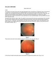
A typical clinical picture has three stages. Behavioral changes <strong>and</strong> intellectual deterioration are present in the firststage. The second stage is characterized by extrapyramidal signs <strong>and</strong> cortical blindness. Dementia develops in thethird stage <strong>and</strong> the course of the disease is terminated by death within 1 to 3 years.<strong>Ocular</strong> symptomsThirty to fifty percent of patients with SSPE develop ocular symptoms. These include maculopathy (Figures 2 to6– fundus pictures <strong>and</strong> fluorescein angiogram from one patient taken 18 hours <strong>and</strong> 11 days apart showing rapidprogression of the lesion), disc edema, papillitis, optic atrophy, retinitis, serous retinal detachment, chorioretinitis,<strong>and</strong> vasculitis. The <strong>measles</strong> virus primarily affects the retina with secondary involvement of the retinal pigmentepithelium <strong>and</strong> choroid. The eye involvement starts as a focal area of retinitis in the macula <strong>and</strong> over time it canspread to the posterior pole <strong>and</strong> peripheral retina as well. Characteristically, there is little or no vitreousinflammation. Retinitis resolves leaving retinal pigment epithelium mottling <strong>and</strong> varying degrees of scarring (Figures6 through 8).PATHOGENESISThe classic <strong>measles</strong> virus replicates by budding <strong>and</strong> fusion. SSPE is caused by <strong>measles</strong> virus deficient of certainvirion polypeptides, such as matrix (M), hemagglutinin (H), <strong>and</strong> fusion (F) proteins. These proteins are necessaryfor alignment of the virus along the host-cell plasma membrane <strong>and</strong> subsequent budding <strong>and</strong> release of the virusfrom the host cell. Defects in these proteins <strong>and</strong> host’s factors may explain the prolonged viral persistence (Figure9). The disease is a true panencephalitis affecting both gray <strong>and</strong> white matter.DiagnosisThe diagnosis is made by the presence of three of these five criteria: 1) clinical course, 2) biopsy or necropsyresults, 3) EEG pattern, 4) elevated globulin levels in cerebrospinal fluid, <strong>and</strong> 5) elevated levels of IgG <strong>measles</strong>antibodies in serum <strong>and</strong> cerebrospinal fluid. The negative IgM <strong>measles</strong> antibodies provide evidence against a new,acute-onset infection.TREATMENTThere is no definitive treatment for SSPE. However, inosiplex <strong>and</strong> intracameral or intrathecal interferon may induceremission or stabilize the clinical course. ButSSPE is, generally a lethal disease. A spontaneous remission is reported in 5% of patients. The survival rate differsfrom 5 to 50% according to various studies. Early diagnosis <strong>and</strong> treatment are essential to improve the prognosis.Reading listBergstrom TJ: Measles infection of the eye. In Viral diseases of the eye. Ed Darrell RW; Lea & Febiger 1985; 233-250.Culbertson WW: Viral retinitis. In Infections of the eye. Ed Tabbara KF, Hyndiuk RA; Little, Brown <strong>and</strong> Company1996; 506.Bloch-Michel E, Helleboid L, Hill C et al.: Measles virus antibody in aqueous humor of patients with uveitisassociated with multiple sclerosis. Lancet 1992; 339:750-751.Park DW, Boldt HC, Massicotte SJ et al.: SSPE manifesting as viral retinitis: Clinical <strong>and</strong> histological findings. Am JOphthalmol 1997; 123:533-542.Salmon JF, Pan EL, Murray AD: Visual loss with dancing extremities <strong>and</strong> mental disturbances. Surv Ophthalmol1991; 35:299-306.Yalaz K, Anlar B, Oktem F et al.: Intraventricular interferon <strong>and</strong> oral inosiplex in the treatment of SSPE. Neurology1992; 42:488-491.