MORPHOLOGICAL OBSERVATIONS ON THE TESTES ... - Unesp
MORPHOLOGICAL OBSERVATIONS ON THE TESTES ... - Unesp
MORPHOLOGICAL OBSERVATIONS ON THE TESTES ... - Unesp

You also want an ePaper? Increase the reach of your titles
YUMPU automatically turns print PDFs into web optimized ePapers that Google loves.
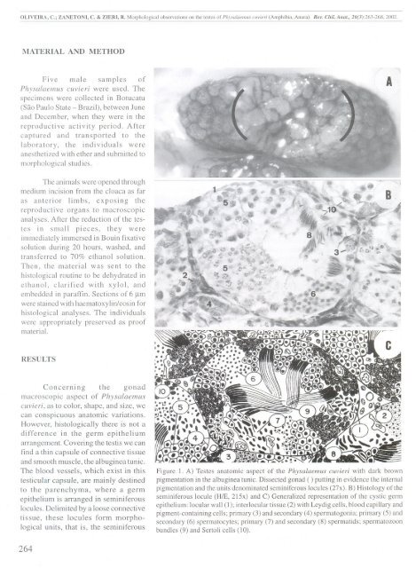
OLIVEIRA, c.; ZANET<strong>ON</strong>I, C. & ZIE)H, R. Morphological observations on the testes of Physalaemus euvieri (Amphibia, Anura). Rev. Chil. Anaf., 20(3):263-268, 2002.MATERIAL AND METHODFive male samples ofPhysalaemus cuvieri were used. Thespecimens were collected in Botucatu(São Paulo State - Brazil), between Juneand December, when they were in thereproductive activity period. Aftercaptured and transported to thelaboratory, the individuaIs wereanesthetized with ether and submitted tomorphologicalstudies.AThe animaIs were opened throughmedium incision from the cloaca as faras anterior limbs, exposing thereproductive organs to macroscopicanalyses. After the reduction of the testesin small pieces, they wereimmediately immersed in Bouin fixativesolution during 20 hours, washed, andtransferred to 70% ethanol solution.Then, the .material was sent to thehistological routine to be dehydrated inethanol, clarified with xylol, andembedded in paraffin. Sections of 6 ~mwere stained with haematoxylin/eosin forhistological analyses, The individuaIswere appropriately preserved as proofmaterial.RESULTSConcerning the gonadmacroscopic aspect of Physalaemuscuvieri, as to color, shape, and size, wecan conspicuous anatomic variations.However, histologically there is not adifference in the germ epitheliumarrangement. Covering the testis we canfind a thin capsule of connective tissueand smooth muscle, the albuginea tunic.The blood vessels, which exist in thistesticular capsule, are maínly destinedto the parenchyma, where a germepithelium is arranged in seminiferouslocules, Delimited by a loose connectivetissue, these locules form morphologicalunits, that is, the seminiferousFigure I, A) Testes anatomic aspect of the Physalaemus cuvieri with dark brownpigmentation in the albuginea tunic, Dissected gonad ( ) putting in evidence the internalpigmentation and the units denominated seminiferous [ocules (27x). B) Histology of theseminiferous locule (HlE, 215x) and C) Generalized representation of the cystic germepithelium: locular wall (I); interlocular tissue (2) with Leydig cells, blood capillary andpigment-containing cells; primary (3) and secondary (4) spermatogonia; primary (5) andsecondary (6) spermatocytes; primary (7) and secondary (8) spermatids; spermatozoonbundles (9) and Sertoli cells (10).264