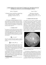
vari<strong>an</strong>ce: 3´ 8 iterations0.9h−line 1200.80.70.60.50.40.30.20.100 20 40 60 80 100 120 140 160 180Figure 4: A post-diffusion vari<strong>an</strong>ce imagepost−diffusion segmentationFigure 6: A cross-sectional view along a selected horizontalline <strong>of</strong> the image <strong>of</strong> Figure 4 before <strong>an</strong>d after the<strong>an</strong>isotropic diffusionA good illustration <strong>of</strong> the operation <strong>of</strong> the <strong>an</strong>isotropicdiffusion equation is given in Figure 6. This is a crosssectionalview along a selected horizontal line <strong>of</strong> the image<strong>of</strong> Figure 4, before <strong>an</strong>d after the diffusion. It c<strong>an</strong> be noticedthat the location <strong>of</strong> the strong edges characterised bylarge values <strong>of</strong> the gradient are preserved. Their strengthis also preserved. Areas between such edges are beingsmoothed out. This nonlinear processing is the foundation<strong>of</strong> the <strong>an</strong>isotropic diffusion algorithm. In addition, theme<strong>an</strong> value <strong>of</strong> the image intensity (vari<strong>an</strong>ce, in the example)is also preserved.Concluding remarksFigure 5: A segmented PCO image after diffusionstarting the algorithm after every 8 iterations. The restartinghelps to maintain the strong edges [6]. The vari<strong>an</strong>ceimage after the <strong>an</strong>isotropic diffusion is presented in Figure4. The resulting segmented image is given in Figure 5.In comparison with the previous segmentation results (Figure3), it c<strong>an</strong> be observed that the incorrectly <strong>class</strong>ified area(upper left part <strong>of</strong> the image) is signific<strong>an</strong>tly reduced, whilethe correctly <strong>class</strong>ified area is unch<strong>an</strong>ged.The <strong>an</strong>isotropic diffusion equation is one example <strong>of</strong> a richbody <strong>of</strong> variational methods which have a long history inimage processing. In application to processing the PCO<strong>images</strong>, the algorithm that we study in this paper is used toimprove segmentation <strong>of</strong> these <strong>images</strong>. The segmentationmethods are currently based on the directional vari<strong>an</strong>ce operator.Applying the <strong>an</strong>isotropic diffusion equation to thevari<strong>an</strong>ce <strong>images</strong> reduces the area <strong>of</strong> incorrectly <strong>class</strong>ifiedparts <strong>of</strong> the difficult-to-segment PCO <strong>images</strong>.AcknowledgmentThe authors would like to express their gratitude to Mr D.J. Spalton Clinical Director <strong>of</strong> the Department <strong>of</strong> Ophthalmology<strong>of</strong> St Thomas’ Hospital for the specification <strong>of</strong> theproblem, the provision <strong>of</strong> the image data <strong>an</strong>d m<strong>an</strong>y useful
discussions.References[1] A. P. Papliński <strong>an</strong>d J. F. Boyce, “Segmentation <strong>of</strong> a<strong>class</strong> <strong>of</strong> <strong>ophthalmological</strong> <strong>images</strong> <strong>using</strong> a directionalvari<strong>an</strong>ce operator <strong>an</strong>d co-occurrence arrays,” OpticalEngineering, vol. 36, pp. 3140–3147, November1997.[2] A. P. Papliński, “Directional filtering in edge detection,”IEEE Tr<strong>an</strong>s. Image Proc., vol. 7, pp. 611–615,April 1998.[3] A. P. Papliński <strong>an</strong>d J. F. Boyce, “Co-occurrence arrays<strong>an</strong>d edge density in segmentation <strong>of</strong> a <strong>class</strong> <strong>of</strong><strong>ophthalmological</strong> <strong>images</strong>,” in Proceedings <strong>of</strong> the 4rdConference on Digital Image Computing: Techniques<strong>an</strong>d Applications, DICTA97, (Auckl<strong>an</strong>d,), pp. 521–528, December 1997.Signal <strong>Processing</strong> <strong>an</strong>d Communication Systems (IS-PACS’98), (Melbourne, Australia), November 1998.[11] S. A. Barm<strong>an</strong>, J. F. Boyce, D. J. Spalton, P. G. Ursell,<strong>an</strong>d E. J. Hollick, “Measurement <strong>of</strong> posterior capsuleopacification,” in Proceedings <strong>of</strong> the Conferenceon Medical Image Underst<strong>an</strong>ding <strong>an</strong>d Analysis,MIUA97, July 1997. Oxford, U.K.[12] M. V. P<strong>an</strong>de, P. G. Ursell, D. J. Spalton, <strong>an</strong>dS. Kundaiker, “High resolution digital retroilluminationimaging <strong>of</strong> the posterior lens capsule aftercataract surgery,” J. Cataract Refract. Surg., vol. 23,pp. 1521–1527, 1997.[13] D. S. Friedm<strong>an</strong>, D. D. Dunc<strong>an</strong>, M. Beatriz, <strong>an</strong>d S. K.West, “Digital image capture <strong>an</strong>d automated <strong>an</strong>alysis<strong>of</strong> posterior capsular opacification,” Invest. OphthalmologyVis. Sci., vol. 40, pp. 1715–1726, July 1999.[4] A. P. Papliński <strong>an</strong>d J. F. Boyce, “Tri-directional filteringin processing a <strong>class</strong> <strong>of</strong> <strong>ophthalmological</strong> <strong>images</strong>,”in Proceedings <strong>of</strong> the IEEE Region 10 AnnualConference, TENCON’97, (Brisb<strong>an</strong>e), pp. 687–690,December 1997.[5] V. Caselles, J.-M. Morel, G. Sapiro, <strong>an</strong>d A. T<strong>an</strong>nenbaum,“Introduction to the special issue on partial differentialequations <strong>an</strong>d geometry driven diffusion inimage processing,” IEEE Tr<strong>an</strong>sactions on Image <strong>Processing</strong>,vol. 7, pp. 269–273, March 1998.[6] G. H. Cottet <strong>an</strong>d M. El Ayyadi, “A Voltera type modelfor image processing,” IEEE Tr<strong>an</strong>sactions on Image<strong>Processing</strong>, vol. 7, pp. 292–303, March 1998.[7] S. T. Acton, “Multigrid <strong>an</strong>isotropic diffusion,” IEEETr<strong>an</strong>sactions on Image <strong>Processing</strong>, vol. 7, pp. 280–291, March 1998.[8] J. Weickert <strong>an</strong>d B. M. ter Haar Romney, “Efficient<strong>an</strong>d reliable schemes for nonlinear diffusion filtering,”IEEE Tr<strong>an</strong>sactions on Image <strong>Processing</strong>, vol. 7,pp. 399–410, March 1998.[9] A. P. Papliński <strong>an</strong>d J. F. Boyce, “<strong>Processing</strong> a <strong>class</strong><strong>of</strong> <strong>ophthalmological</strong> <strong>images</strong> <strong>using</strong> <strong>an</strong> <strong>an</strong>isotropic diffusionequation,” in Proceedings <strong>of</strong> the 2nd AnnualIASTED International Conference on ComputerGraphics <strong>an</strong>d Imaging (CGIM’99), (Palm Springs,California, USA), October 1999. Accepted for presentation.[10] A. P. Papliński <strong>an</strong>d J. F. Boyce, “A first-order waveequation in modelling the behaviour <strong>of</strong> epithelialcells in <strong>an</strong> eye posterior capsule,” in Proceedings <strong>of</strong>the 6th IEEE International Workshop on Intelligent