Soft Matter - User Web Pages - Monash University
Soft Matter - User Web Pages - Monash University
Soft Matter - User Web Pages - Monash University

You also want an ePaper? Increase the reach of your titles
YUMPU automatically turns print PDFs into web optimized ePapers that Google loves.
<strong>Soft</strong> <strong>Matter</strong><br />
PAPER<br />
View Article Online<br />
View Journal | View Issue<br />
Downloaded by <strong>Monash</strong> <strong>University</strong> on 13 March 2013<br />
Published on 22 February 2013 on http://pubs.rsc.org | doi:10.1039/C3SM00016H<br />
Cite this: <strong>Soft</strong> <strong>Matter</strong>, 2013, 9, 3631<br />
Received 2nd January 2013<br />
Accepted 30th January 2013<br />
DOI: 10.1039/c3sm00016h<br />
www.rsc.org/softmatter<br />
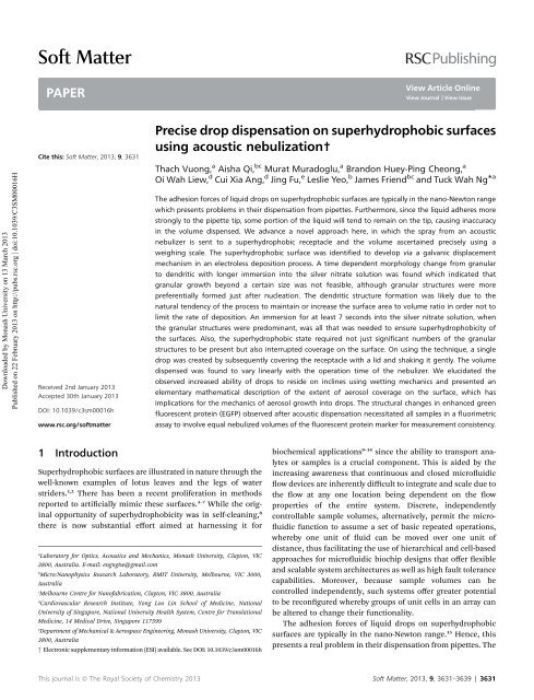
Precise drop dispensation on superhydrophobic surfaces<br />
using acoustic nebulization†<br />
Thach Vuong, a Aisha Qi, bc Murat Muradoglu, a Brandon Huey-Ping Cheong, a<br />
Oi Wah Liew, d Cui Xia Ang, d Jing Fu, e Leslie Yeo, b James Friend bc and Tuck Wah Ng* a<br />
The adhesion forces of liquid drops on superhydrophobic surfaces are typically in the nano-Newton range<br />
which presents problems in their dispensation from pipettes. Furthermore, since the liquid adheres more<br />
strongly to the pipette tip, some portion of the liquid will tend to remain on the tip, causing inaccuracy<br />
in the volume dispensed. We advance a novel approach here, in which the spray from an acoustic<br />
nebulizer is sent to a superhydrophobic receptacle and the volume ascertained precisely using a<br />
weighing scale. The superhydrophobic surface was identified to develop via a galvanic displacement<br />
mechanism in an electroless deposition process. A time dependent morphology change from granular<br />
to dendritic with longer immersion into the silver nitrate solution was found which indicated that<br />
granular growth beyond a certain size was not feasible, although granular structures were more<br />
preferentially formed just after nucleation. The dendritic structure formation was likely due to the<br />
natural tendency of the process to maintain or increase the surface area to volume ratio in order not to<br />
limit the rate of deposition. An immersion for at least 7 seconds into the silver nitrate solution, when<br />
the granular structures were predominant, was all that was needed to ensure superhydrophobicity of<br />
the surfaces. Also, the superhydrophobic state required not just significant numbers of the granular<br />
structures to be present but also interrupted coverage on the surface. On using the technique, a single<br />
drop was created by subsequently covering the receptacle with a lid and shaking it gently. The volume<br />
dispensed was found to vary linearly with the operation time of the nebulizer. We elucidated the<br />
observed increased ability of drops to reside on inclines using wetting mechanics and presented an<br />
elementary mathematical description of the extent of aerosol coverage on the surface, which has<br />
implications for the mechanics of aerosol growth into drops. The structural changes in enhanced green<br />
fluorescent protein (EGFP) observed after acoustic dispensation necessitated all samples in a fluorimetric<br />
assay to involve equal nebulized volumes of the fluorescent protein marker for measurement consistency.<br />
1 Introduction<br />
Superhydrophobic surfaces are illustrated in nature through the<br />
well-known examples of lotus leaves and the legs of water<br />
striders. 1,2 There has been a recent proliferation in methods<br />
reported to articially mimic these surfaces. 3–7 While the original<br />
opportunity of superhydrophobicity was in self-cleaning, 8<br />
there is now substantial effort aimed at harnessing it for<br />
a<br />
Laboratory for Optics, Acoustics and Mechanics, <strong>Monash</strong> <strong>University</strong>, Clayton, VIC<br />
3800, Australia. E-mail: engngtw@gmail.com<br />
b<br />
Micro/Nanophysics Research Laboratory, RMIT <strong>University</strong>, Melbourne, VIC 3000,<br />
Australia<br />
c<br />
Melbourne Centre for Nanofabrication, Clayton, VIC 3800, Australia<br />
d<br />
Cardiovascular Research Institute, Yong Loo Lin School of Medicine, National<br />
<strong>University</strong> of Singapore, National <strong>University</strong> Health System, Centre for Translational<br />
Medicine, 14 Medical Drive, Singapore 117599<br />
e<br />
Department of Mechanical & Aerospace Engineering, <strong>Monash</strong> <strong>University</strong>, Clayton, VIC<br />
3800, Australia<br />
† Electronic supplementary information (ESI) available. See DOI: 10.1039/c3sm00016h<br />
biochemical applications 9–14 since the ability to transport analytes<br />
or samples is a crucial component. This is aided by the<br />
increasing awareness that continuous and closed microuidic<br />
ow devices are inherently difficult to integrate and scale due to<br />
the ow at any one location being dependent on the ow<br />
properties of the entire system. Discrete, independently<br />
controllable sample volumes, alternatively, permit the micro-<br />
uidic function to assume a set of basic repeated operations,<br />
whereby one unit of uid can be moved over one unit of<br />
distance, thus facilitating the use of hierarchical and cell-based<br />
approaches for microuidic biochip designs that offer exible<br />
and scalable system architectures as well as high fault tolerance<br />
capabilities. Moreover, because sample volumes can be<br />
controlled independently, such systems offer greater potential<br />
to be recongured whereby groups of unit cells in an array can<br />
be altered to change their functionality.<br />
The adhesion forces of liquid drops on superhydrophobic<br />
surfaces are typically in the nano-Newton range. 15 Hence, this<br />
presents a real problem in their dispensation from pipettes. The<br />
This journal is ª The Royal Society of Chemistry 2013 <strong>Soft</strong> <strong>Matter</strong>, 2013, 9, 3631–3639 | 3631
<strong>Soft</strong> <strong>Matter</strong><br />
View Article Online<br />
Paper<br />
Downloaded by <strong>Monash</strong> <strong>University</strong> on 13 March 2013<br />
Published on 22 February 2013 on http://pubs.rsc.org | doi:10.1039/C3SM00016H<br />
use of exible pipette tips 11 permits drop volumes down to<br />
around 10 mL to be deposited, albeit this requires careful<br />
execution in order for them not to contact (from the tip<br />
springing back aer dispensation) the surface and thus damage<br />
the nano and micro features that endow superhydrophobicity.<br />
It should also be noted that since the liquid adheres more<br />
strongly to the pipette tip, an unknown portion of the liquid will<br />
remain within the tip, causing substantial inaccuracy in the<br />
dispensed volume.<br />
The quest to develop bioanalysis tools based on superhydrophobicity<br />
is also founded on the issues of availability and<br />
cost. Bioanalytical chemists in well endowed laboratories are<br />
oen accustomed to having a plethora of convenient consumables<br />
and sophisticated instrumentation at their disposal. Many<br />
researchers in resource-limited developing countries, or eld<br />
workers in remote locations far from modern conveniences, have<br />
been unable to take advantage of modern bioanalytical techniques<br />
due to a lack of infrastructure. This is unfortunate as it is<br />
these same researchers that usually have the greatest need for<br />
bioanalytical tools that will help diagnose diseases such as<br />
tuberculosis or malaria. Not surprisingly, there have been recent<br />
efforts expended to use alternative materials such as paper to<br />
serve as the analyte handling media. 16 This approach, while<br />
useful for methods based on electrochemical detection, is not as<br />
effective for methods that are based on optical detection, that<br />
arguably offer the highest versatility and sensitivity. Alternative<br />
cost effective approaches have since been reported. 17,18 Despite<br />
this, paper remains indispensable as a relatively cheap, compact<br />
and robust reservoir for test samples and biochemical analytes.<br />
In this work, we seek to develop a technique that will allow us<br />
to deliver drops of specic volumes on superhydrophobic<br />
surfaces from cost effective storage media such as paper so that<br />
they can in turn be harnessed to be developed into cost effective<br />
devices that permit transport. In the process, we will study the<br />
nature of how small aerosols form on these surfaces before<br />
evolving into single drops.<br />
2 Materials and methods<br />
We advance the approach depicted in Fig. 1 to circumvent this<br />
problem. A liquid supply chain was created out of a reservoir<br />
that delivers to a short capillary tube section, whose tip is placed<br />
in contact with a surface acoustic wave (SAW) nebulizer running<br />
at 30 MHz frequency using a small piece of tissue paper that<br />
constituted a capillary wick. 19 The SAW device was constructed<br />
out of a low-loss piezoelectric substrate, specically, a 127.86 <br />
Y–X-rotated single-crystal lithium niobate (LiNbO 3 ) substrate,<br />
with pairs of chromium–aluminum interdigital transducers<br />
fabricated on one side via standard UV photolithography. When<br />
an AC signal is supplied to the transducer at its resonant<br />
frequency, the SAW in the form of a Rayleigh wave propagates<br />
along the LiNbO 3 surface from the transducer at about 3900 m<br />
s 1 . Although the surface displacement amplitudes are only in<br />
the 1–10 nm range, the accelerations are extremely high (about<br />
10 7 ms 2 ) due to excitation at frequencies over 10 MHz. These<br />
huge surface accelerations are transmitted into the liquid<br />
placed on the substrate, inducing acoustic streaming. 20 When<br />
Fig. 1 Schematic description of the scheme to obtain precise volumes of drops<br />
on superhydrophobic surfaces. A surface acoustic wave nebulizer delivers a spray<br />
of aerosol droplets onto the receptacle in which the exact volume is determined<br />
using an accurate weighing scale. By covering with a superhydrophobic lid and<br />
gentle shaking, a single drop is created.<br />
the energy is sufficient (i.e., electrical power supplied in the 1–<br />
3 W range), destabilization of the liquid's free surface occurs.<br />
This leads to a breakup of capillary waves, generating a spray of<br />
aerosol droplets through a process known as SAW atomization<br />
or nebulization. 21,22 When this spray of aerosol is channeled<br />
onto a semi-spherical superhydrophobic receptacle, larger<br />
drops develop on the receptacle surface from multiple coalescence<br />
events that are inuenced by gravity (which tend to draw<br />
them towards the receptacle trough).<br />
The receptacle was fashioned out of a copper sheet (1 mm<br />
thickness), polished earlier to remove all visible scratches using<br />
silicon carbide electro-coated waterproof abrasive paper<br />
(KMCA, WET/DRY S85 P600), by an 8.5 mm radius ball indenter.<br />
Prior to use, the receptacle surface was rst cleaned using<br />
absolute ethanol, allowed to air dry, and then immersed in a<br />
24.75 mM aqueous solution of AgNO 3 for 1 minute to form the<br />
micro and nano structures. Aer this, the surface was rinsed<br />
with copious amounts of distilled water followed by absolute<br />
ethanol before being allowed to air dry. Once dried, it was<br />
immersed in a 1 mM solution of the surface modier<br />
CF 3 (CF 2 ) 7 CH 2 CH 2 SH in absolute ethanol for 5 minutes. Aer<br />
removal, it was again rinsed with copious amounts of distilled<br />
water, followed by absolute ethanol, and then air dried.<br />
For a side experiment to study the nano and microstructures<br />
forming in relation to wetting, we created coupons 20 20 mm<br />
in size out of the same copper sheet (1 mm thickness) and using<br />
the same polishing and cleaning process as previously used.<br />
The coupons were then immersed in the 24.75 mM aqueous<br />
solution of AgNO 3 for selected periods ranging from 2 seconds<br />
to 120 seconds before being removed to air dry. Aer drying, the<br />
3632 | <strong>Soft</strong> <strong>Matter</strong>, 2013, 9, 3631–3639 This journal is ª The Royal Society of Chemistry 2013
View Article Online<br />
Paper<br />
<strong>Soft</strong> <strong>Matter</strong><br />
Downloaded by <strong>Monash</strong> <strong>University</strong> on 13 March 2013<br />
Published on 22 February 2013 on http://pubs.rsc.org | doi:10.1039/C3SM00016H<br />
morphology of the superhydrophobic surface was characterized<br />
with an FEI Quanta 3D FEG scanning electron microscope<br />
(SEM). The elemental composition was characterized by an X-<br />
ray energy dispersive spectrometer (EDS) associated with the<br />
SEM. The wetting characteristics were evaluated by placing 5 mL<br />
of sessile drops of water on each surface (aer immersing into<br />
the surface modifying solution) and then determining the<br />
contact angle.<br />
In the technique, the weight of the liquid delivered to the<br />
receptacle, which could be correlated to volume, was determined<br />
using a weighing scale (A&D GR-200 with 0.0001 g<br />
precision) on which the receptacle is placed on. Once the<br />
required volume was achieved, aerosol delivery from the<br />
nebulizer was terminated, a superhydrophobic lid placed over<br />
the receptacle, and the assembly shaken gently to merge the<br />
drops together into one. Apart from ensuring that no liquid<br />
spilled out of the receptacle, the lid also served to limit the<br />
effects of evaporation, which is signicant for small drop<br />
volumes. Experiments were conducted to establish the liquid<br />
(Milli-Q water) delivery characteristics when the power to the<br />
nebulizer was kept constant at 2 W.<br />
Lastly, the effect of nebulization on the structural functionality<br />
of enhanced green uorescent protein (EGFP) was evaluated.<br />
This C-terminally His 6 -tagged uorescent protein was isolated<br />
from genetically modied Escherichia coli and puried to near<br />
homogeneity by automated affinity chromatography using 1 mL<br />
bed volume Mini Pronity IMAC cartridges on the Pronia<br />
Protein Purication System (Bio-Rad) under the default program<br />
settings of the Native IMAC method with integrated desalting<br />
into sodium phosphate buffer (pH 7.4). The His 6 -tagged EGFP<br />
was checked for purity by SDS-PAGE (see ESI†) andquantied<br />
using the BCA protein assay (Pierce, USA). Samples of the protein<br />
were nebulized at different powers, collected in capillary tubes,<br />
imaged together using a uorescence microscope (Olympus<br />
BX61), and the intensities extracted using the ImageJ soware.<br />
3 Results and discussion<br />
The micro- and nano-structures developed by immersing the<br />
copper coupons into the aqueous solution of AgNO 3 were formed<br />
by an electroless deposition process. This process is similar to<br />
electrolytic plating except that no external electrodes are needed. 23<br />
In electroless deposition, the metal ions are typically reduced into<br />
metals by the introduction of a reducing agent. A variety of<br />
procedures with different reagents have been demonstrated for<br />
electroless deposition of silver. 24,25 In the process conducted here,<br />
however, the deposition was able to proceed without any external<br />
reducing agent. Rather, a galvanic displacement mechanism<br />
occurred in which the silver cations in solution were reduced just<br />
as copper from the surface was oxidized.<br />
The SEM micrographs of structures formed in association<br />
with various immersion times of the copper coupons into the<br />
silver nitrate solution are presented in Fig. 2. The background<br />
striated structures seen at 2 seconds were formed due to the<br />
polishing process. On the substrate, very small granules started<br />
to be deposited at numerous nucleation sites. These granules<br />
Fig. 2 Scanning electron micrographs obtained from immersing the copper coupons into the silver nitrate solution for different lengths of time followed by air drying.<br />
It can be seen that granular structures developed with short period immersion, whilst dendritic structures formed with longer period immersion.<br />
This journal is ª The Royal Society of Chemistry 2013 <strong>Soft</strong> <strong>Matter</strong>, 2013, 9, 3631–3639 | 3633
<strong>Soft</strong> <strong>Matter</strong><br />
View Article Online<br />
Paper<br />
Downloaded by <strong>Monash</strong> <strong>University</strong> on 13 March 2013<br />
Published on 22 February 2013 on http://pubs.rsc.org | doi:10.1039/C3SM00016H<br />
Fig. 3 Scanning electron micrographs at higher magnifications that provide a<br />
clearer picture of the (a) granular and (b) dendritic structures developed.<br />
grew in size with longer immersion times until about 10–20<br />
seconds. At the 20 seconds mark, the granules appeared to<br />
shrink slightly in size whilst exhibiting greater dendritic growth<br />
from the surface of each granule. From then onwards, the<br />
dendritic structures began to proliferate on the existing granular<br />
structures, developing later into fern-like foliage. The<br />
distinct differences between these two types of structures<br />
(granular and dendritic) are shown more clearly in the higher<br />
magnication micrographs provided in Fig. 3.<br />
The time dependent morphology changes appear to indicate<br />
that granular growth beyond a certain size was not feasible,<br />
although granular structures were more preferentially formed<br />
just aer nucleation. This is likely due to the natural tendency<br />
of the process to maintain or increase the surface area to<br />
volume ratio in order not to limit the rate of deposition. Taking<br />
a sphere for example, the surface area to volume ratio scales<br />
according to 3/r, where r is the radius, inferring that the surface<br />
area to volume ratio reduces as the sphere increases in size.<br />
Thus, the formation of dendritic structures offers an avenue by<br />
nature to circumvent this obstacle. This argument is supported<br />
somewhat by the XRD maps obtained that revealed no signicant<br />
elemental changes in the structures.<br />
Aer the surfaces were treated with the modier, we found<br />
that immersion for at least 7 seconds into the silver nitrate<br />
solution was all that was needed to ensure superhydrophobicity<br />
of the surfaces. At this stage, the structures appeared to be<br />
predominantly granular. Previous studies have shown that<br />
granular structures alone were sufficient to cause superhydrophobicity.<br />
26 Hence, we were able to conclude that the<br />
subsequent dendritic structures were not needed to maintain<br />
the non-wetting characteristic, although they seemed not to<br />
have a role in modifying it. The micrograph at 5 seconds<br />
immersion also showed signicant coverage of granular structures<br />
over the substrate. This presented an interesting conundrum<br />
as to why superhydrophobicity could not be sustained at<br />
this stage. On more careful examination, we found that there<br />
were regions on the substrate surface where the granular<br />
structures were not fully developed. It appears then that the<br />
superhydrophobic state requires not just signicant numbers of<br />
the granular structures to be present on the surface but also<br />
uninterrupted structure coverage.<br />
We move now to discuss the experimentation results in<br />
obtaining the drops. During each run on liquid deposition, the<br />
nebulizer was cyclically pulsed on for 5 seconds and off for 5<br />
seconds. This was done to accommodate the response time of<br />
the weighing scale. Fig. 4 presents results of the mass recorded<br />
in relation to time in which the nebulizer was operated for three<br />
typical runs. Highly linear trends are observed, indicating that<br />
xed quanta of liquid were dispensed with each pulsed operation<br />
of the nebulizer for a specic run. While the data for two of<br />
the runs were almost identical, the gradient for a third run was<br />
signicantly altered. This was due to the process that happens<br />
in the tissue as it served to draw liquid out from the reservoir<br />
before perturbations from the SAW device are able to dislodge it<br />
for delivery. In the course of this process, factors that affect the<br />
transfer of liquid in and out of the tissue (such as temperature<br />
and airborne particles attaching to the bers) likely caused the<br />
ow rate to vary with each run.<br />
This result implied that an open-loop operation using precalibration<br />
without using the weighing scale was not feasible.<br />
Due to the ability of the SAW driven nebulizer to operate nearly<br />
instantly, from zero to full power and to zero power again in<br />
Fig. 4 Plots of the readings from the weighing scale against the operation time of the nebulizer. The trends from each run are highly linear, albeit the slope variation<br />
indicates that pre-calibrated operation without the weighing scale was not feasible.<br />
3634 | <strong>Soft</strong> <strong>Matter</strong>, 2013, 9, 3631–3639 This journal is ª The Royal Society of Chemistry 2013
Paper<br />
View Article Online<br />
<strong>Soft</strong> <strong>Matter</strong><br />
Downloaded by <strong>Monash</strong> <strong>University</strong> on 13 March 2013<br />
Published on 22 February 2013 on http://pubs.rsc.org | doi:10.1039/C3SM00016H<br />
Fig. 5 Images of (a) multiple nebulized droplets formed on the surface of the<br />
receptacle and (b) a single drop that results after shaking the receptacle. Fig. 6 Illustrations depicting predominantly high (a) Cassie and (b) Wenzel<br />
wetting states of relatively larger drops and aerosols residing respectively on<br />
superhydrophobic surfaces. (c) When further aerosols arrive at the surface, they<br />
approximately 1 microsecond, there was no ‘lagging volume’<br />
delivered when the nebulizer was turned off. Hence, the<br />
limiting factor for accurate volume dispensation was only that<br />
dictated by the mass resolution of the weighing scale. In the<br />
current case, the volume resolution was 1 mL based on the<br />
density of water being 1000 kg m 3 and the weighing scale's<br />
mass resolution being 0.0001 g. The response of the weighing<br />
scale also determined the time needed, since the off times could<br />
be shortened if it settled faster. We have also found that good<br />
isolation from dra and ambient vibrations was crucial to<br />
maintaining accuracy.<br />
The formation of multiple drops in the receptacle (Fig. 5(a))<br />
of up to 3 mL by volume (by estimation) before the gentle<br />
shaking operation was applied to dislodge them to form a nal<br />
single drop (Fig. 5(b)) presents an interesting conundrum.<br />
Experiments with drops of this volume typically show that they<br />
move easily when dispensed on superhydrophobic surfaces. In<br />
fact, earlier dynamical studies conducted show that very small<br />
forces (in the nano-Newton range) are needed to move water<br />
drops on this surface. 15 Coupled with the curvature of the<br />
receptacle, this should then result in a single drop forming all<br />
the time even when no shaking was introduced. This apparent<br />
anomalous behavior can be explained by the Cassie and Wenzel<br />
wetting states of superhydrophobic surfaces. 27 When a drop<br />
impinges on a wetting surface, it is known that it will rst<br />
expand rapidly. 28 With sufficient momentum of the drop, the<br />
surface microstructures are able to impale the liquid surface. As<br />
the liquid loses kinetic energy, the drop will eventually settle<br />
into a static state, leading to the observation of stickiness. On a<br />
non-wetting surface, alternatively, stronger capillary and<br />
hydrodynamic forces develop to impede this impalement<br />
process. Consequently, the drop is able to bounce off and lose<br />
energy through a succession of bounces.<br />
When drops of larger sizes impinge on a superhydrophobic<br />
surface, there is very high likelihood that the impalement<br />
process will not occur. Upon settlement from bouncing, they are<br />
expected to develop a high proportion of Cassie states at the<br />
three-phase contact line, facilitating easy sliding and rolling of<br />
the liquid body along the surface (Fig. 6(a)). We, of course,<br />
ignore for convenience the situation where the bouncing drops<br />
collide with each other in mid air. With individual aerosol drops<br />
(which are smaller in size) landing on the surface, however, the<br />
merge with existing aerosols on the surface to create larger volumes that coalescence<br />
with other surface bound aerosols. Due to the lack of a sufficiently large<br />
perturbation, the predominant Wenzel states cannot convert to Cassie states,<br />
allowing the liquid body to stay on the inclined surface with larger volumes.<br />
probability of impalement is increased since higher Laplace<br />
pressures develop on them. 27,28 The impalement process<br />
essentially develops high degrees of the Wenzel wetting state on<br />
the surface as liquid lls into the crevices between the microstructures<br />
(Fig. 6(b)). As more aerosols arrive, they either merge<br />
with those already on the surface or grow to the extent of coalescence<br />
with other surface-residing aerosols. In the absence of<br />
sufficiently large perturbations to convert the predominant<br />
Wenzel states into Cassie states, 29 the drop coalesced from<br />
aerosols remains adherent on the inclined surface even at larger<br />
volumes for which an equivalent volume drop deposited upon<br />
the surface would be in the predominant Cassie state from the<br />
start (Fig. 6(c)). The formation of multiple drops on the surface<br />
(Fig. 5(a)) appears to indicate some links with the process of<br />
condensation. However, previous studies conducted with<br />
condensation have shown a tendency for surfaces to lose their<br />
superhydrophobicity, 30 likely arising from damages to the<br />
surface structures during the process. That a single drop could<br />
be attained here (aer shaking) with no apparent loss in<br />
superhydrophobic behavior (Fig. 5(b)) shows differences in the<br />
underlying mechanisms.<br />
While the drops are attached to the surface with a predominant<br />
Wenzel wetting state, the shaking of the receptacle<br />
imbues them with energy (see Fig. 7). With sufficient<br />
momentum, the drop will be able to dislodge from the surface<br />
to leave behind a thin lm of liquid. Due to the direction of the<br />
shaking, this will occur more like a shearing operation, tearing<br />
the drop from the liquid embedded in the microstructures. 15<br />
The very small volume of the thin liquid lm le behind renders<br />
it easily evaporable while the drop now functions in a<br />
predominant Cassie state. We contend that the ability of the<br />
liquid lm to evaporate quickly plays a role in the conversion<br />
process, since a previous study using lotus leaves has shown<br />
that extensive pre-wetting using condensation over the surface<br />
(which creates a thin lm of liquid in the Wenzel state) will<br />
cause a loss in superhydrophobic behavior of drops locating<br />
later over it. 31 Strictly speaking then, the description of the<br />
This journal is ª The Royal Society of Chemistry 2013 <strong>Soft</strong> <strong>Matter</strong>, 2013, 9, 3631–3639 | 3635
<strong>Soft</strong> <strong>Matter</strong><br />
View Article Online<br />
Paper<br />
Downloaded by <strong>Monash</strong> <strong>University</strong> on 13 March 2013<br />
Published on 22 February 2013 on http://pubs.rsc.org | doi:10.1039/C3SM00016H<br />
Fig. 7 Illustrations depicting the initial predominantly high (a) Wenzel wetting<br />
state of a drop on the surface in which with sufficient momentum developed (in<br />
the direction of the arrow) will (b) cause the drop to dislodge and leave behind a<br />
thin film of liquid. The very small volume of the latter renders it easily evaporable<br />
while the former now functions as a drop in the Cassie state.<br />
wetting state change here does not refute the notion that the<br />
Wenzel state is strongly irreversible. Wetting state changes are<br />
oen thought of as pertaining to the entire body of liquid. The<br />
ability of the liquid body to separate hence imbues the “liberated”<br />
component with the capacity to seek another predominant<br />
wetting state. Evidence of the ability of drops to separate<br />
on superhydrophobic surfaces has been reported, albeit in a<br />
different context. 32<br />
It is also apt at this point to mention that the conception of a<br />
fully Cassie state is not viable due to the heterogeneity of the<br />
microstructures developed (see Fig. 2). It has been previously<br />
established that the spacing between protruding microstructures<br />
and the height of the protruding microstructures dictate<br />
whether a droplet will assume Cassie or Wenzel states. 33 An<br />
interesting rumination relates to the interesting result of Jin<br />
et al. 34 that showed the possibility for superhydrophobic<br />
surfaces to possess strong adhesive forces by virtue of high van<br />
der Waals forces acting. Will it be possible to achieve the<br />
Wenzel to Cassie state changes with lowered movement of the<br />
drop during the shaking process This has advantageous<br />
implications in terms of practical device development.<br />
An ability to mathematically model the formation of a single<br />
drop from the spray of aerosol droplets will be instructive,<br />
although likely an involved undertaking due to the stochastic<br />
and dynamical nature of the mechanisms involved in (i) aerosols<br />
arriving at the receptacle surface, (ii) aerosols growing into<br />
drops, (iii) drops detaching from the surface under gravity, and<br />
(iv) drops coalescing. In the context of (iv), the unexpected<br />
drop–drop bouncing behavior recently uncovered on superhydrophobic<br />
surfaces 35 portends greater complexity in the<br />
modeling. We present here an elementary description of the<br />
extent of aerosol coverage on the surface that has implications<br />
for the mechanics of aerosols growing into drops.<br />
A single aerosol that arrives as a sphere with radius r 0 (which<br />
can be estimated to a high degree of accuracy using optical<br />
methods 36,37 ) on a semi-spherical surface of radius R will result<br />
in a liquid body that is governed by the equilibrium three-phase<br />
contact angle q. This can be described using a model<br />
comprising two spheres that intersect with each other. The<br />
parameters r 0 , R, and q can be related to the solid angle U via<br />
equations (outlined in the Appendix) that can be solved. The<br />
solid angle provides a convenient depiction of the extent of<br />
coverage taken from an assumed point source (the nebulizer).<br />
This is rather akin to the delivery of light from a point source in<br />
radiometry. 38<br />
Fig. 8 presents plots of U against r 0 /R for various values of q.<br />
As r 0 and R were 5 mm and 8.5 mm, respectively, the abscissa<br />
values were normalized to O(10 3 ). The values were found using<br />
Fig. 8 Plots of the solid angle U subtended by an aerosol droplet that arrives as a sphere with radius r 0 on a semi-spherical surface of radius R for various equilibrium<br />
three-phase contact angles q. The residence of the aerosol droplet on the surface can be described using a model comprising two spheres that intersect with each other.<br />
The relevant parameters can be related, via equations that can be solved, to the solid angle.<br />
3636 | <strong>Soft</strong> <strong>Matter</strong>, 2013, 9, 3631–3639 This journal is ª The Royal Society of Chemistry 2013
Paper<br />
View Article Online<br />
<strong>Soft</strong> <strong>Matter</strong><br />
Downloaded by <strong>Monash</strong> <strong>University</strong> on 13 March 2013<br />
Published on 22 February 2013 on http://pubs.rsc.org | doi:10.1039/C3SM00016H<br />
derivations given in the ESI.† The denition of superhydrophobicity<br />
is loosely correlated to a value of q ranging from<br />
120 to 180 . In the gure, the variations in the solid angles<br />
calculated based on this are signicant. If we consider the case<br />
of q ¼ 120 , changing r 0 /R from 2 10 3 to 4 10 3 increases<br />
the solid angle by 4.2 times. An increase in the solid angle is<br />
generally favorable as it implies a greater probability for the<br />
aerosols that arrive later to impinge on those already on the<br />
surface. This improves the chance of growth towards drops and<br />
thus also the propensity for them to detach and roll towards the<br />
base of the receptacle. If the aerosol radius were to be kept<br />
constant, reducing R would achieve this. It should be noted,<br />
however, that too small a value of R will increase the chances of<br />
the spray envelope to fall outside the receptacle, thereby causing<br />
material loss. The solid angle values with q ¼ 170 alternatively<br />
are small, which is seemingly negative in terms of increasing<br />
the probability of aerosol coalescence on the surface. Nevertheless,<br />
the adhesion forces of drops with higher q values are<br />
also typically smaller, enabling even a small aerosol drop to roll<br />
down to the base. In addition, the values of U remain relatively<br />
invariant with r 0 /R when q is closer to 180 .<br />
With nebulizer powers of 1.1 W, 1.3 W, 1.52 W, and 2.05 W,<br />
the uorescence intensity readings, normalized to the reading<br />
without nebulization, were 0.81, 0.77, 0.80, and 0.84 respectively.<br />
This indicated some expected loss in uorescence, not<br />
inconsistent with previous results assessing post-nebulized<br />
protein viability, 19 although different levels of power in the<br />
range used did not seem to have a varying effect. Samples that<br />
were nebulized also retained their uorescence activity with no<br />
signs of any post deterioration in emission intensity aer<br />
storage for a week at 4 C. GFP uorescence is known to be<br />
affected by pH, 39 dissolved oxygen, 40 and high temperatures. 41 It<br />
is possible that a slight disruption of the structural integrity of<br />
EGFP, in particular its uorophore, may have been caused by<br />
strong initial perturbations delivered to the sample. All samples<br />
in a uorimetric assay, therefore, should comprise equal<br />
volumes of EGFP or uorescent protein marker equivalently<br />
nebulized to ensure consistency in measurements made.<br />
4 Conclusions<br />
We have identied that a galvanic displacement mechanism in<br />
an electroless deposition process occurred in which the silver<br />
cations in solution were reduced just as copper from the surface<br />
was oxidized and was responsible for creating the micro- and<br />
nano-scaled structures that endow superhydrophobicity on the<br />
copper substrate. A time dependent morphology change from<br />
granular to dendritic with longer immersion into the silver<br />
nitrate solution was found. This indicated that granular growth<br />
beyond a certain size was not feasible, although granular<br />
structures were more preferentially formed just aer nucleation.<br />
The dendritic structure formation was likely due to the<br />
natural tendency of the process to maintain or increase the<br />
surface area to volume ratio in order not to limit the rate of<br />
deposition. An immersion for at least 7 seconds into the silver<br />
nitrate solution was all that was needed to ensure superhydrophobicity<br />
of the surfaces. This allowed for the deduction<br />
that the dendritic structures were not needed to maintain the<br />
non-wetting characteristic, although they seemed not to have a<br />
role in modifying it. Also, the superhydrophobic state required<br />
not just signicant numbers of the granular structures to be<br />
present, but also interrupted coverage on the surface. In using<br />
the proposed technique, having the nebulizer cyclically pulsed<br />
on for 5 seconds and off for 5 seconds was needed to accommodate<br />
the response time of the weighing scale. Highly linear<br />
trends were observed, indicating that xed quanta of liquid<br />
were dispensed with each pulsed operation of the nebulizer for<br />
a specic run. However, the ow rate may be altered and this<br />
was due to factors that affected the transfer of liquid in and out<br />
of the tissue (such as temperature and airborne particles<br />
attaching to the bers). With individual aerosols landing on the<br />
receptacle surface, the probability of impalement was increased<br />
since higher Laplace pressures developed on them. The<br />
impalement process then developed high degrees of the Wenzel<br />
state on the surface. With sufficient momentum from shaking,<br />
the drop was able to dislodge from the surface leaving behind a<br />
thin lm of liquid. The very small volume of the thin liquid lm<br />
rendered it easily evaporable while the drop then functioned in<br />
a predominant Cassie state. In using EGFP samples for verication,<br />
uorescence emission could be retained to about 80% of<br />
its original level and was not affected by different levels of power<br />
used on the SAW device. In summary, we have developed a<br />
practical approach to deposit micro-liter volume drops on<br />
superhydrophobic surfaces stably and precisely. This is expected<br />
to facilitate biochemical applications using these surfaces.<br />
Appendix<br />
Analysis of aerosol formation on a semi-spherical surface<br />
The residence of a single aerosol on a semi-spherical surface<br />
can be depicted by the intersection of two spheres (smaller one<br />
of the drop, and larger one of the surface) as shown in Fig. S1.†<br />
Since :OAB ¼ :O 0 AB ¼ 90 , the contact angle q is given by<br />
q ¼ 180 f. (A1)<br />
This is related to r (radius of the drop on the surface), R<br />
(radius of the surface), and a (distance between centers) via<br />
a 2 ¼ R 2 + r 2 2rRcos f ¼ R 2 + r 2 +2rRcos q. (A2)<br />
We next seek to establish the volume of liquid residing on<br />
the spherical surface. If the larger sphere is centered at (0,0,0)<br />
and the smaller sphere at (a,0,0) in Cartesian coordinates, we<br />
have<br />
Solving for x, we have<br />
(x a) 2 +(R 2 x 2 ) ¼ r 2 . (A3)<br />
x ¼ a2 r 2 þ R 2<br />
: (A4)<br />
2a<br />
If we apply this to the equation of the larger sphere, we have<br />
This journal is ª The Royal Society of Chemistry 2013 <strong>Soft</strong> <strong>Matter</strong>, 2013, 9, 3631–3639 | 3637
<strong>Soft</strong> <strong>Matter</strong><br />
View Article Online<br />
Paper<br />
Downloaded by <strong>Monash</strong> <strong>University</strong> on 13 March 2013<br />
Published on 22 February 2013 on http://pubs.rsc.org | doi:10.1039/C3SM00016H<br />
y 2 þ z 2 ¼ R 2 x 2 ¼ 4a2 R 2 ða 2 r 2 þ R 2 Þ 2<br />
: (A5)<br />
4a 2<br />
Hence, at the point of intersection, we have a circle of radius<br />
b given by<br />
b ¼ 1 qffiffiffiffiffiffiffiffiffiffiffiffiffiffiffiffiffiffiffiffiffiffiffiffiffiffiffiffiffiffiffiffiffiffiffiffiffiffiffiffiffiffiffiffiffiffiffiffi<br />
4a<br />
2a<br />
2 R 2 ða 2 r 2 þ R 2 Þ 2 : (A6)<br />
This creates two caps of respective heights<br />
h R ¼ R x ¼<br />
ðr R þ aÞðr þ R aÞ<br />
; (A7)<br />
2a<br />
ðR r þ aÞðr þ R aÞ<br />
h r ¼ r a þ x ¼ : (A8)<br />
2a<br />
Since the equation of volume of a spherical cap is known, we<br />
have<br />
V ¼ VðR; h R ÞþVðr; h r Þ<br />
¼ pðR þ r aÞ2 ða 2 þ 2ar 3r 2 þ 2aR þ 6rR 3R 2 Þ<br />
: (A9)<br />
12a<br />
The extent of coverage of a single aerosol drop on the<br />
substrate surface can be conveniently depicted by the solid<br />
angle U in which<br />
pffiffiffiffiffiffiffiffiffiffiffiffiffiffiffi!<br />
R<br />
U ¼ 2pð1 cos fÞ ¼2p 1<br />
2 a 2<br />
: (A10)<br />
R<br />
The maximum solid angle that can be subtended from a<br />
point source is 4p radians. If the radius r 0 of aerosol delivered is<br />
known, this parameter can be related to the volume V by<br />
assuming the aerosol to be a sphere using V ¼ 4p(r 0 ) 3 /3. In (A9)<br />
then, r 0 with R will relate to a and r. Using (A2) and (A10), we can<br />
then relate r 0 and R instead to q and U.<br />
Verication of protein purity<br />
The quality of C-terminal His 6 -tagged EGFP purication was<br />
determined using SDS-PAGE analysis by immobilized metal<br />
affinity chromatography (IMAC). The crude bacterial lysate (CL)<br />
containing His 6 -tagged EGFP (indicated by the red arrow) was<br />
loaded onto an IMAC column prepacked with UNOsphereÔ<br />
beads containing the chelating ligand iminodiacetic acid<br />
charged with nickel. The strong affinity of the His 6 -tag for the<br />
transition metal results in efficient binding of the recombinant<br />
EGFP onto the column resin as indicated by the loss of the EGFP<br />
band in the ow through fraction (FT). Two wash buffer cycles<br />
(W1 and W2) effectively removed most of the contaminating<br />
proteins bound on the column. Finally, puried His 6 -tagged<br />
EGFP (E + D) was eluted from the IMAC column by displacement<br />
with 250 mM imidazole and desalted into sodium phosphate<br />
buffer as a near homogeneous product (indicated by the<br />
red arrow in Fig. S2†).<br />
Acknowledgements<br />
TW, OW and Jing F. appreciate funding support from the<br />
Australian Research Council Grant DP120100583. James F.<br />
wishes to acknowledge nancial support from the Australian<br />
Research Council via grants DP120100013 and DP120100835,<br />
and the provision of facilities and equipment from the Melbourne<br />
Centre for Nanofabrication.<br />
References<br />
1 W. Barthlott and C. Neinhuis, Planta, 2001, 202, 1.<br />
2 X. Gao and L. Jiang, Nature, 2004, 432, 36.<br />
3 L. Y. L. Wu, Q. Shao, X. C. Wang, H. Y. Zheng and C. C. Wong,<br />
So <strong>Matter</strong>, 2012, 8, 6232.<br />
4 Y. Lai, X. Gao, H. Zhuang, J. Huang, C. Lin and L. Jiang, Adv.<br />
Mater., 2009, 21, 3799.<br />
5 H. Zhou, H. Wang, H. Niu, A. Gestos, X. Wang and T. Lin,<br />
Adv. Mater., 2012, 24, 2409.<br />
6 S. M. Kang, I. You, W. K. Cho, H. K. Shon, T. G. Lee, I. S. Choi,<br />
J. M. Karp and H. Lee, Angew. Chem., Int. Ed., 2012, 49, 9401.<br />
7 Y. Li, L. Li and J. Sun, Angew. Chem., Int. Ed., 2010, 49,<br />
6129.<br />
8 R. Blossey, Nat. Mater., 2003, 2, 301.<br />
9 A. I. Neto, C. A. Custódio, W. Song and J. F. Mano, So<br />
<strong>Matter</strong>, 2011, 7, 4147.<br />
10 X. Li, Y. Liu, A. Zhu, Y. Luo, Z. Deng and Y. Tian, Anal. Chem.,<br />
2010, 82, 6512.<br />
11 F. Shao, T. W. Ng, O. W. Liew, J. Fu and T. Sridhar, So<br />
<strong>Matter</strong>, 2012, 8, 3563.<br />
12 J. Ballester-Beltrán, P. Rico, D. Moratal, W. Song, J. F. Mano<br />
and M. Salmerón-Sánchez, So <strong>Matter</strong>, 2011, 7, 10803.<br />
13 T. Vuong, B. H. P. Cheong, J. K. K. Lye, O. W. Liew and<br />
T. W. Ng, Anal. Biochem., 2012, 430, 53.<br />
14 F. Gentile, G. Das, M. L. Coluccio, F. Mecarini, A. Accardo,<br />
L. Tirinato, R. Tallerico, G. Cojoc, C. Liberale,<br />
P. Candeloro, P. Decuzzi, F. De Angelis and E. Di Fabrizio,<br />
Microelectron. Eng., 2010, 87, 798.<br />
15 T. W. Ng and Y. Panduputra, Langmuir, 2012, 28, 453.<br />
16 E. Carrilho, S. T. Phillips, S. J. Vella, A. W. Martinez and<br />
G. M. Whitesides, Anal. Chem., 2009, 81, 5990.<br />
17 B. H.-P. Cheong, V. Diep, T. W. Ng and O. W. Liew, Anal.<br />
Biochem., 2012, 422, 39.<br />
18 X. Y. Li, B. H.-P. Cheong, A. Somers, O. W. Liew and T. W. Ng,<br />
Langmuir, 2013, 29, 849.<br />
19 A. Qi, L. Yeo, J. Friend and J. Ho, Lab Chip, 2010, 10, 470.<br />
20 J. Friend and L. Yeo, Rev. Mod. Phys., 2011, 83, 647.<br />
21 A. Qi, L. Yeo and J. Friend, Phys. Fluids, 2008, 20, 074103.<br />
22 L. Yeo and J. Friend, Biomicrouidics, 2009, 3, 012002.<br />
23 G. O. Mallory, J. B. Hajdu, Electroless Plating: Fundamentals<br />
and Applications, AESF, Orlando, FL, 1990.<br />
24 S. G. Warrier and R. Y. Lin, J. Mater. Sci., 1993, 28, 4868.<br />
25 H. Chang, C. H. Pitt and G. B. Alexander, J. Mater. Sci., 1993,<br />
28, 5207.<br />
26 J. T. Han, D. H. Lee, C. Y. Ryu and K. Cho, J. Am. Chem. Soc.,<br />
2004, 126, 4796.<br />
27 A. Lafuma and D. Quéré, Nat. Mater., 2003, 2, 457.<br />
28 D. Bartolo, F. Bouamrirene, E. Verneuil, A. Buguin,<br />
P. Silberzan and S. Moulinet, Europhys. Lett., 2006, 74, 299.<br />
29 J. B. Boreyko and C.-H. Chen, Phys. Rev. Lett., 2009, 103,<br />
174502.<br />
3638 | <strong>Soft</strong> <strong>Matter</strong>, 2013, 9, 3631–3639 This journal is ª The Royal Society of Chemistry 2013
Downloaded by <strong>Monash</strong> <strong>University</strong> on 13 March 2013<br />
Published on 22 February 2013 on http://pubs.rsc.org | doi:10.1039/C3SM00016H<br />
Paper<br />
30 B. Mockenhaupt, H.-J. Ensikat, M. Spaeth and W. Barthlott,<br />
Langmuir, 2008, 24, 13591.<br />
31 Y.-T. Cheng and D. E. Rodak, Appl. Phys. Lett., 2005, 86,<br />
144101.<br />
32 J. W. Krumpfer, P. Bian, P. Zheng, L. Gao and T. J. McCarthy,<br />
Langmuir, 2011, 27, 2166.<br />
33 J. B. Lee, H. R. Gwon, S. H. Lee and M. Cho, Mater. Trans.,<br />
2010, 51, 1709.<br />
34 M. Jin, X. Feng, L. Feng, T. Sun, J. Zhai, T. Li and L. Jiang,<br />
Adv. Mater., 2005, 17, 1977.<br />
35 H. Mertaniemi, R. Forchheimer, O. Ikkala and R. H. A. Ras,<br />
Adv. Mater., 2012, 24, 5738.<br />
View Article Online<br />
<strong>Soft</strong> <strong>Matter</strong><br />
36 H. Zuo, Q. Liu, J. Wang, L. Yang and S. Luo, Opt. Lett., 2010,<br />
35, 1380.<br />
37 Y. Wang, S. Fan, X. Feng, G. Yan and Y. Guan, Appl. Opt.,<br />
2006, 45, 7456.<br />
38 R. W. Boyd, Radiometry and the Detection of Optical Radiation,<br />
John Wiley & Sons, 1983.<br />
39 S. Enoki, K. Saeki, K. Maki and K. Kuwajima, Biochemistry,<br />
2004, 43, 14238.<br />
40 C. Zhang, X. H. Xing and K. Lou, FEMS Microbiol. Lett., 2005,<br />
249, 211.<br />
41 C. Zhang, M.-S. Liu and X.-H. Xing, Appl. Microbiol.<br />
Biotechnol., 2009, 84, 511.<br />
This journal is ª The Royal Society of Chemistry 2013 <strong>Soft</strong> <strong>Matter</strong>, 2013, 9, 3631–3639 | 3639