Processing a class of ophthalmological images using an anisotropic ...
Processing a class of ophthalmological images using an anisotropic ...
Processing a class of ophthalmological images using an anisotropic ...
You also want an ePaper? Increase the reach of your titles
YUMPU automatically turns print PDFs into web optimized ePapers that Google loves.
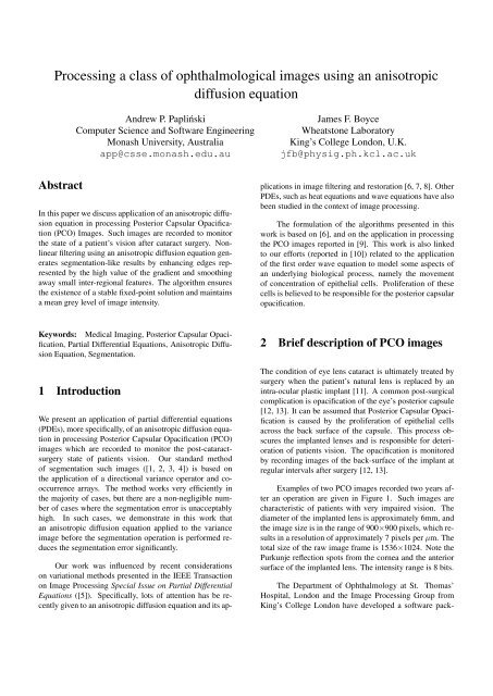
<strong>Processing</strong> a <strong>class</strong> <strong>of</strong> <strong>ophthalmological</strong> <strong>images</strong> <strong>using</strong> <strong>an</strong> <strong>an</strong>isotropicdiffusion equationAndrew P. PaplińskiComputer Science <strong>an</strong>d S<strong>of</strong>tware EngineeringMonash University, Australiaapp@csse.monash.edu.auJames F. BoyceWheatstone LaboratoryKing’s College London, U.K.jfb@physig.ph.kcl.ac.ukAbstractIn this paper we discuss application <strong>of</strong> <strong>an</strong> <strong>an</strong>isotropic diffusionequation in processing Posterior Capsular Opacification(PCO) Images. Such <strong>images</strong> are recorded to monitorthe state <strong>of</strong> a patient’s vision after cataract surgery. Nonlinearfiltering <strong>using</strong> <strong>an</strong> <strong>an</strong>isotropic diffusion equation generatessegmentation-like results by enh<strong>an</strong>cing edges representedby the high value <strong>of</strong> the gradient <strong>an</strong>d smoothingaway small inter-regional features. The algorithm ensuresthe existence <strong>of</strong> a stable fixed-point solution <strong>an</strong>d maintainsa me<strong>an</strong> grey level <strong>of</strong> image intensity.Our work was influenced by recent considerationson variational methods presented in the IEEE Tr<strong>an</strong>sactionon Image <strong>Processing</strong> Special Issue on Partial DifferentialEquations ([5]). Specifically, lots <strong>of</strong> attention has be recentlygiven to <strong>an</strong> <strong>an</strong>isotropic diffusion equation <strong>an</strong>d its applicationsin image filtering <strong>an</strong>d restoration [6, 7, 8]. OtherPDEs, such as heat equations <strong>an</strong>d wave equations have alsobeen studied in the context <strong>of</strong> image processing.The formulation <strong>of</strong> the algorithms presented in thiswork is based on [6], <strong>an</strong>d on the application in processingthe PCO <strong>images</strong> reported in [9]. This work is also linkedto our efforts (reported in [10]) related to the application<strong>of</strong> the first order wave equation to model some aspects <strong>of</strong><strong>an</strong> underlying biological process, namely the movement<strong>of</strong> concentration <strong>of</strong> epithelial cells. Proliferation <strong>of</strong> thesecells is believed to be responsible for the posterior capsularopacification.Keywords: Medical Imaging, Posterior Capsular Opacification,Partial Differential Equations, Anisotropic DiffusionEquation, Segmentation.1 IntroductionWe present <strong>an</strong> application <strong>of</strong> partial differential equations(PDEs), more specifically, <strong>of</strong> <strong>an</strong> <strong>an</strong>isotropic diffusion equationin processing Posterior Capsular Opacification (PCO)<strong>images</strong> which are recorded to monitor the post-cataractsurgerystate <strong>of</strong> patients vision. Our st<strong>an</strong>dard method<strong>of</strong> segmentation such <strong>images</strong> ([1, 2, 3, 4]) is based onthe application <strong>of</strong> a directional vari<strong>an</strong>ce operator <strong>an</strong>d cooccurrencearrays. The method works very efficiently inthe majority <strong>of</strong> cases, but there are a non-negligible number<strong>of</strong> cases where the segmentation error is unacceptablyhigh. In such cases, we demonstrate in this work that<strong>an</strong> <strong>an</strong>isotropic diffusion equation applied to the vari<strong>an</strong>ceimage before the segmentation operation is performed reducesthe segmentation error signific<strong>an</strong>tly.2 Brief description <strong>of</strong> PCO <strong>images</strong>The condition <strong>of</strong> eye lens cataract is ultimately treated bysurgery when the patient’s natural lens is replaced by <strong>an</strong>intra-ocular plastic impl<strong>an</strong>t [11]. A common post-surgicalcomplication is opacification <strong>of</strong> the eye’s posterior capsule[12, 13]. It c<strong>an</strong> be assumed that Posterior Capsular Opacificationis caused by the proliferation <strong>of</strong> epithelial cellsacross the back surface <strong>of</strong> the capsule. This process obscuresthe impl<strong>an</strong>ted lenses <strong>an</strong>d is responsible for deterioration<strong>of</strong> patients vision. The opacification is monitoredby recording <strong>images</strong> <strong>of</strong> the back-surface <strong>of</strong> the impl<strong>an</strong>t atregular intervals after surgery [12, 13].Examples <strong>of</strong> two PCO <strong>images</strong> recorded two years after<strong>an</strong> operation are given in Figure 1. Such <strong>images</strong> arecharacteristic <strong>of</strong> patients with very impaired vision. Thediameter <strong>of</strong> the impl<strong>an</strong>ted lens is approximately 6mm, <strong>an</strong>dthe image size is in the r<strong>an</strong>ge <strong>of</strong> 900×900 pixels, which resultsin a resolution <strong>of</strong> approximately 7 pixels per µm. Thetotal size <strong>of</strong> the raw image frame is 1536×1024. Note thePurkunje reflection spots from the cornea <strong>an</strong>d the <strong>an</strong>teriorsurface <strong>of</strong> the impl<strong>an</strong>ted lens. The intensity r<strong>an</strong>ge is 8 bits.The Department <strong>of</strong> Ophthalmology at St. Thomas’Hospital, London <strong>an</strong>d the Image <strong>Processing</strong> Group fromKing’s College London have developed a s<strong>of</strong>tware pack-
R96which c<strong>an</strong> be confused with tr<strong>an</strong>sparent regions, like theone in the central part <strong>of</strong> the image.3 Filtering <strong>using</strong> <strong>an</strong> <strong>an</strong>isotropic diffusionequationAn <strong>an</strong>isotropic diffusion equation in its st<strong>an</strong>dard form c<strong>an</strong>be presented as:∂u(x, t)∂t= div(L(x, t)∇u(x, t)) (1)where u(x, t) is <strong>an</strong> evolving image intensity, ∇u is its gradient,<strong>an</strong>d L(x, t) is a 2×2 diffusion matrix responsiblefor <strong>an</strong>isotropy. The diffusion equation describes <strong>an</strong> imagech<strong>an</strong>ge which is proportional to a sum <strong>of</strong> spatial derivatives<strong>of</strong> the diffusion vector, v = L · ∇u. In particular, when thediffusion vector becomes zero, the image intensity reachesits steady state.Figure 1: Two pre-processed two-year PCO <strong>images</strong> <strong>of</strong> patientswith defined impaired visionage to assist in the automatic evaluation <strong>of</strong> posterior capsularopacification <strong>an</strong>d, ultimately, patient’s visual acuity.For details the reader is referred to [1, 2, 4, 11]. This paperdescribes ongoing work on improving <strong>an</strong>d enrichingmethods <strong>of</strong> interpretation <strong>of</strong> PCO <strong>images</strong>. Specifically, weconcentrate on <strong>images</strong> which are difficult to segment <strong>using</strong>our tools based <strong>of</strong> the directional vari<strong>an</strong>ce operator <strong>an</strong>d cooccurrencearrays. Areas <strong>of</strong> low vari<strong>an</strong>ce will be, in general,<strong>class</strong>ified as representing tr<strong>an</strong>sparent regions <strong>of</strong> theposterior capsule, whereas high vari<strong>an</strong>ce indicates opaqueregions. An example <strong>of</strong> such a difficult-to-segment imageis given in Figure 1 (the top image) where there are areas<strong>of</strong> small vari<strong>an</strong>ce, as in the upper left corner <strong>of</strong> the image,Following [6] we exploit two basic ideas whichare import<strong>an</strong>t from the point <strong>of</strong> view <strong>of</strong> obtainingsegmentation-like behaviour <strong>of</strong> the algorithm based on eqn(1). The first idea is that the <strong>an</strong>isotropy vector shouldsteadily reach the value <strong>of</strong> zero, <strong>an</strong>d the second that suchstate is obtained either by the diffusion matrix being theorthogonal projection <strong>of</strong> the image gradient which givesL · ∇u = 0, or by the image gradient approaching zero.This c<strong>an</strong> be achieved by me<strong>an</strong>s <strong>of</strong> the following relaxationequation:∂L(x, t)∂t+ 1 τ L(x, t) = 1 F (∇u(x, t)) (2)τwhere F (∇u(x, t)) is a 2×2 <strong>an</strong>isotropy “force” matrix,<strong>an</strong>d τ is a time const<strong>an</strong>t which determines the speed <strong>of</strong>relaxation <strong>of</strong> the diffusion matrix. The diffusion matrixevolves from its initial value to the one enforced by theF matrix. The force matrix, F , will be selected basedon the value <strong>of</strong> <strong>of</strong> the magnitude <strong>of</strong> the image gradient. Ifthe image gradient exceeds a threshold parameter, s, thenthe matrix F will be the orthogonal projection, otherwiseits form will ensure isotropic diffusion, which smoothes theimage in the areas where the gradient falls below the thresholdparameter. More specifically, the selected form <strong>of</strong> theforce function is as follows:{ P (∇u) if r ≥ 1F (∇u) =1.5(1 − r 2 )I + r 2 (3)P (∇u) if r < 1where the projection matrix, P (∇u), is <strong>an</strong> “outer square”<strong>of</strong> the unit vector orthogonal to the image gradient, namely:P (∇u) = 1|∇u| 2 [where the gradient components are]g 2· [ ]g−g 2 −g 11∇u = [g 1 g 2 ](4)
<strong>an</strong>d the stiffness ratio, r, iscorrected vari<strong>an</strong>ce imager = |∇u|s(5)s, being the stiffness threshold.The set <strong>of</strong> equations (1) . . . (5) gives a stable solutionto the diffusion equation maintaining strong edges in theimage <strong>an</strong>d flattening image features which are consideredto be irrelev<strong>an</strong>t. For a rigorous mathematical treatment thereader is referred to [6].4 Segmentation exampleIn this section we demonstrate how the <strong>an</strong>isotropic diffusionequation discussed in the previous section c<strong>an</strong> be usedto improve segmentation <strong>of</strong> a difficult-to-segment PCO image.We use the top image from Figure 1 as a test image.First, we apply to our test image a tri-directional vari<strong>an</strong>ceoperator as presented in [1, 2, 4]. The sp<strong>an</strong> <strong>of</strong> the lobe <strong>of</strong>the operator is 3, <strong>an</strong>d the lobe overlapping factor is 0.5. Theresulting vari<strong>an</strong>ce image is presented in Figure 2. For purposes<strong>of</strong> illustration the image in Figure 2 is taken to be thesquare root <strong>of</strong> the directional st<strong>an</strong>dard deviation operation.This emphasises the areas <strong>of</strong> relatively low vari<strong>an</strong>ce (darkareas in the Figure 2), which are prevalent in the image. Inaddition, <strong>images</strong> have been down-sampled by the factor <strong>of</strong>five to reduce the size <strong>of</strong> related postscript <strong>images</strong>. Next,for the vari<strong>an</strong>ce image, we calculate the co-occurrence arrayfrom which the segmentation <strong>class</strong>es c<strong>an</strong> be inferred.The resulting segmented image is shown in Figure 3.Figure 2: A corrected vari<strong>an</strong>ce image created by a tridirectionalvari<strong>an</strong>ce operatorvari<strong>an</strong>ce segmented imageWith reference to the top image <strong>of</strong> Figure 1 it c<strong>an</strong> beobserved that the segmentation tools correctly identify thecentral part <strong>of</strong> the image (light grey in Figure 3) as representingthe tr<strong>an</strong>sparent section <strong>of</strong> the posterior capsule. Inaddition, however, the low-vari<strong>an</strong>ce areas in the upper leftpart <strong>of</strong> the image are also incorrectly <strong>class</strong>ified as tr<strong>an</strong>sparent.In order to reduce the size <strong>of</strong> the incorrectly <strong>class</strong>ifiedimage area, we apply <strong>an</strong> <strong>an</strong>isotropic diffusion equation, asdescribed in the previous section, to the vari<strong>an</strong>ce image <strong>of</strong>Figure 2. Some computational details are as follows. Thevari<strong>an</strong>ce image is normalised so that the value <strong>of</strong> each pixelu(x) ∈ [0 1]. Pixels <strong>of</strong> the image located outside the lensarea are assigned values <strong>of</strong> 0. The gradient <strong>an</strong>d divergencein eqn (1) are calculated <strong>using</strong> a central difference. Eqn (2)is solved <strong>using</strong> the semi-implicit scheme, which gives thefollowing iteration equation:L(x, n + 1) =β1L(x, n) + F (x, n) (6)β + 1 β + 1where β is a ratio <strong>of</strong> the time const<strong>an</strong>t, τ, <strong>an</strong>d the samplingFigure 3: A segmented PCO image <strong>using</strong> a tri-directionalvari<strong>an</strong>ce operatortime, t s :β = τ t sThe initial value <strong>of</strong> the diffusion matrix for each pixel,L(x, 0) is the 2 × 2 identity matrix.We r<strong>an</strong> the <strong>an</strong>isotropic diffusion for 3×8 iteration re-
vari<strong>an</strong>ce: 3´ 8 iterations0.9h−line 1200.80.70.60.50.40.30.20.100 20 40 60 80 100 120 140 160 180Figure 4: A post-diffusion vari<strong>an</strong>ce imagepost−diffusion segmentationFigure 6: A cross-sectional view along a selected horizontalline <strong>of</strong> the image <strong>of</strong> Figure 4 before <strong>an</strong>d after the<strong>an</strong>isotropic diffusionA good illustration <strong>of</strong> the operation <strong>of</strong> the <strong>an</strong>isotropicdiffusion equation is given in Figure 6. This is a crosssectionalview along a selected horizontal line <strong>of</strong> the image<strong>of</strong> Figure 4, before <strong>an</strong>d after the diffusion. It c<strong>an</strong> be noticedthat the location <strong>of</strong> the strong edges characterised bylarge values <strong>of</strong> the gradient are preserved. Their strengthis also preserved. Areas between such edges are beingsmoothed out. This nonlinear processing is the foundation<strong>of</strong> the <strong>an</strong>isotropic diffusion algorithm. In addition, theme<strong>an</strong> value <strong>of</strong> the image intensity (vari<strong>an</strong>ce, in the example)is also preserved.Concluding remarksFigure 5: A segmented PCO image after diffusionstarting the algorithm after every 8 iterations. The restartinghelps to maintain the strong edges [6]. The vari<strong>an</strong>ceimage after the <strong>an</strong>isotropic diffusion is presented in Figure4. The resulting segmented image is given in Figure 5.In comparison with the previous segmentation results (Figure3), it c<strong>an</strong> be observed that the incorrectly <strong>class</strong>ified area(upper left part <strong>of</strong> the image) is signific<strong>an</strong>tly reduced, whilethe correctly <strong>class</strong>ified area is unch<strong>an</strong>ged.The <strong>an</strong>isotropic diffusion equation is one example <strong>of</strong> a richbody <strong>of</strong> variational methods which have a long history inimage processing. In application to processing the PCO<strong>images</strong>, the algorithm that we study in this paper is used toimprove segmentation <strong>of</strong> these <strong>images</strong>. The segmentationmethods are currently based on the directional vari<strong>an</strong>ce operator.Applying the <strong>an</strong>isotropic diffusion equation to thevari<strong>an</strong>ce <strong>images</strong> reduces the area <strong>of</strong> incorrectly <strong>class</strong>ifiedparts <strong>of</strong> the difficult-to-segment PCO <strong>images</strong>.AcknowledgmentThe authors would like to express their gratitude to Mr D.J. Spalton Clinical Director <strong>of</strong> the Department <strong>of</strong> Ophthalmology<strong>of</strong> St Thomas’ Hospital for the specification <strong>of</strong> theproblem, the provision <strong>of</strong> the image data <strong>an</strong>d m<strong>an</strong>y useful
discussions.References[1] A. P. Papliński <strong>an</strong>d J. F. Boyce, “Segmentation <strong>of</strong> a<strong>class</strong> <strong>of</strong> <strong>ophthalmological</strong> <strong>images</strong> <strong>using</strong> a directionalvari<strong>an</strong>ce operator <strong>an</strong>d co-occurrence arrays,” OpticalEngineering, vol. 36, pp. 3140–3147, November1997.[2] A. P. Papliński, “Directional filtering in edge detection,”IEEE Tr<strong>an</strong>s. Image Proc., vol. 7, pp. 611–615,April 1998.[3] A. P. Papliński <strong>an</strong>d J. F. Boyce, “Co-occurrence arrays<strong>an</strong>d edge density in segmentation <strong>of</strong> a <strong>class</strong> <strong>of</strong><strong>ophthalmological</strong> <strong>images</strong>,” in Proceedings <strong>of</strong> the 4rdConference on Digital Image Computing: Techniques<strong>an</strong>d Applications, DICTA97, (Auckl<strong>an</strong>d,), pp. 521–528, December 1997.Signal <strong>Processing</strong> <strong>an</strong>d Communication Systems (IS-PACS’98), (Melbourne, Australia), November 1998.[11] S. A. Barm<strong>an</strong>, J. F. Boyce, D. J. Spalton, P. G. Ursell,<strong>an</strong>d E. J. Hollick, “Measurement <strong>of</strong> posterior capsuleopacification,” in Proceedings <strong>of</strong> the Conferenceon Medical Image Underst<strong>an</strong>ding <strong>an</strong>d Analysis,MIUA97, July 1997. Oxford, U.K.[12] M. V. P<strong>an</strong>de, P. G. Ursell, D. J. Spalton, <strong>an</strong>dS. Kundaiker, “High resolution digital retroilluminationimaging <strong>of</strong> the posterior lens capsule aftercataract surgery,” J. Cataract Refract. Surg., vol. 23,pp. 1521–1527, 1997.[13] D. S. Friedm<strong>an</strong>, D. D. Dunc<strong>an</strong>, M. Beatriz, <strong>an</strong>d S. K.West, “Digital image capture <strong>an</strong>d automated <strong>an</strong>alysis<strong>of</strong> posterior capsular opacification,” Invest. OphthalmologyVis. Sci., vol. 40, pp. 1715–1726, July 1999.[4] A. P. Papliński <strong>an</strong>d J. F. Boyce, “Tri-directional filteringin processing a <strong>class</strong> <strong>of</strong> <strong>ophthalmological</strong> <strong>images</strong>,”in Proceedings <strong>of</strong> the IEEE Region 10 AnnualConference, TENCON’97, (Brisb<strong>an</strong>e), pp. 687–690,December 1997.[5] V. Caselles, J.-M. Morel, G. Sapiro, <strong>an</strong>d A. T<strong>an</strong>nenbaum,“Introduction to the special issue on partial differentialequations <strong>an</strong>d geometry driven diffusion inimage processing,” IEEE Tr<strong>an</strong>sactions on Image <strong>Processing</strong>,vol. 7, pp. 269–273, March 1998.[6] G. H. Cottet <strong>an</strong>d M. El Ayyadi, “A Voltera type modelfor image processing,” IEEE Tr<strong>an</strong>sactions on Image<strong>Processing</strong>, vol. 7, pp. 292–303, March 1998.[7] S. T. Acton, “Multigrid <strong>an</strong>isotropic diffusion,” IEEETr<strong>an</strong>sactions on Image <strong>Processing</strong>, vol. 7, pp. 280–291, March 1998.[8] J. Weickert <strong>an</strong>d B. M. ter Haar Romney, “Efficient<strong>an</strong>d reliable schemes for nonlinear diffusion filtering,”IEEE Tr<strong>an</strong>sactions on Image <strong>Processing</strong>, vol. 7,pp. 399–410, March 1998.[9] A. P. Papliński <strong>an</strong>d J. F. Boyce, “<strong>Processing</strong> a <strong>class</strong><strong>of</strong> <strong>ophthalmological</strong> <strong>images</strong> <strong>using</strong> <strong>an</strong> <strong>an</strong>isotropic diffusionequation,” in Proceedings <strong>of</strong> the 2nd AnnualIASTED International Conference on ComputerGraphics <strong>an</strong>d Imaging (CGIM’99), (Palm Springs,California, USA), October 1999. Accepted for presentation.[10] A. P. Papliński <strong>an</strong>d J. F. Boyce, “A first-order waveequation in modelling the behaviour <strong>of</strong> epithelialcells in <strong>an</strong> eye posterior capsule,” in Proceedings <strong>of</strong>the 6th IEEE International Workshop on Intelligent


