
Create successful ePaper yourself
Turn your PDF publications into a flip-book with our unique Google optimized e-Paper software.
1920Enhancing <strong>Breast</strong> CancerFeatures of <strong>Breast</strong> Malignancy:MorphologyKineticsCourtesy: Dr. Bruce Porter2122BI-RADSGrading BI-RADS ®•Published by ACR•Mammography•Ultrasound•<strong>MRI</strong>•0 - Incomplete exam•1 - Normal•2 - Benign•3 - Probably Benign•4 – Probably Malignant•5 – Malignant•6 – Biopsy ProvenCancer• 1 Normal Benign• 2 Benign• 3 Probably Benign Imaging follow up or tissue• 4 Probably Malignant TISSUE• 5 Malignant TISSUEBI-RADS <strong>MRI</strong> Scoring23<strong>MRI</strong> Scoring System (Gottingen score)24•<strong>MRI</strong> Artefact Category•Density Type•1-4•1-4Points 0 1 2Shape -Margins Well -defined Poorly definedContrast Homogenous Inhomogeneous RingInitial Inc 100%Post Initial Increase Plateau Washout
3132<strong>MRI</strong> <strong>Breast</strong> Screening<strong>MRI</strong> <strong>Breast</strong> Screening• Sensitivity• Clinical 17.9• Mammography 33.3• <strong>MRI</strong> 79.5• Specificity• Clinical 98.1• Mammography 95.0• <strong>MRI</strong> 79.53334American Cancer Society Guidelines for<strong>Breast</strong> Screening with <strong>MRI</strong> as an Adjunctto Mammography• March 2007• Screening <strong>MRI</strong> is recommended:• for women with an approximately 20–25% or greater lifetimerisk of breast cancer,• including women with a strong family history of breast orovarian cancer and• women who were treated for Hodgkin disease.Non-Screening Clinical Indications for Contrast<strong>Breast</strong> MR• Lobular Cancer• Occult <strong>Breast</strong> Cancer• Close or positive surgical margins• Post-operative scar vs. tumor recurrence• Neo-adjuvant chemotherapy or brachytherapy• Suspected multiple or bilateral cancers• Implants and known or suspected cancer• Problematic Mammogram• Assessment of all diagnosed breast cancers.3536<strong>MRI</strong> in unilateral breast cancer<strong>MRI</strong> in diagnosed breast cancer• NEMJ March 2007• 969 with diagnosis of unilateral breast cancer• Other breast normal clinically and mammo• Occult cancer in second breast 3.1%• April 2008• European <strong>Breast</strong> Cancer Conference in Germany• 249 patients who had malignancy on biopsy had <strong>MRI</strong>• Altered management in 13%• 20 additional cancers – 8%• Better than mammo or US in assessing tumour size• “We would like to see <strong>MRI</strong> become a standard preoperativetreatment for breast cancer, along with biopsy,mammography, and ultrasound”
37 38Analysis3940Unilateral vs. Bilateral• Cancers arebilateral in up to10%Multiple lesionsInfiltrating ductal CaFibroadenoma• Exclusion ofcontra-lateral tumoris of great clinicaland personal valueCourtesy: Dr. Bruce PorterCourtesy of FHDI4142DCIS
4950Contrast-enhanced <strong>Breast</strong> MR: AccuracyStaging• Varies significantly with technique and experience.• Sensitivity: > 94 – 98 %• Negative Predictive Value: > 95 %• Specificity: 37 – 97 %• Positive Predictive Value: ~ 65- 75 %• For DCIS sensitivity is lower, especially for low orintermediate grades; also for some lobular, low grade, orless angiogenic invasive carcinomas.Courtesy: Dr. Bruce Porter5152Chest STIR: Aids in cancer stagingMR - guided breast biopsies• Look for highest positive lymph node• Use body coil, supine placement• Some lesions are not visible with mammography orultrasound (US)• After identified with MR (diagnosis)• Many lesions can be found using US• US is interactive, cheaper, faster• MR-guided BB needed for those lesions which cannot be seenin retrospect on US or mammaographyCourtesy of FHDI5354<strong>MRI</strong> <strong>Breast</strong> Biopsy Device<strong>Breast</strong> Biopsy• Lateral and medial access• Needle or wire placement(core, fna)• Vacuum assisted biopsyextraction• Software controlledguidance• Comfortable patientsupport
5556Spectroscopy - MRSMRS monitoring of Chemotherapy• Early results show specificity• > 95%• Choline peak at 3.6 ppm• Single voxel studiesBeforeChemotherapy1 stcycle2 ndcycle3 rdcycle5758MRS monitoring of ChemotherapySilicone Implant Rupture <strong>MRI</strong>• MR is the definitive exam for implantassessment.• Non-contrast unless a question ofcancer.4 th cycle 5 th cycle 6 th cycleSiliconeCourtesy of FHDI and USCD5960Implant ruptureConclusion• <strong>MRI</strong> is the ‘gold standard’ if resources are available.• Combined approach with Mammogaphy and Ultrasound.• Should be used for screening high risk cases.• Should be used to assess all diagnosed breast cancer.• Use scoring system and BI-RADS• You will get false positives• No false negatives• Don’t screen unless you can biopsy.
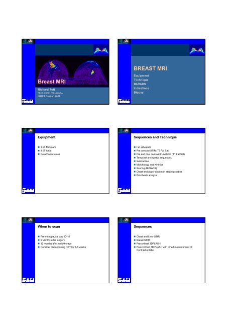
61Thank you!Richard TuftFRCS, FRCR, FFRad(D)(SA)<strong>ISRRT</strong> Durban 2008