Issue 33 Autumn 2012 - Bases
Issue 33 Autumn 2012 - Bases
Issue 33 Autumn 2012 - Bases
- No tags were found...

Create successful ePaper yourself
Turn your PDF publications into a flip-book with our unique Google optimized e-Paper software.
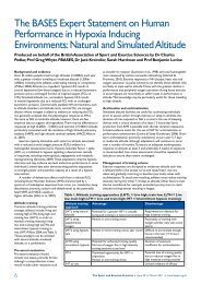
Dual-Energy X-ray Absorptiometry: Thescience behind the scanAdam Hawkey has analysed the bones of professional sports performers, clinical patients and NASA astronauts,in an attempt to limit injury, reduce the onset of osteoporosis and improve crew health during long-durationspaceflight. In this article he explains how x-ray technology is used to monitor bone health and reduce fracturerisk.While early observations of bone mineral density(BMD) were based on the use of densitometryof plain x-rays, clinical diagnosis of osteoporosisis currently based on BMD measurement ofthe hip, and/or lumbar spine, using Dual-EnergyX-ray Absorptiometry (DXA: see Figure 1).Developed in the late 1980s, DXA works bydifferentiating the absorption of X-rays from twodifferent energies, enabling the determinationof the amount of mineral in a designated area.Measurement of BMD is critical to the earlydetection of osteoporosis as the lower theBMD, the higher the risk of fracture; for eachstandard deviation decrease in BMD, fracturerisk increases by approximately 200-300%. BMDmeasurement for assessing osteoporosis riskis therefore comparable to measuring bloodpressure to predict stroke and substantially betterthan measuring serum cholesterol to predictcardiovascular disease (Anderson & Delmas,2002).Figure 1. Example of a DXA scan,Courtesy GE HealthcareBMD is derived from the bone mineralcontent (BMC); as bone mass is very highlycorrelated to its mineral composition. By dividingthe BMC (g) by the area of bone being measured(cm 2 ) a value of BMD (g/cm 2 ) can be obtained.This is not a density in the traditional/engineeringsense, that is, total mass divided by volume (g/cm 3 ),but instead an areal density of mineral, sometimesmore accurately referred to as a BMD. DXA isalso able to measure the depth and compositionof adjacent soft tissue to establish levels oflean and fat mass. This is important as overlyingdepths of fat, water, and air, and the thicknessand composition of tissue surrounding the bonewill have an effect on measured BMD. This alsomeans that DXA can be used for accurate andwords: Adam HawkeyAdam is Director of the Centre forOsteoporosis and Obesity Researchand Education at the University ofWolverhampton. He is also Chairof the BASES Biomechanics InterestGroup and a Professional Memberof the National OsteoporosisSociety. A.Hawkey@wlv.ac.ukReferencesAnderson, M. & Delmas,P.D. (2002). Osteoporosis: anunderdiagnosed and undertreatedpublic health issue. Karger Gazette,65, 3-5.Hawkey, A. (<strong>2012</strong>).Understanding the causes,prevention and treatment ofosteoporosis (part 2): detectingand diagnosing the disease. Journalof Sports Therapy, 5(1), 7-14.Kalpakcioglu, B.B.,Morshed, S., Engelke, K.& Genent, H.K. (2008).Advanced imaging of bonemacrostructure and microstructurein bone fragility and fracture repair.Journal of Bone and Joint Surgery,90 (Suppl. 1), 68-78.Kaul, S. et al. (<strong>2012</strong>). Dual-Energy X-ray Absorptiometryfor quantification of visceral fat.Obesity, 20(6), 1313-18.Micklesfield, L.K. et al.(<strong>2012</strong>). Dual-Energy X-rayperforms as well as clinicalcomputed tomography for themeasurement of visceral fat.Obesity, 20(5), 1109-1114.World Health Organisation(1994). Assessment of fracturerisk and its application to screeningfor postmenopausal osteoporosis.Report of a WHO Study Group.Geneva: World Health Organization(WHO Technical Report Series,No. 843).reliable body composition analysis; with somesystems now being able to distinguish betweensub-cutaneous and visceral fat (Kaul et al., <strong>2012</strong>;Micklesfield et al., <strong>2012</strong>). To enable effectiveosteoporosis diagnosis, values obtained from DXAscans are compared to reference data prescribedby the World Health Organisation (WHO, 1994).Osteoporosis is defined as a BMD of 2.5standard deviations (SD) or more below theaverage for a young healthy female population:T-scores of -1.0 or greater are classified as normalwith a low fracture risk; scores between -1.0 and-2.5 are classified as osteopenia and come witha moderate risk of fracture; while scores lowerthan -2.5 are classed as osteoporosis and carrya high risk of future fracture risk. Z-scores canalso be determined, which allow comparison ofan individual’s BMD against corresponding normalvalues for matched age, sex and ethnic origin.Recently, other methods of analysis havebecome available, including micro-computedtomography (microCT) and quantitativecomputed tomography (QCT). While thesetechniques offer more detailed three-dimensionalmeasurement (g/cm 3 ) of bone health, enhancinginvestigations into bone fragility, defining skeletalresponses to innovative therapies, and assessingbiomechanical alterations (Kalpakcioglu et al.,2008), DXA is currently considered the goldstandard for BMD measurement as it is accurate,precise, requires only a short scanning period, andhas a limited radiation dose. The relative radiationexposure risk from DXA is very low, with valuesapproximating 0.02 - 5 microsieverts (µSv) for thewhole body and 0.4 - 4 µSv for the lumbar spine;compared to 12 - 20 µSv for a chest x-ray and0.8 - 5 millisieverts (mSv) and 6 - 18 mSv for CTscans of the brain and chest respectively (Hawkey,<strong>2012</strong>).The National Osteoporosis Society (NOS)estimate that 1 in 2 women and 1 in 5 men areaffected by osteoporosis. There are reportedly300,000 fractures annually in the UK (comparedto 275,000 heart attacks and 110,000 strokes)leading to 1,150 deaths each month. Withthese shocking statistics, the effective diagnosis,prevention, and treatment of osteoporosis isnow a public health priority. It would appearthat the measurement of BMD, through the goldstandard of DXA, is a crucial factor in reducingthe significant physical, psychosocial and financialconsequences of the disease.22 The Sport and Exercise Scientist n <strong>Issue</strong> <strong>33</strong> n <strong>Autumn</strong> <strong>2012</strong> n www.bases.org.uk