Full text - PDF - The National Cancer Institute
Full text - PDF - The National Cancer Institute
Full text - PDF - The National Cancer Institute

You also want an ePaper? Increase the reach of your titles
YUMPU automatically turns print PDFs into web optimized ePapers that Google loves.
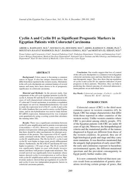
Journal of the Egyptian Nat. <strong>Cancer</strong> Inst., Vol. 14, No. 4, December: 289-301, 2002Cyclin A and Cyclin D1 as Significant Prognostic Markers inEgyptian Patients with Colorectal CarcinomaABEER A. BAHNASSY, M.D. 1 ; SOUMAYA EL-HOUSSINI, M.D. 2 ; ABDEL-RAHMAN N. ZEKRI, Ph.D. 3 ;MOUSTAFA RAAFAT MAHMOUD, Ph.D. 1 ; BASMAA GOMAA, M.D. 5 and MOSTAFA EL-SERAFI, M.D. 4Tissue Culture and Cytogenetics Unit1; Surgical Pathology Unit2; Pathology Department; Virology and Immunology Unit3;<strong>Cancer</strong> Biology Department; Medical Oncology Department4, <strong>National</strong> <strong>Cancer</strong> <strong>Institute</strong> and Microbiology and ImmunologyDepartment5; Kasr El-Aini School of Medicine, Cairo University, Cairo Egypt.ABSTRACTBackground: Colon cancer is becoming a commoncancer in Egypt. It also has unique characteristics thatdiffer from those reported in the western society. Aberrationsin the cell cycle checkpoints are common features of severaltumors and many have been shown to be of prognosticsignificance in colorectal carcinoma.Material and Methods: In the present study, fourcomponents of the cell cycle regulator proteins [cyclin D1,cyclin A, histone H3 and the Ki-67] were examined in 60cases of well-characterized colorectal adenocarcinomas,47 colon and 13 rectal carcinomas, to ascertain co-regulationand impact on survival. Immunohistochemistry was usedto study the expression level of Ki-67, cyclin A and cyclinD1 while Southern blot was used to assess cyclin D1genestatus, and in situ hybridization was used to study histoneH3 mRNA expression. Immunoreactivity was evaluatedsemi quantitatively using a scoring system that calculatesthe staining index (SI).Results: <strong>The</strong>re was a significant correlation betweencyclin D1 gene amplification and its protein overexpression(concordance = 63.6%). However, some cases revealedprotein overexpression without gene amplification indicatingthat there should be other mechanisms for aberrant accumulationof the protein. <strong>The</strong> expression of Ki-67 significantlycorrelated with the other 3 proteins as well as withcyclin D1 amplification. <strong>The</strong> SIs for Ki-67, cyclin A andcyclin D1 were higher in large-sized, poorly differentiatedtumors. Furthermore, the SIs of Ki-67 and histone H3 werehigher in patients > 50 years of age. <strong>The</strong> SI of cyclin D1was significantly higher in cases with deeply invasivetumors and those with positive lymph node metastasis. <strong>The</strong>overall survival was significantly lower in patients whosetumors showed cyclin A overexpression as well as thosewith cyclin D1 amplification and/or overexpression. Multivariateanalysis showed that up-regulation of cyclin D1and cyclin A were two independent prognostic factors incolorectal carcinoma patients.Conclusion: Our results suggest that loss of controlof the cell cycle checkpoints is a common event in Egyptiancolorectal carcinoma cases and may therefore be an importanttherapeutic target. <strong>The</strong>y also show that up-regulationof cyclin A and cyclin D1 are superior indicators of poorprognosis in these patients. <strong>The</strong>refore, they can help inpredicting the clinical outcome of colorectal adenocarcinomapatients on an individual basis.Key Words: Colorectal carcinoma - Cyclin A - cyclin D1- Histone H3 - Ki-67 - Survival.INTRODUCTIONColorectal cancer (CRC) is the third mostcommon cancer in western countries [17]. InEgypt, CRC has unique characteristics that differfrom those reported in other countries of thewestern society. Unlike western countries whereCRC is prevalent among olderly people, 35%of the Egyptian patients with CRC are under 40years of age [31]. Moreover, the histopathologicalcriteria and the mutational profile of the tumorsdiagnosed in Egypt are different from those ofthe western countries. In general, they are ofhigh histological grade and stage and they carrymore mutations. It was therefore mentioned thatthe uniquely high proportion of early-onset CRC,the early and continuous exposure to hazardousenvironmental agents, the different mutationalspectrum and the prevalent consanguinity inEgypt justify further studies [32].It is now commonly believed that most cancersresult from accumulation of genetic alterationsinvolving certain groups of genes, that289
290Cyclin A and Cyclin D1 as Significant Prognostic Markershave recently been defined as cancer-causinggenes. <strong>The</strong> majority of these genes are cell cycleregulators that either stimulate or inhibit the cellcycle progression [17].Cell proliferation allows orderly progressionthrough the cell cycle, which is governed by anumber of proteins including cyclins (cyclin A,B1, B2, C, D1-3 and E) and cyclin dependentkinases [6,14]. <strong>The</strong> cyclins belong to a superfamilyof genes whose products complex with variouscyclin-dependent kinases (CDKs) to regulatetransitions through key checkpoints of the cellcycle [2]. Abnormalities of several cyclins inneoplastic cells have been reported, implicating,in particular, cyclin A, cyclin E and cyclin D[2,24].Cyclin D1 is a G1 cyclin since its peak leveland maximum activity are reached during theG1 phase of the cell cycle, hence it apparentlyregulates the transition from G1 to S phase. Onthe other hand, cyclin A reaches its maximumlevel during the S and G2 phases, thus it isregarded as a regulator of the transition to mitosis[11]. <strong>The</strong> mechanisms likely to activate the oncogenicproperties of the cyclins include chromosomaltranslocation in certain B cell lymphomas,gene amplification [8] and aberrant proteinoverexpression [8,24].Recent studies have shown that histone H3mRNA expression can be used to identify the Sphase fraction through the in situ hybridization(ISH) technique [10,25]. <strong>The</strong> level of histone H3mRNA reaches its peak during the S phase andthen drops rapidly at the G2 phase [5]. To thebest of our knowledge, there is only a singlereport that investigated histone H3 mRNA expressionin CRC [11].In face of the increasing, relatively highincidence of CRC and its peculiar pattern in theEgyptian population, the present study was conductedto assess the relationship between theproliferative activity of Ki-67 (pan-cell cyclemarker), cyclin D1 (G1 phase marker), histoneH3 mRNA (S phase marker), cyclin A (S to G2phase marker) and the clinicopathologic featuresof CRC. <strong>The</strong> expression level of these markerswas also correlated with the clinical outcome ofpatients in terms of disease free and overallsurvival.MATERIAL AND METHODSTissue samples:Paraffin-embedded tumor tissues were obtainedfrom 60 CRC patients (47 colon and 13rectal carcinomas) who were diagnosed andunderwent resection at the <strong>National</strong> <strong>Cancer</strong><strong>Institute</strong>, Cairo Egypt, during the period fromJanuary 1997 to June 2002. Clinicopathologicdata of the studied cases are illustrated in table1. None of the patients received any chemotherapyor irradiation prior to surgery. Histologicaldiagnosis of all cases was done by 2 independentpathologists according to the WHO histologicalclassification [16], and tumors were pathologicallystaged according to the TNM staging system[30]. Fifteen patients had well-differentiated adenocarcinomas,21 had moderately differentiatedadenocarcinomas and 24 revealed poorlydifferentiatedadenocarcinomas. <strong>The</strong> depth oftumor invasion was classified as invasion of themucosa including muscularis mucosa (m), invasionof the submucosa (sm), or invasion beyondthe submucosa [11]. Normal colonic tissues wereobtained from autopsy specimens (n=10) andfrom the resection margin of carcinomas (n=10)and were used as a control. <strong>The</strong> actual survivalrate of the patients was calculated from the dateof resection to the date of death.Immunohistochemistry:Four micron sections of each normal andtumor specimen were cut onto positive-chargedslides, air dried overnight, de-paraffinized inxylene, hydrated through a series of gradedalcohol and washed in distilled water and 0.01PBS (pH 7.4). Slides were then processed forIHC as previously described by Handa et al.,[11] using the following antibodies: Ki-67 (MIB-1, Dako), cyclin A (6E6; Novocastra, Newcastle-Upon-Tyne, UK) and cyclin D1 (DCS-6, Dako).A case of invasive breast cancer was used as apositive control for Ki-67 and cyclin A and acase of mantle cell lymphoma was used as acontrol for cyclin D1. Negative controls wereobtained by replacing the primary antibody bynon-immunized rabbit or mouse serum.Brown nuclear staining was regarded as apositive result for all studied markers. <strong>The</strong> proportionof positively-stained cells and the intensityof staining were scored in tumor and normalcolorectal mucosal sections at medium power
Abeer A. Bahnassy, et al. 291(X200). <strong>The</strong> degree of positive tumor staining(percentage of positive tumor cells in the examinedsection) was scored from 1-6 and the stainingintensity was scored from 0-6 according tothe pattern of staining in the examined section.Staining index (SI) was calculated by multiplyingthe cellularity and staining scores as describedby King et al. [18].In situ hybridization:All tumor samples and 5 normal controlswere assessed for histone H3 mRNA by ISHusing the commercially available 550 base fluorescein-labeledDNA probe (Dako, Carpinteria,CA) as described by Nagao et al. [25]. This probehybridizes to the whole mRNA transcript of thehuman H3gene including the5’ and 3’ untranslatedregions. Scoring of histone H3 mRNAwas performed as for immunohistochemistry,but hybridization signals were detected in thecytoplasm.Molecular detection of cyclin D1 gene amplification:High molecular weight DNA was extractedfrom paraffin-embedded tissues of the tumorand normal colorectal mucosal samples as previouslydescribed [28]. <strong>The</strong> proportion of neoplasticand normal cells was determined in eachtumor sample by examining hematoxylin andeosin- stained slides obtained from the edge ofthe specimen used for DNA extraction. Tumorsamples were evaluated for amplification ofcyclin D1 if more than 75% of the examinedsections were formed of neoplastic cells. Accordingly,50 cases were eligible for the analysis.Ten micrograms of the extracted DNA was digestedwith EcoR1. DNA from selected caseswas also digested with BglII and HindIII. Sampleswere separated on 0.8% agarose gels andtransferred to Hybond-N membranes (AmershamInt., Amersham, UK). <strong>The</strong> membranes werehybridized with 50% formamide, 5X SSC, 5XDenhardt’s, 500Mg/ml denatured salmon spermDNA, 10% dextran sulphate and 106cpm/ml of32 P-labeled PRAD-1 probe for 24 h. After hybridization,membranes were washed with 2XSSC, 0.1% SDS at room temperature for 30 minfollowed by 2X SSC, 0.1% SDS at 60ºC for 30min, and 0.1 X SSC, 0.1% SDS at 60ºC for 1 h.<strong>The</strong> filters were then autoradiographed using anintensifying screen at -70ºC for 24-72 h. Afterbeing stripped free of the PRAD-1 probe, thesame blots were hybridized with 32 P-labeled β-actin probe to normalize against possible variationsin the loading or transfer of DNA. <strong>The</strong>autoradiograms were analyzed using a densitometer.Intensities of PRAD-1/cyclin D1 werenormalized to the β-actin control bands. <strong>The</strong>degree of amplification was calculated fromthese normalized values. Amplification wasconsidered when the signal of the tumor bandwas ≥ 2-fold the value of the matched normalmucosa [15].Statistical analysis:<strong>The</strong> Mann-Whitney non-parametric test wasused to compare the SIs of pairs of subjectswhereas the Kruskal-Wallis was used for categoricaldata. Correlation between indices wasperformed using a simple linear regression test.<strong>The</strong> Kaplan-Meier method was used to generatesurvival curves which were analyzed by the logranktest. <strong>The</strong> impact of different variables onsurvival was determined using the Cox proportionalhazards model. p.values less than 0.05were considered significant.RESULTSNormal colorectal mucosa revealed positiveimunostaining for Ki-67 in the lower half of thecrypts only. A heterogeneous staining patternwas detected in the neoplastic cells of well- andmoderately-differentiated adenocarcinomaswhereas a diffuse homogeneous staining patternwas detected in poorly-differentiated carcinomas(Fig. 1). <strong>The</strong> staining index (SI) of Ki-67 rangedfrom 10-40.2 (mean: 24.6 ± 6.5).Immunostaining for cyclin D1 was predominantlynuclear, however cytoplasmic stainingwas also detected in some cases. However, unlessa nuclear staining was also detected, cases withcytoplasmic staining only were considered negative.Normal colorectal mucosal samples werealmost negative for cyclin D1. Marked heterogeneitywas observed in well- and moderatelydifferentiatedadenocarcinomas even within thesame tumor. However, poorly-differentiatedcarcinomas revealed a diffuse staining patternwith more darkly-stained nuclei (Fig. 2). <strong>The</strong> SIof the cyclin D1 ranged from 0.5-28.6 (mean:9.3 ± 4.2).Positive nuclear staining for cyclin A was
292Cyclin A and Cyclin D1 as Significant Prognostic Markersdetected in all cases of CRC and in nonneoplasticcontrol samples. <strong>The</strong> staining patternwas similar to that of Ki-67 with positivelystainednuclei confined to the lower half of thecrypts in normal colonic mucosa and diffuselydispersedin carcinomas (Fig.3). <strong>The</strong> SI of cyclinA ranged from 3.3-30.2 (mean: 15.1 ± 6.6).Histone H3 mRNA was intensely expressedin the cytoplasm of all examined samples whetherneoplastic or non-neoplastic (Fig.4). <strong>The</strong> distributionof histone H3 mRNA was similar to thatof cyclin A and Ki-67. However, the proportionof histone H3 mRNA positive cells was less thanthat for Ki-67. <strong>The</strong> SI of histone H3 ranged from1.8-24.2 (mean: 12.4 ± 5.3).<strong>The</strong> PRAD-1 probe detected 3 EcoRI fragmentsof 4.0, 2.2 and 2.0 and 1 BglII fragmentof 15 Kb [15]. PRAD-1/cyclin D1 gene amplificationwas detected in 22/50 (44%) cases analyzed.<strong>The</strong> degree of amplification was heterogeneouswith 2-10 fold increase when comparedto normal mucosal samples. Amplification wasconfirmed by other restriction enzymes (Fig. 5).Correlations:Linear regression analysis of SIs revealed asignificant correlation between Ki-67 and cyclinD1, cyclin A, histone H3 as well as between theSIs of cyclin A and histone H3 (p = 0.008, 0.0001and 0.0001, respectively) (Fig. 6).Table (1) shows a statistically significantrelationship between SI of both Ki-67 and cyclinA and the degree of differentiation of the tumoras well as the size of the tumor (p < 0.001 andp < 0.01 respectively). In addition, Ki-67 SI washigher in patients
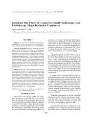
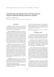
Abeer A. Bahnassy, et al. 293Fig. (1): Positive nuclear staining for Ki-67 in normal colonic mucosa showing positively-stained nuclei in the lower half of the crypts(a), well differentiated (b), moderately differentiated (c), and poorly differentiated (d) adenocarcinomas. Adenocarcinomas show diffusestrong immunostaining. <strong>The</strong> staining index is higher in poorly differentiated tumors than in other carcinomas. Scale bars: 50Mm (X200).Fig. (2): Positive nuclear immunostaining for cyclin D1 in normal colonic mucosa (a), well differentiated (b), moderately differentiated(c), and poorly differentiated (d) adenocarcinomas. <strong>The</strong> arrows indicate the positive nuclear staining which is frequent and strong inpoorly differentiated tumors but infrequent or absent in other carcinomas. Scale bars: 50Mm (X200).
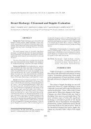
294 Cyclin A and Cyclin D1 as Significant Prognostic MarkersFig. (3): Nuclear staining for cyclin A in normal colonic mucosa (a), well differentiated (b), moderately differentiated (c), and poorlydifferentiated (d) adenocarcinomas. Poorly differentiated carcinomas show more positively-stained nuclei than moderately orwell-differentiated tumors. Scale bars: 50Mm (X200).Fig. (4): ISH of histone H3 mRNA in normal colonic mucosa (a), well differentiated (b), moderately differentiated (c) and poorly differentiated(d) adenocarcinomas. Histone H3 mRNA in expressed in the normal colonic mucosa as well as in carcinomas. Scale bars: 50Mm(X200).
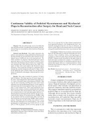
Abeer A. Bahnassy, et al. 295Fig. (5-A)PRAD-1N T N T N T N T N T N T N T15 kbB-ActinFig. (5-B)Fig. (5-A): Southern blot analysis of seven cases of colonicadenocarcinomas (T1-T7), poorly (1,2,4,5) moderatelydifferentiated (3,6,7), and normal mucosa (N). GenomicDNA was digested with BglII, fractionated by electrophoresisin agarose gel, transferred onto membranes and hybridizedwith PRAD1 and β-actin probes for loading control. Tumorsnumber 1-6 (Lanes 1-6) show different degrees ofPRAD1/cyclin D1 amplification, tumor number 7 (lane 7)was not amplified. B: Southern blot analysis of 3 cases ofadenocarcinomas (T) and matched normal colonic mucosa(N). Genomic DNA was digested with EcoRI, fractionatedby electrophoresis in agarose gel, transferred onto membranesand hybridized with PRAD1 and B-actin probes for loadingcontrol. <strong>The</strong> identification of the 3 tumors is the same as inFig. 5A with amplification of PRAD1/cyclin D1 in tumorsnumber 4,5 (Lanes 1,2) but not 7 (Lane 3).PRAD -14 kb2.2 kb2 kbB-ActinN T N T N T30r = 0.339, p = 0.008320r = 0.732, p = 0.0001Cyclin D1 SI2010Histone H3 SI1000 10 20 30Fig. (6-A) Ki-67 SI00 10 20 30Fig. (6-B) Ki-67 SI30r = 0.713, p = 0.000120r = 0.947, p = 0.0001Cyclin A SI2010Histone H3 SI1000 10 20 30Ki-67 SIFig. (6-C)00 10 20 30Cyclin A SIFig. (6-D)Fig. (6): Correlation between the staining intensity of Ki-67 vs. cyclin D1 (a), Ki-67 vs. histone H3 (b), Ki-67 vs. cyclin A (c) and cyclinA vs. histone H3 mRNA expression.
296 Cyclin A and Cyclin D1 as Significant Prognostic MarkersSurvlval rate10.80.60.40.2Ki-67 highLog Rank: p = 0.2562Survlval rate10.80.60.40.2p = 0.005Cyclin D1 lowCyclin D1 highKi-67 low Log Rank: p = 0.057400 20 40 60 80MonthsFig. (7-A)00 20 40 60 80MonthsFig. (7-B)110.80.8Survlval rate0.60.40.2Histone H3 highSurvlval rate0.60.40.2Cyclin A highCyclin A lowHistone H3 low Log Rank: p = 0.020800 20 40 60 80Fig. (7-C)Months00 20 40 60 80Fig. (7-D)MonthsFig. (7): Kaplan-Meier survival curves for colorectal carcinoma. Overall survival is significantly lower in patients with cyclin A and cyclinD1 overexpression. Patients with high SI for histone H3 mRNA have poorer prognosis but this was not statistically significant.No significant difference was present between patients with high Ki-67 SI and those with low Ki-67 SI.Table (I): Correlation between the staining index of Ki-67, cyclin DI , cyclin A, histone H3 and the clinicopathologicalfeatures of the patients.No. ofSI (mean + SD)Variables cases Ki –67 Cylcin DI Cylcin A Histone H3Sex:MaleFemaleAge (years):≥ 50< 50Tumor size (cm):< 5.0≥ 5.0Histology:NormalCarcinomaGIGIIGIIILymph node:NegativePositiveDepth of invasion:m,smbeyond smStage:IIIIIIIV* p. value < 0.05 (significant)362441193327206015212433271743627121518.0 ± 6.420.1 ± 5.811.7 ± 6.0*23.8 ± 5.612.2 ± 6.3*30.1 ± 6.23.5 ± 2.0*30.3 ± 6.211.7 ± 6.211.8 ± 5.630.0 ± 4.319.5 ± 7.021.3 ± 4.920.7 ± 6.721.9 ± 6.220.6 ± 6.720.8 ± 6.922.0 ± 5.424.7 ± 6.16.7 ± 4.38.8 ± 8.45.6 ± 5.27.7 ± 6.85.3 ± 3.8*22.8 ± 7.20.6 ± 0.2*24.9 ± 6.36.6 ± 4.08.9 ± 3.622.0 ± 8.15.4 ± 5.3*20.6 ± 6.93.1 ± 3.1*12.4 ± 6.55.7 ± 6.95.3 ± 4.37.7 ± 6.011.3 ± 9.612.7 ± 5.710.0 ± 6.010.0 ± 5.313.6 ± 5.711.5 ± 6.1*28.6 ± 5.62.3 ± 1.1*27.2 ± 5.810.0 ± 5.412.3 ± 6.527.0 ± 4.911.9 ± 6.512.5 ± 5.011.9 ± 7.212.2 ± 5.624.2 ± 6.924.6 ± 6.027.1 ±5.227.5 ± 5.510.7 ± 5.310.7 ± 5.46.0 ± 5.0*22.0 ± 5.210.3 ± 4.9*24.0 ± 5.62.2 ± 0.910.7 ± 5.311.4 ± 4.97.8 ± 5.411.5 ± 5.412.3 ± 5.514.2 ± 5.010.4 ± 5.110.7 ± 5.411.1 ± 5.310.4 ± 5.710.4 ± 4.912.3 ± 6.2
Abeer A. Bahnassy, et al. 297Table (2): Uunivariate analysis of the relationship between survival and the tested markers.Predictive VariablesKi –67:< 11.5≥ 11.5MedianSurvival HR CI p36321.8260.636-5.2430.26Cyclin D1:< 6.1≥ 6.135187.2461.007-45.1500.03*Histone H3:< 8.2≥ 8.235294.6390.854-25.1960.07Cyclin A:< 10.5≥ 10.535157.8201.017-60.1220.02*Histological grade:LowHigh38107.3312.696-19.9400.0001*Lymph node:NegativePositive38156.8261.973-23.6210.002*Stage:I,II,IIIIV38126.3781.842-22.0830.001*Tumor size (cm):< 5.0≥ 5.035134.8351.386-16.8680.01*Depth of invasion:T1,T2T3,T436207.7591.024-58.7890.04*Age (years):< 50≥ 5038282.8020.988-7.9430.0526Sex:MaleFemale38360.69600.274-1.7660.4449* p. value < 0.05 (significant) HR: Hazard Ratio CI: 95% confidence IntervalTable (3): Multivariate Analysis of the Relationship between Survival and the Tested Markers.PredictiveVariables HR CI pCyclin D1:(baseline < 6.1)Cyclin A:(baseline < 10.5)Positive Lymph node metastasisStage IVDepth of invasion:T3, T4Age (years):≥ 50Sex10.864—13.886—3.9213.4115.4081.9960.9101.055-86.250—1.012-190.579—1.057-14.4721.048-12.0830.449-65.0800.678-5.8780.315-2.3580.03*—0.0490*—0.0410*0.03*0.18360.23100.8453p. value < 0.05 (significant) HR: Hazard Ratio CI: 95% confidence Interval
298Cyclin A and Cyclin D1 as Significant Prognostic MarkersDISCUSSION<strong>The</strong> proliferative activity of colorectal cancercells has been investigated in several studieseither by immunohistochemical determinationof cell proliferation index using antibodies tosome types of cyclins or by flow cytometricdetermination of the S phase fraction of the cellcycle [11].Although Leach et al. [20] did not find cyclinDI gene amplification in a panel of 47 CRC celllines, its protein was overexpressed in about30% of colon carcinoma cases that were includedin the studies of Bartakova et al. [2] and Arberet al. [1]. In the former study, the authors statedthat cyclin D1is aberrantly accumulated in asignificant subset of human CRC cases and thatthe cell lines derived from colon cancer aredependent on this cyclin protein in their cellcycle progression. Moreover, Arber et al. [1]demonstrated that overexpression of cyclin DIwas detected in about 30% of adenomatouspolyps of the colon indicating that this overexpressionis a relatively early event in coloncarcinogenesis that could possibly be responsiblefor the occurrence of pathological changes inthe mucosa that precedes neoplastic transformation.More recently, Holland et al. [13], Pasz-Walczak et al. [27] and Utsonomiya et al.[34]reported up-regulation of cyclin D1 in 58.7%,100% and 43% of their studied cases, respectively,using IHC techniques and/or westernblotting.In the present study, up-regulation of cyclinDI was detected in 68% of the studied cases.Cyclin D1SI was significantly higher in carcinomasthan in normal colorectal mucosa, and inpoorly-differentiated adenocarcinomas it wasapproximately twice that of other histologicaltypes. Amplification and/or overexpression ofcyclin D1 correlated well with deeply invasivetumors and positive lymph node metastasis. Ourresults in this regards are consistent with mostof the available reports in this con<strong>text</strong> [11,21]. In2001, Holland et al., [13] studied 126 cases ofCRC and demonstrated that deregulation ofcyclin D1 and p21WAF proteins are importantin colorectal tumorigenesis and have implicationsfor patient prognosis. Similarly, McKay et al.[23] studied some cell cycle-checkpoint proteinsincluding cyclin D1, p53, p16, Rb-1, PCNA andp27 in CRC. <strong>The</strong>y found that cyclin D1 was theonly protein examined that was related to improvedoutcome in these patients. In addition toour present work, the results of these previouslymentioned studies lead us to assume that cyclinD1 is a valuable prognostic marker in CRCpatients. However, few studies failed to detectany correlation between cyclin DI overexpressionand the clinicopathological factors in CRC [1,2].<strong>The</strong> controversy in the results of different studiescould partially be explained by the difference inthe sampling of studied cases. <strong>The</strong> present studyincluded 24 cases of poorly differentiated adenocarcinoma,which was not common in otherstudies of colorectal carcinoma in western countries.This was possible because unlike CRC ofthe western countries, which is usually of lowhistological grade, the majority of CRC casesdiagnosed in Egypt are of high histological grade[32]. However, our results are consistent withthose of Handa et al. [11] since they were ableto include an appreciably large number of poorlydifferentiatedadenocarcinoma cases in theirstudy. <strong>The</strong> correlation between cyclin D1 overexpressionand the high histological grade wasalso reported in other tumor types. In non-smallcell lung carcinomas, cyclin DI immunostainingwas prominent in poorly differentiated areas butabsent in the well-differentiated ones [22]. Inaddition, PRAD-1/cyclin DI gene amplificationand overexpression of its mRNA have beenfound in squamous cell carcinomas of the larynxin association with invasive, high grade tumorswith positive lymph node metastasis [15]. Anotherpossible explanation for the observed controversyin the results obtained from different studies isthe difference in the detection method used sinceIHC, western blotting, Southern blotting; northernblotting and flowcytometry were all usedfor studying cyclins.In the present work, overexpression of cyclinD1 was detected in a higher percentage thanamplification of the PRAD-1/cyclin DI genewith a 63.6% concordance. This was similarlyreported by Bartakova et al. [2] who mentionedthat although immunohistochemical detectionof cyclin D1distinguishes tumors with the 11q13amplification (<strong>The</strong> PRAD-1/cyclin DI gene locus)from non-amplified cases, it also appearsto identify a subset of cases in which cyclin D1is overexpressed in the absence of gene amplification.Consistent with this hypothesis arereports of elevated cyclin D1 mRNA levels [3]and immunohistochemically detectable accumulationof the protein [8] in over one-third of breast
Abeer A. Bahnassy, et al. 299cancer cases at a frequency significantly higherthan that deduced from DNA amplification studies.<strong>The</strong>se data imply that mechanisms otherthan gene amplification can also lead to deregulationand accumulation of cyclin D1 in commonsolid tumors.So far, studies have been done to reveal theprognostic significance of cyclin DI overexpressionin various carcinomas, including CRC [21].However, the studies yielded conflicting results.This may indicate that organ heterogeneity playsa role in the involvement of cyclin DI in thedevelopment and progression of cancer. In ourstudy, patients with tumors that exhibited cyclinDI overexpression tended to have poor prognosis.It is well known that cyclin A is involved inthe onset of DNA replication [9] and is requiredfor G2-M transition in mammalian cells [26].Overexpression of cyclin A would, therefore,contribute to the high proliferative activity incancer cells. Cyclin A achieves its maximal levellate in the cycle (S to G2 phase) leading to directentry of cancer cells into mitosis and may, thus,reflect the high proliferative activity of cancercells more sharply than Ki- 67. It was reportedthat 75% of non-small cell lung carcinoma patientsexhibited immunohistochemical stainingfor cyclin A and that patients with cyclin Apositive carcinomas had significantly shortermedian survival times [35]. In addition, Chao etal. [4] found that 39% of patients with humanhepatoecellular carcinoma exhibited cyclin Aoverexpression in tumorous tissue by westernblot analysis. <strong>The</strong>y mentioned that overexpressionof cyclin A could be an independent prognosticfactor in hepatocellular carcinoma. Incontrast, Wang et al. [36] reported that cyclin Aexpression in normal colon mucosa was higherthan that in cancer tissue in 63 % of their studiedcases, and only 10 % of the carcinomas hadhigher cylcin A expression. However, Handa etal. [11] were able to detect cyclin A overexpressionin 77% of their 73 cases of CRC. <strong>The</strong>y alsodemonstrated that cylcin A could be used as aprognostic factor of CRC. More recently, Hebermannet al. [12] studied cases of ulcerative colitiswith and without an associated adenocarcinomafor the presence of cyclin A overexpression.<strong>The</strong>y found that cyclin A overexpression washigher in cases of ulcerative colitis with adenocarcinomasthan those without adenocarcinomas.Consequently, they concluded that cyclin A couldbe used for monitoring ulcerative colitis patientsand for the early detection of an emerging carcinomain this high risk group of patients.In our study, cyclin A was detected in 80%of the patients and Cox regression analysisshowed that in addition to cyclin D1, cyclin Acould be used as a prognostic marker in colorectalcarcinoma cases. Our results regarding the prognosticvalue of cyclin A were in agreement withthose of Handa et al. [11].It is difficult to determine the exact reasonfor the controversy in results between the presentwork or the study of Handa et al. [11] and Wanget al. [36]. However, this controversy in resultscould be partially attributed to the inclusion ofa larger number of high-grade tumors in thepresent study and that of Handa et al.[11] or tothe sensitivity of the detection method [36].It would have been useful if we assessed theexpression level of cyclin A by another technique(DNA amplification). This would have addedmore information regarding the gene status onone hand and confirmed the results of IHC onthe other. Unfortunately, this was not possiblebecause in most of the cases included in thepresent work, the extracted DNA was not sufficientto study cyclin A amplification after theassessment of cyclin D1.Nagao et al. [25] reported that histone H3labeling index significantly correlated with ki-67 immunostaining and was high in poorly differentiatedhuman hepatocellular carcinoma. Inour study, histone H3 SI significantly correlatedwith the SI of Ki–67. However, no statisticallysignificant correlation was found between histoneH3 SI and any of the studied clinicopathologicalfactors, although it was higher in adenocarcinomasthan in normal mucosa.Although Ki-67 immunostaining reflects theproliferative activity of CRC, it has not beenrecognized as a significant prognostic factor inthis type of tumors [7,19,29]. However, Suzuki atal. [33] reported that the Ki-67 labeling indexcorrelated with local invasion of colorectal carcinomabut not with the metastatic potential. Inthe present study, there was a significant relationshipbetween the SI of Ki-67 and the degreeof differentiation of the tumors as well as theirsize. However, Kaplan-Meier survival curvesshowed no significant difference in survival rates
300Cyclin A and Cyclin D1 as Significant Prognostic Markersbetween patients with and without overexpressionof Ki-67.In conclusion, our results demonstrate thatcyclin D1, cyclin A, histone H3 and Ki-67 areoverexpressed in a subset of CRC cases. Overexpressionof both cyclin D1 and cyclin A correlateswith poor differentiation and tumor progression.Our results also indicate the superiorityof cylcin A and cyclin D1 as indicators of poorprognosis compared to Ki-67 and histone H3mRNA in colorectal carcinoma. Overexpressionof cyclin A and D1 is thus identified as a significant,independent prognostic factor for colorectalcarcinoma. Our findings in this regard are especiallyimportant in stage I and II patients sincethose patients usually have a poor prognosis inspite of being node-negative. Hence, in thisgroup of patients there is a great demand formore accurate, individually-based, biologicalprognostic factors that help in accurate predictionof the clinical outcome of the disease as well asin categorizing patients for appropriate therapeuticmodalities. If these findings are confirmedin a larger study, evaluation of cyclin A and D1may be applicable to clinical management ofCRC, allowing the identification of patients withpoor prognosis. It will also be valuable to comparethe prognostic impact of these two parametersin two separate groups including metastaticand non-metastatic CRC.REFERENCES1- Arber N., Hibshoosh,H., Moss S., Sutter T., Zhang Y.,Begg M., Wang S., Weinstein I. and Holt P.: Increasedexpression of cyclin D1 is an early event in multistagecolorectal carcinogenesis. Gastroenterol., 110: 669-674, 1996.2- Bartkova J., Lukas J., Strauss, M., and Bartek J.: <strong>The</strong>PRAD-1/cyclin D1 oncogene product accumulatesaberrantly in a subset of colorectal carcinomas. Int J.<strong>Cancer</strong>, 58: 568-573, 1994.3- Buckley M., Sweeney K., Hamilton J., Sini R., ManningD., Nicholson R., DeFazio A., watts C., Musgrove E.,and Sutherland R.: Expression and amplification ofcyclin genes in human breast cancer. Oncogene, 8:2127-2133, 1993.4- Chao Y., Shih Y., Chiu J., Chau G., Lui W., Yang W.,Lee S. and Huang T.: Overexpression of cyclin A butnot SKP 2 correlates with the tumor relapse of humanhepatocellular carcinoma. <strong>Cancer</strong> Res., 58: 985-990.1998.5- Chou M., Chang A., McBride J., Donoff B., GallagherG. and Wong D. A.: Rapid method to determine proliferationpatterns of normal and malignant tissues byH3 mRNA in situ hybridization. Am. J. Pathol., 136:729-733. 1990.6- Cordon-Cardo C.: Mutations of cell cycle regulators.Biological and clinical implications for human neoplasia.Am.r J. Pathol., 147: 545-560, 1995.7- Gerdes J., Lemke H., Baisch H., Wacker H., SchwabU., and Stein H.: Cell cycle analysis of a cell proliferation-associatedhuman nuclear antigen defined by themonoclonal antibody Ki-67. Immunol., 133: 1710-1715. 1984.8- Gillett C., Fantl V., Smith R., Fisher C., Bartek J.,Dickson C., Barnes D., and Peters G. Amplificationand overexpression of cyclin D1 in breast cancerdetected by immunohistochemical staining. <strong>Cancer</strong>Res., 54: 1812-1817, 1994.9- Girard F., Strausfield U., Fernandez A. and Lamb N.:Cyclin A is required for the onset of DNA replicationin mammalian fibroblasts. Cell, 67: 1169-1179, 1991.10- Gown A., Jiang J., Matles H., Skelly M., GoodpasterT., Cass L., Reshatof M., Spaulding D. and ColtreraM.: Validation of the S-phase specificity of histone(H3) in situ hybridization in normal and malignantcells. J. Histochem Cytochem., 44: 221-226, 1996.11- Handa K., Yamakaua M., Takeda H., Kimura S. andTakahashi T.: Expression of the cell cycle markers incolorectal carcinoma: Superiority of cyclin A as anindicator of poor prognosis. Int J. <strong>Cancer</strong>, 84: 225-233, 1999.12- Hebermann J., Lenander C., Roblick U.J., Kruger S.,Ludwig D., Alaiya A., Freitag S., Dumbgen L., BruchHP., Stange E., Salo S., Tryggvason K., Auer G. andSchimmelpenning H.: Ulcerative colitis and colorectalcarcinoma: DNA profile, laminin-5 gamma 2 chainand cyclin A expression as early markers for riskassessment. Scand J. Gastroenterol., 36 (7): 751-758,2001.13- Holland T., Elder J., McCloud J., Hall C., Deakin M.,Fryer A., Elder G. and Hoban P.: Subcellular localizationof cyclin D1 protein in colorectal tumors is associatedwith p21 (WAF1/CIP1) expression and correlateswith patient survival. Int J. <strong>Cancer</strong>, 95 (5): 302-306,2001.14- Hunter T. and Pines J.: Cyclins and cancer. II. CyclinD and CDK inhibitors come of age. Cell, 79: 573-5781994.15- Jares P., Fernandez P., Campo E., Nadal A., BoschoF., Aiza G., Nayach I., Traserra J. and Cardesa A.:PRAD-1/cyclin D1 gene amplification correlates withmessenger RNA overexpression and tumor progressionin human laryngeal carcinomas. <strong>Cancer</strong> Res., 54: 4813-4817, 1994.16- Jass J. and Sobin L.H.: World Health Organization.International classification of tumors: histologicaltyping of intestinal tumors, Springer, New York., 1989.17- Jiang G-L. and Huang S.: Adenovirus expressing RIZ1in tumor suppressor gene therapy of microsatelliteunstable colorectal cancers. <strong>Cancer</strong> Res., 61:1796-1798, 2001.
Abeer A. Bahnassy, et al. 30118- King R., Coffer A., Gilbert J., Lewis K., Nash R., MillisR., Raju S. and Taylor R.: Histochemical studies witha monoclonal antibody raised against a partially purifiedsoluble estradiol receptor preparation from humanmyometrium. <strong>Cancer</strong> Res., 45: 5728-5733, 1985.19- Kubota Y., Petras R., Easley K., Bauer T., Tubbe R.and Fazio V.: Ki-67-determined growth fraction versusstandard staging and grading parameters in colorectalcarcinoma. A multivariate analysis. <strong>Cancer</strong>, 70: 2602-2609, 1992.20- Leach F., Elledge S., Willson J., Markowitz S., KinzlerK. and Vogelstein B. Amplification of cyclin genes incolorectal carcinomas. <strong>Cancer</strong> Res., 53: 1986-1989.1993.21- Maeda K., Chung Y., Kang S., Ogawa M., Onoda N.,Nakata B., Nishiguchi Y., Okuno M., and Sowa M.Overexpression of cyclin D1 and p53 is associatedwith disease recurrence in colorectal adenocarcinoma.Int J. <strong>Cancer</strong>, 74: 310-315, 1997.22- Mate J., Ariza A., Arcil C., Lopez D., Isamat M., Perez-Piteira J. and Navas- Palacios J.: Cyclin D1 overexpressionin non- small cell lung carcinoma: correlationwith Ki-67 labeling index and poor cytoplasmic differentiation.J. Pathol., 180: 395- 399, 1996.23- McKay J., Douglas J., Ross V., Curran S., Loane J.,Ahmed F., Cassidy J.,McLeod H. and Murray G.:Analysis of key cell cycle checkpoint proteins incolorectal tumors. J. Pathol., 196 (4): 386-393, 2002.24- Motokura T. and Arnold A.: Cyclins and oncogenesis.Biochim. Biophys. Acta., 1155: 63-78, 1993.25- Nagao T., Ishida Y. and Kondo Y.: Determination ofS-phase cells by in situ hybridization for histone H3mRNA in hepatocellular carcinoma: correlation withhistological grade and other cell proliferative markers.Mod Pathol., 9: 99-104, 1996.26- Pagano M., Pepperkok R., Verde F., Ansorge W. andDraetta G. Cyclin A is required at two points in thehuman cell cylce. EMBO J., 11: 961-971, 1992.27- Pasz-Walczak G., Kordek R. and faflik M.: P21 (WAF1)expression in colorectal cancer: correlation with p53and cyclin D1 expression, clinicopathological parametersand prognosis. Pathol Res Pract., 197 (10): 683-689, 2001.28- Robert J.C., Boerrigter L., Evers S.G., Wisman P.,Mooiw J. and Rodenhuis S. A rapid and simple procedurefor the routine detection of ras point mutationsin formalin-fixed, paraffin-embedded tissues. DigMolec. Path., 1 (2):136-139, 1992.29- Shain A., Ro I., Brown R., Ordonez N., Cleary K., El-Naggar A., Wilson P. and Ayala A.: Assessment of Ki-67- derived tumor proliferative activity in colorectaladenocarcinomas. Mod Pathol., 7: 17-22, 1994.30- Sobin L. and Wittekind C.: TNM classification ofmalignant tumors (5 th ed). John Wiley, New York.,1997.31- Soliman A., Bondy M., Levin B., Hamza M.R., IsmailK., Ismail S., Hammam H.M., El-Hattab O., KamalS.M., Soliman A.A., Dorgham L.A., McPherson R.S.,and Beasley P.R.: Colorectal cancer in Egyptian patientsunder 40 years of age. Int J. <strong>Cancer</strong>, 71: 26-30, 1997.32- Soliman A., Bondy M., Guan Y., El-Badawi S., MokhtarN., Bayomi S., Raouf AA., Ismail S., McPherson RS.,Abdel-Hakim TF., Beasley PR. Levin B. and Wei Q.:Reduced expression of mismatch repair genes in colorectalcancer patients in Egypt. Int J. Oncology, 12:1315-1319, 1998.33- Suzuki H., Matsumoto K. and Terabe M.: Ki-67antibody labeling index in colorectal carcinoma. J.Clin Gastroenterol., 15: 317-320, 1992.34- Utsunomiya T., Doki Y., Takemoto H., Shiozaki H.,Yano M., Sekimoto M., Tamura S., Yasuda T., FujiwaraY. and Monden M.: Correlation of beta-catenin andcyclin D1 expression in colon cancers. Oncology, 61(3): 226-233, 2001.35- Volm M., Koomagi R., Mattern J. and Stammler G.:Cyclin A in lung carcinomas. Br. J. <strong>Cancer</strong>, 75: 1774-1778, 1997.36- Wang A., Yoshimi N., Suzui M., Yamauch1 A., TaraoM. and Mori H.: Different expression patterns of cyclinsA, D1 and E in human colorectal cancer. J. <strong>Cancer</strong>Res. Clin. Oncol., 122: 122-126, 1996.37- Zhuai Y.L., Nikaido T., Shiozawa T., Orii A. and FujiS.: Expression of cyclins and cyclin dependant kinasesin smooth muscle tumors of the uterus. Int J. <strong>Cancer</strong>(pred. Oncol.), 84: 244-250, 1999.