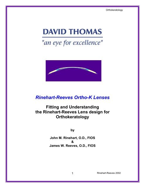
Rinehart-Reeves Ortho-K Lenses Fitting and Understanding the ...
Rinehart-Reeves Ortho-K Lenses Fitting and Understanding the ...
Rinehart-Reeves Ortho-K Lenses Fitting and Understanding the ...

Create successful ePaper yourself
Turn your PDF publications into a flip-book with our unique Google optimized e-Paper software.
<strong>Ortho</strong>keratology<strong>Rinehart</strong>-<strong>Reeves</strong> <strong>Ortho</strong>-K <strong>Lenses</strong><strong>Fitting</strong> <strong>and</strong> Underst<strong>and</strong>ing<strong>the</strong> <strong>Rinehart</strong>-<strong>Reeves</strong> Lens design for<strong>Ortho</strong>keratologybyJohn M. <strong>Rinehart</strong>, O.D., FIOS&James W. <strong>Reeves</strong>, O.D., FIOS1<strong>Rinehart</strong>-<strong>Reeves</strong> 2002
<strong>Ortho</strong>keratologyIntroduction<strong>Ortho</strong>keratology is a non-surgical application of rigid contact lenses for <strong>the</strong>purpose of controlling <strong>the</strong> progression or temporarily reducing or eliminatingmyopia <strong>and</strong> astigmatism. <strong>Ortho</strong>K is a <strong>the</strong>rapeutic application of speciallydesigned rigid contact lenses to produce a temporary improvement in unaidedvisual acuity. Once maximum improvement has occurred, retainer lenses areworn part-time during <strong>the</strong> day or while sleeping, <strong>the</strong>n removed for waking hours.The earliest forms of orthokeratology came from corneal contact lensesbeing fit slightly flatter than K. This resulted in flattening of <strong>the</strong> cornea <strong>and</strong> doctorsof <strong>the</strong> late 50’s <strong>and</strong> early 60’s noticed patients were becoming slightly lessnearsighted upon removing <strong>the</strong>ir lenses.In <strong>the</strong> early 1960’s several optometrists developed orthokeratologytechniques of <strong>the</strong>ir own. I believe <strong>the</strong> most prominent to be Dr’s. Charles May <strong>and</strong>Stuart Grant. Their methods used large, flat lenses with large optical zones. Theironly choice of material was PMMA.The 1970’s gave us <strong>the</strong> birth of new rigid materials, which are gaspermeable. This development has greatly improved safety <strong>and</strong> efficacy forconventional contact lens wear <strong>and</strong> orthokeratology. Gas permeable <strong>Ortho</strong>Klenses allow large diameter lenses to be fit; this aids centration <strong>and</strong>, <strong>the</strong>refore,yields better results. The 1970’s gave birth to reverse geometry lenses for<strong>Ortho</strong>keratology. Alfred A. Fontana, O.D., FAAO first described <strong>the</strong> use of suchlenses <strong>and</strong> began personally using this design in <strong>the</strong> early 70’s.The 1980’s produced <strong>the</strong> first marketed reverse geometry lenses.Dr. Richard Wlodya conceived <strong>the</strong> OK 3 design, <strong>and</strong> Mr. Nick Stoyan was <strong>the</strong> firstto fabricate this design in his lab.So far in <strong>the</strong> 1990’s we have reverse geometry lenses with multiple curvessteeper than <strong>the</strong> base curve <strong>and</strong> reverse geometry lenses with aspheric basecurves.<strong>Ortho</strong>keratology has been a consistently changing science <strong>and</strong> will continueto evolve as long as bright imaginative doctors strive to improve <strong>the</strong>ir patient’svision.2<strong>Rinehart</strong>-<strong>Reeves</strong> 2002
<strong>Ortho</strong>keratologyGetting Started in <strong>Ortho</strong>keratologyPractitioners interested in developing an <strong>Ortho</strong>keratology practice willrequire virtually <strong>the</strong> same equipment as is needed for <strong>the</strong> fitting of traditional gaspermeable lenses.• Corneal Topography (DO NOT PERFORM ORTHOKERATOLOGYWITHOUT A TOPOGRAPHER) to:• Rule out pathological corneal disorders such as keratoconus orpellucid marginal degeneration.• Possibly provide a value for corneal eccentricity.• Monitor corneal changes, <strong>and</strong> determine <strong>the</strong> location <strong>and</strong> size of <strong>the</strong>treatment zone.O<strong>the</strong>r useful equipment (optional only) higher quality manufacturing st<strong>and</strong>ardsshould make <strong>the</strong>se unnecessary.• Lens Diameter Gauge, to verify lens diameter.• H<strong>and</strong>-held Magnifier with Reticule, to measure <strong>the</strong> widths of <strong>the</strong> variouszones on <strong>the</strong> lens.• Thickness Gauge, to measure centre <strong>and</strong> edge thickness.• Focimeter, to verify <strong>the</strong> lens power <strong>and</strong> assess <strong>the</strong> quality of <strong>the</strong> lens optics.• Radiuscope, to verify <strong>the</strong> base curve radius <strong>and</strong> assess <strong>the</strong> quality of <strong>the</strong>optics of <strong>the</strong> base curve.• Edge Analyzer, to assess <strong>the</strong> contour of <strong>the</strong> lens edge.• Yellow Wratten Filter, to enhance fluorescein pattern evaluation.• Lens Modification Equipment to:• to resurface (polish) <strong>the</strong> front <strong>and</strong> back surfaces of <strong>the</strong> lenses• to make power adjustments• to recontour edges• to modify <strong>the</strong> lens fit by making changes in or near <strong>the</strong> peripheralcurveSTAFF TRAININGAll staff members need a working knowledge of orthokeratology <strong>and</strong>how best to present it to <strong>the</strong> patients in your office. Everyone must be able toanswer inquiries regarding fees, office policy concerning refunds, appointmentscheduling. This can be done both with written communications <strong>and</strong>/or in staffmeetings.Staff members should be enthusiastic about <strong>Ortho</strong>K. They also need toconvey that excitement to <strong>the</strong> patients. Patients love to hear comments about howwell <strong>the</strong> procedure is working for <strong>the</strong>m. This reinforces <strong>the</strong> practitioner’scomments.3<strong>Rinehart</strong>-<strong>Reeves</strong> 2002
<strong>Ortho</strong>keratologyPATIENT & FAMILY COMMUNICATIONSIt is beneficial to have informational pamphlets <strong>and</strong>/or h<strong>and</strong>outs to enhanceour presentation of <strong>Ortho</strong>keratology as a treatment option. This reinforces mycomments <strong>and</strong> may stimulate questions <strong>and</strong> additional interest.The International <strong>Ortho</strong>keratology Section of National Eye ResearchFoundation has established guidelines for <strong>the</strong> advertising of <strong>Ortho</strong>keratology. Werecommend that you study whatever national guidelines exist regarding yourpromotion of <strong>Ortho</strong>keratology in your practice.CONTRACTSIt is generally best to put your policies concerning orthokeratology in writing.They should state <strong>the</strong> length of <strong>the</strong> treatment period that is included in <strong>the</strong> originalfee. Include a statement about <strong>the</strong> cost to replace lost or damaged lenses. Statevery clearly your refund policy <strong>and</strong> that it is impossible to guarantee a specificresult.WHO BENEFITS FROM ORTHOKERATOLOGYThe number one group of people that benefit most from <strong>Ortho</strong>keratologyare young progressive myopes. We hate <strong>the</strong> idea of st<strong>and</strong>ing idly by <strong>and</strong> watchinga 2.00D myope become a -3.00 <strong>and</strong> -4.00. Parents who are myopic usuallyunderst<strong>and</strong> <strong>and</strong> are most often receptive to <strong>the</strong> idea of <strong>Ortho</strong>K when it ispresented as a <strong>the</strong>rapeutic option for <strong>the</strong>ir child.If <strong>the</strong> parents aren’t willing to have <strong>the</strong> procedure performed at this time,suggest fitting <strong>the</strong> child with rigid gas permeable lenses to at least slow <strong>the</strong>progression.Ano<strong>the</strong>r group that will want <strong>Ortho</strong>K are those whose careers requireunaided acuity better than <strong>the</strong>y currently have, such as pilots, policemen <strong>and</strong>firemen. There are some careers that may specifically exclude people who havehad refractive surgery or orthokeratology. Do your best to make <strong>the</strong> patient awarethat this could be a problem.Some patients want <strong>Ortho</strong>keratology so <strong>the</strong>y can be somewhat lessdependent on our glasses <strong>and</strong> contact lenses, examples, <strong>the</strong> weekend athlete whofinds dust irritating with his contact lenses on, or <strong>the</strong> swimmer who wants to beable to see <strong>the</strong> timing clock. Sometimes it’s just nice to be able to watch TV in bedknowing you can fall asleep without having to remove your lenses or risk bendingyour glasses4<strong>Rinehart</strong>-<strong>Reeves</strong> 2002
<strong>Ortho</strong>keratologyPATIENT SELECTIONFirst it is imperative that <strong>the</strong> patient is in good health, both general <strong>and</strong>ocular. Medications that have a drying effect on <strong>the</strong> eyes may reduce lens comfort<strong>and</strong> if dry enough could compromise corneal health. Be sure to clear up anyocular surface disease <strong>and</strong> meibomian gl<strong>and</strong> dysfunction prior to beginning<strong>Ortho</strong>keratology on any patient.Age is seldom a consideration for <strong>Ortho</strong>keratology. The patient must have<strong>the</strong> dexterity to h<strong>and</strong>le lenses <strong>and</strong> <strong>the</strong> commitment to properly maintain <strong>the</strong> lenses.Patients up to -4.50 D of myopia <strong>and</strong> up to 1.50 D WTR astigmatism aregood c<strong>and</strong>idates for <strong>Ortho</strong>keratology. It is best if topography shows <strong>the</strong>astigmatism is confined to <strong>the</strong> central 2.0-2.5 mm of <strong>the</strong> cornea. You can expectto reduce approximately 50-60% of <strong>the</strong> astigmatism. Patients with ATRastigmatism greater than 0.75 D must be informed that <strong>the</strong>ir vision may not be asgood as <strong>the</strong>y desire. Also consider <strong>the</strong> potential for residual astigmatism. Thiscan be very annoying for many patients <strong>and</strong> you must counsel <strong>the</strong> patient prior to<strong>the</strong>rapy.Evaluate <strong>the</strong> pupil diameter in dim illumination. Those with large pupilsshould be discouraged because <strong>the</strong>y will very likely suffer from ghost images atnight.Corneal eccentricity may be a factor in predicting <strong>the</strong> amount of change thatcan be expected. In general, <strong>the</strong> greater <strong>the</strong> eccentricity, <strong>the</strong> greater <strong>the</strong> amountof refractive change. One estimate is that for each dioptre of refractive change<strong>the</strong>re must be 0.21 eccentricity.Perhaps <strong>the</strong> most difficult criteria to evaluate are patient motivation <strong>and</strong>expectations. Considerable time must be devoted to informing <strong>the</strong> patient of <strong>the</strong>positives as well as <strong>the</strong> limitations of <strong>Ortho</strong>keratology. The limitations should bepresented in a positive light. The fact that <strong>Ortho</strong>keratology is not permanent is nota negative, think of it as <strong>Ortho</strong>keratology is reversible.5<strong>Rinehart</strong>-<strong>Reeves</strong> 2002
<strong>Ortho</strong>keratologyORTHOKERATOLOGY EXAMINATION1. Case HistoryA. Includes ocular <strong>and</strong> medical history.B. Inquire as to <strong>the</strong> patient’s reasons for wanting <strong>Ortho</strong>K.2. Visual AcuitiesA. AidedB. UnaidedC. Best corrected3. BiomicroscopyA. Inspect <strong>the</strong> cornea for signs of trauma, dry eyesyndrome <strong>and</strong> limbal staining.B. Assess <strong>the</strong> tears both quantity <strong>and</strong> quality.C. Look for abnormal lid structures that may interfere with blinking<strong>and</strong>/or tear exchange.D. Determine <strong>the</strong> rate of <strong>the</strong> patient’s blink <strong>and</strong> its quality.Too slow a rate or incomplete closure will likely yield poorcontact lens wear.E. Inspect <strong>the</strong> endo<strong>the</strong>lial layer of <strong>the</strong> cornea for defects.F. Look for corneal elevations <strong>and</strong> neovascularization.4. Ophthalmoscopy5. Keratometry <strong>and</strong> Corneal Topography6. Tonometry7. Visual Analysis8. Patient/Parent Consultation, this includes in depth discussion of <strong>the</strong>pros <strong>and</strong> cons of <strong>Ortho</strong>K for this patient.9. Assess <strong>the</strong> Patient’s MotivationA. Are <strong>the</strong>ir goals realistic for <strong>the</strong>ir correction?B. Do <strong>the</strong>y have a positive attitude, are <strong>the</strong>y enthusiastic?C. Does <strong>the</strong>ir schedule allow <strong>the</strong>m to keep <strong>the</strong> appointments?(This can be a time consuming procedure for <strong>the</strong> first threeWeeks)10. Discuss Fees with Patient/ParentsA. Total Fee <strong>and</strong> How it is to be PaidB. Refund PolicyC. Guarantee PolicyDo Not Make Any Guarantees Beyond You Will AlwaysAttempt to Get <strong>the</strong> Best Possible Results.6<strong>Rinehart</strong>-<strong>Reeves</strong> 2002
<strong>Ortho</strong>keratologyFollow up VisitsWhen <strong>the</strong> patient is fit with <strong>the</strong> more aggressive reverse geometry designs,whe<strong>the</strong>r for day or night wear, <strong>the</strong> patient must be seen <strong>the</strong> very next day. Thesubsequent evaluations will be at <strong>the</strong> end of one week; at <strong>the</strong> end of 2 weeks ofwear, at <strong>the</strong> end of 1 month of wear, <strong>the</strong>n every month until <strong>the</strong> result is stable.Expect stability within <strong>the</strong> first month on most patients.Each time a new lens design is dispensed, see <strong>the</strong> patient <strong>the</strong> next day.You do not want <strong>the</strong> patient wearing a lens that has become too tight. A tight lenswill cause deleterious consequences.The typical follow up visit will include:1. Questioning <strong>the</strong> patient about <strong>the</strong>ir comfort <strong>and</strong> improvement of unaidedvisual acuity.2. Measuring <strong>and</strong> recording acuity with <strong>and</strong> without <strong>the</strong> lenses.3. Performing an over refraction4. Evaluating <strong>the</strong> fluorescein pattern.5. Evaluating corneal health.6. Doing a subjective refraction7. Taking keratometric readings <strong>and</strong>/or topography8. Inspecting <strong>the</strong> lenses, including verification of all lens parameters9. Cleaning <strong>the</strong> lenses as necessary10. Modifying or ordering new lenses as necessary to maintain <strong>the</strong> desiredfitting characteristics.11. Reviewing, as necessary, <strong>the</strong>ir progress <strong>and</strong> <strong>the</strong> intended wearingschedule.Wearing SchedulePatient using <strong>the</strong> nightwear orthokeratology lenses, should remove <strong>the</strong>lenses prior to leaving <strong>the</strong> office. He/She should put <strong>the</strong> lenses on 10 to 15minutes before retiring for <strong>the</strong> evening <strong>and</strong> should remove <strong>the</strong> lenses 15-30minutes after <strong>the</strong>y rise in <strong>the</strong> morning. If necessary to maintain good acuity, have<strong>the</strong> patient wear <strong>the</strong> lenses about 1 hour in <strong>the</strong> mid-afternoon.For patients wearing <strong>the</strong> lenses during <strong>the</strong>ir waking hours, you may check<strong>the</strong>m at <strong>the</strong> end of one day’s wear. If all is fine at that time, see <strong>the</strong>m on <strong>the</strong> samefollow up schedule as your nightwear patients.7<strong>Rinehart</strong>-<strong>Reeves</strong> 2002
<strong>Ortho</strong>keratologyAny time <strong>the</strong> fit is less than ideal, you MUST make a change to restoreoptimal fitting characteristics. If you do not make changes, problems will arisewhich will prolong <strong>the</strong> process <strong>and</strong> reduce <strong>the</strong> success rateMaterialsIf <strong>the</strong> lenses are to be used as night wears; be sure to use a material that isapproved for extended wear. This will provide <strong>the</strong> best possible oxygen supply to<strong>the</strong> cornea during sleep. High Dk materials are ideal for <strong>Ortho</strong>keratology.New information is appearing that seems to indicate that <strong>the</strong> very high Dklenses reduce <strong>the</strong> occurrence of lens adhesions on overnight wear lenses.The approved material for <strong>the</strong> <strong>Rinehart</strong>-<strong>Reeves</strong> <strong>Ortho</strong>keratology lensdesign is Boston XO by Polymer Technology Corporation.Base Curve Radius (Back Optical Zone Diameter)6.0 mm wideReverse/Relief Zone0.6 mm wideAlignment/<strong>Fitting</strong> ZoneRange 0.7 to 1.5 mmPeripheral Curve Zone0.4 mm wideSchematic Drawing of a 4 Curve Reverse Geometry Lens8<strong>Rinehart</strong>-<strong>Reeves</strong> 2002
<strong>Ortho</strong>keratology<strong>Rinehart</strong>-<strong>Reeves</strong> Lens Design for <strong>Ortho</strong>keratologyReverse Geometry <strong>Lenses</strong> (4 <strong>and</strong> 5 Curve Designs)This lens design will frequently reduce up to 3.50 to 4.25 D of myopia. Thisis going to be your lens of choice for most of your patients. With this fittingtechnique your initial lens is expected to be your final lens.The base curve will generally be a 6.0 mm diameter. The first steep curve(reverse/relief curve) will be <strong>the</strong> steepest portion of <strong>the</strong> lens. This curve is mostfrequently 0.6 mm wide. The next curve (alignment/fitting curve) will range from0.7 to 1.5 mm wide. Diagnostic fitting is <strong>the</strong> best method to determine <strong>the</strong> idealalignment/fitting curve radius. This will be described later in <strong>the</strong> manual. Theperipheral curve is typically .4 mm wide with a radius of 10.50 to 12.25 mm.The initial base curve will be 0.75 D flatter than <strong>the</strong> amount of myopia youare planning to reduce. For example, a 2.50 D myope will be fit 3.25 D flatter than<strong>the</strong> flattest K-reading. It is recommended to go no more than 5.00 D flatter thanflat K.There are many methods to determine <strong>the</strong> radius of <strong>the</strong> reverse/relief curve.The exact method used to determine <strong>the</strong> radius of this steepest curve is tocalculate a curve that results in a lens with a sagittal depth equal to <strong>the</strong> sagittaldepth of <strong>the</strong> ideal diagnostic lens. Diagnostic fitting procedures are discussedlater in <strong>the</strong> manual.The alignment/fitting curve is designed to aid in lens centration. It is best tofit this curve in alignment with <strong>the</strong> midperipheral cornea. This is <strong>the</strong> curve you willmost likely modify if you have problems with <strong>the</strong> lens centering. Diagnostic fittingmust be used to determine <strong>the</strong> exact configuration of this zone.The peripheral curve is again .4 mm wide with a radius of 10.50 to 12.25 mm.The overall diameter of <strong>the</strong> lens will be ei<strong>the</strong>r 10.0 mm to 11.0 mm.Fluorescein pattern of this design should show a significant central touch(which is not actually apical touch), followed by a narrow area of clearance, <strong>the</strong>n awide zone of mid peripheral alignment <strong>and</strong> lastly peripheral clearance.Typical R & R Fluorescein Pattern9<strong>Rinehart</strong>-<strong>Reeves</strong> 2002
<strong>Ortho</strong>keratologyThe area of central touch (which does not actually touch <strong>the</strong> cornea) mustcentre over <strong>the</strong> pupil. The alignment/fitting curve is manipulated to position <strong>the</strong>lens as near to centre as possible. This can be done by ei<strong>the</strong>r changing <strong>the</strong> radius<strong>and</strong>/or changing <strong>the</strong> width of <strong>the</strong> zone.Once <strong>the</strong> “ideal” relationships are determined, <strong>the</strong> alignment/fitting curveremains <strong>the</strong> same. When <strong>the</strong> base curve is changed, <strong>the</strong> reverse/relief curve mustbe changed such that <strong>the</strong> sagittal depth remains <strong>the</strong> same as <strong>the</strong> diagnostic lens.With a constant sagittal depth <strong>and</strong> no changes in <strong>the</strong> alignment/fitting zone, anynew lens will centre <strong>and</strong> move <strong>the</strong> same as <strong>the</strong> previous lens.Estimating <strong>the</strong> Amount of Myopia ReductionThere are a multitude of <strong>the</strong>ories <strong>and</strong> estimates used to determine <strong>the</strong>amount of myopia that can be reduced on any given patient. This estimate isimportant because it gives guidance when consulting with a potentialorthokeratology patient.One of <strong>the</strong> most aggressive formulas is from Dr. Richard Wlodyga;Myopia reduction = 2 (CK – TK) + 1.00 D. CK = central keratometer readingTK = temporal keratometer reading.At <strong>the</strong> o<strong>the</strong>r end of <strong>the</strong> spectrum is <strong>the</strong> <strong>the</strong>ory that for every 1.00 D ofmyopia reduction <strong>the</strong> cornea must have an eccentricity of 0.21. Therefore, toreduce a 3.00 D myope <strong>the</strong>re must be a corneal eccentricity of at least 0.63.These various <strong>the</strong>ories demonstrate one of <strong>the</strong> problems we have as<strong>Ortho</strong>keratologists. There does not seem to be a method we can use toaccurately <strong>and</strong> consistently determine <strong>the</strong> amount of myopia reduction for anygiven patient. I would expect that over <strong>the</strong> next few years this area would be givena lot of attention.10<strong>Rinehart</strong>-<strong>Reeves</strong> 2002
<strong>Ortho</strong>keratologyDiagnostic Lens <strong>Fitting</strong>The best method to arrive at <strong>the</strong> initial lens for <strong>the</strong> patient is through acombination of diagnostic lens fitting <strong>and</strong> post fit topography. No o<strong>the</strong>r method willyield <strong>the</strong> highly reproducible fitting characteristics necessary for this type ofcontact lens fitting. Topography alone does not provide enough exact informationfor <strong>the</strong> purpose of lens designing. Using only K readings <strong>and</strong> subjective refractionto design lenses is even less adequate. Diagnostic fitting will account for allvariables, such as, corneal eccentricity, lid pressures <strong>and</strong> tear viscosity.When performing <strong>the</strong> diagnostic fitting, it is important to direct all of yourattention towards lens centration <strong>and</strong> movement. The goal is to achieve centrationwith approximately 1.0 – 1.5mm movement. The area beneath <strong>the</strong> optical zone<strong>and</strong> reverse/relief curve are not factors in lens centration <strong>and</strong> movement when youare using <strong>the</strong> <strong>Rinehart</strong>-<strong>Reeves</strong> <strong>Fitting</strong> Philosophy. The alignment/fitting zone is<strong>the</strong> controlling factor for lens centration <strong>and</strong> movement.Diagnostic Lens <strong>Fitting</strong> Procedure1. The initial diagnostic lens is selected based on <strong>the</strong> eccentricity of <strong>the</strong> cornea.• Low eccentricity (0.0 to 0.3) - select an alignment/fitting curve that is 0.50 Dflatter than K.• Normal eccentricity (0.31 to 0.55) - select an alignment/fitting curve that is0.75 D flatter than flat K.• High eccentricity (0.56 to 0.70) – select an alignment/fitting curve that is1.00 D flatter than flat K.Horizontal Corneal MeridianHigh EccentricityNormal EccentricityLow Eccentricity2. Verify <strong>the</strong> base curve radius <strong>and</strong> power of <strong>the</strong> diagnostic lens prior to placing iton <strong>the</strong> patient’s eye.3. Allow <strong>the</strong> lens to settle on <strong>the</strong> eye for 10 to 15 minutes before you evaluate <strong>the</strong>performance.4. Instil a very small amount of fluorescein.5. ,Evaluate <strong>the</strong> lens positioning <strong>and</strong> movement using <strong>the</strong> Yellow Wratten Filter toenhance <strong>the</strong> fluorescein pattern11<strong>Rinehart</strong>-<strong>Reeves</strong> 2002
<strong>Ortho</strong>keratology6. Determine <strong>the</strong> ideal lens which will be <strong>the</strong> flattest Alignment Curve radius thatcentres.• If <strong>the</strong> lens positions too high or moves excessively—steepen <strong>the</strong> AlignmentCurve radius.• If <strong>the</strong> lens positions too low or does not move—flatten <strong>the</strong> Alignment Curveradius.• It is preferable to have <strong>the</strong> lens position slightly low as opposed to slightlyhigh.7. Perform an over-refraction. Use this value to do a calculated central K <strong>and</strong>verify <strong>the</strong> necessary lens power.Once you have determined <strong>the</strong> best fitting Alignment/<strong>Fitting</strong> Curve, youhave completed <strong>the</strong> diagnostic fitting.Ideal Lens Cornea Relationship (perfect Sagittal Depth)Reverse/Relief Curve Too Steep (Sagittal Depth too great)Reverse/Relief Curve Too Flat (Sagittal Depth too low)12<strong>Rinehart</strong>-<strong>Reeves</strong> 2002
<strong>Ortho</strong>keratologyMyopia ReductionThere are times when your patient will not be able to go through <strong>the</strong>complete orthokeratology program but has a desire for a reduction of <strong>the</strong>ir myopia.The obstacle may be time, money or some o<strong>the</strong>r reason. Under <strong>the</strong>secircumstances you may want to consider an instalment program, where <strong>the</strong><strong>the</strong>rapy takes place in a series of smaller steps.This will take more time on your part <strong>and</strong> you will incur additional lab bills;<strong>the</strong>refore, your overall fee will be higher. The advantage to your patient is <strong>the</strong>y getto pay as <strong>the</strong>y go, but <strong>the</strong>y always pay for <strong>the</strong> next step in advance.The program used breaks <strong>the</strong> <strong>the</strong>rapy into 5 steps; <strong>the</strong> fee for each step is¼ of <strong>the</strong> usual global fee. Each step will include care for a finite period of 2 to 3months. The first step needs to show <strong>the</strong> patient significant improvement <strong>and</strong>each subsequent step will be smaller. Generally reduce about 35% to 40% of <strong>the</strong>myopia in step one leaving about 15% to be reduced in each of <strong>the</strong> later steps.This program will not work with a 1.00 D myope; you do not have enoughcorrection available to reduce it in small steps. You might consider offering <strong>the</strong>sepatients a 2 - step program, with each step costing <strong>the</strong> patient 60% of your usualglobal fee. There are a lot of options available; always put <strong>the</strong> program in writing.The patient can cease treatment at any time <strong>and</strong> resume <strong>the</strong>rapy later at <strong>the</strong>irdiscretion.The lens designs principles are <strong>the</strong> same, only less aggressive. Adjust yourtarget correction based on <strong>the</strong> number of steps <strong>the</strong> patient is planning to use tocomplete <strong>the</strong>rapy. The 3.00 D myope may begin with a lens that is 1.25 D flatterthan K; this will give immediate noticeable improvement in vision. Each step afterthat will be about 0.50 D flatter than <strong>the</strong> previous lens. The remaining curves aredesigned using <strong>the</strong> usual formulas.These patients seldom have difficulties because you are performing <strong>the</strong><strong>the</strong>rapy in such a slow, controlled fashion. Consider offering a program of thistype to your patients. It will increase <strong>the</strong> number of cases you are able to initiate.It can be mutually beneficial to both doctor <strong>and</strong> patient.13<strong>Rinehart</strong>-<strong>Reeves</strong> 2002
<strong>Ortho</strong>keratologyNight WearNightwear works well when <strong>the</strong> patient has clear vision for <strong>the</strong>ir wakinghours. If vision decreases significantly too early in <strong>the</strong> day, it may be necessaryfor <strong>the</strong> patient to wear <strong>the</strong> lens for 1 to 2 hours to fine tune <strong>the</strong>ir vision. Yourpatients should be instructed to NOT remove <strong>the</strong> lens immediately upon waking.Lubrication <strong>and</strong> lens wear of about 15 to 30 minutes in <strong>the</strong> morning will improveacuity.It will be impractical for <strong>the</strong> higher myopes to rely solely on nightwear. Theywill not be visually functional. These patients will be able to remove <strong>the</strong>ir lenseslate in <strong>the</strong> day <strong>and</strong> be more functional than prior to orthokeratology.Retainer WearRetainer wear should be dictated by <strong>the</strong> patient’s acuity. Every attemptshould be made to minimize large fluctuations in corneal curvature. If <strong>the</strong> patientis wearing <strong>the</strong> lenses only at night <strong>and</strong> acuity is good in <strong>the</strong> evening, this is <strong>the</strong>proper amount of lens wear. If acuity drops by <strong>the</strong> end of <strong>the</strong> day, have <strong>the</strong> patientput <strong>the</strong> lenses on 1-2 hours before retiring for <strong>the</strong> evening. Some people will onlyneed a few hours of wear to maintain acuity. For <strong>the</strong>m retainer wear should be in<strong>the</strong> early morning so as to provide <strong>the</strong> best possible acuity for <strong>the</strong>ir workday.It is recommended that you not decrease retainer wear more frequentlythan every 2 months. The key for long-term success is to maintain stability. Awell-informed patient will be able to monitor <strong>and</strong> modify <strong>the</strong>ir retainer wear safely.14<strong>Rinehart</strong>-<strong>Reeves</strong> 2002
<strong>Ortho</strong>keratologyTrouble Shooting <strong>the</strong> <strong>Rinehart</strong>-<strong>Reeves</strong> 4 Curve Reverse GeometryLens DesignWhen <strong>the</strong> patient has results that are not what we expected it is necessaryfor us to solve <strong>the</strong> problems in a timely fashion. Troubleshooting requires verycareful analysis of <strong>the</strong> lens <strong>and</strong> eye prior to making lens changes or modifications.The following information must be ga<strong>the</strong>red:• Topography- this will show <strong>the</strong> size <strong>and</strong> location of <strong>the</strong> treatment zone,as well as show central isl<strong>and</strong>s.• Lens Verification- You must know exactly what lens you have on <strong>the</strong>eye NOW in order to solve a problem. Never assume <strong>the</strong> lens is asordered, measure <strong>the</strong> base curve <strong>and</strong> power. If ei<strong>the</strong>r has changed younow do not know <strong>the</strong> parameters of <strong>the</strong> rest of <strong>the</strong> lens.• Fluorescein Pattern Evaluation- This allows you to assess <strong>the</strong> lensmovement <strong>and</strong> centration in an open eye situation.• Slit Lamp Evaluation of Cornea- You are looking for evidence ofphysiological problems.• Refraction- this provides information as to <strong>the</strong> amount of improvementas well as <strong>the</strong> quality of <strong>the</strong> corneal optics.Armed with <strong>the</strong> above information as well as input from your patientinterview you can now systematically approach any problem <strong>and</strong> have a highprobability of solving <strong>the</strong> problem.Troubleshooting in <strong>Ortho</strong>keratology involves three main areas:• Lens Positioning Problems• Vision Related Problems• Physiological Problems1. Lens Positioning Problems:First <strong>and</strong> foremost it is necessary to evaluate <strong>the</strong> fluorescein pattern. Mostof your attention should be given to <strong>the</strong> area of <strong>the</strong> alignment/fitting curve. This is<strong>the</strong> region of <strong>the</strong> lens that most influences <strong>the</strong> lens position.15<strong>Rinehart</strong>-<strong>Reeves</strong> 2002
<strong>Ortho</strong>keratologyLow Riding <strong>Lenses</strong>:This is generally not a significant problem, unless <strong>the</strong> lens is fixedlow or if <strong>the</strong> patient experiences ghost vision. Because <strong>the</strong> peripheralcornea is flatter than <strong>the</strong> central cornea <strong>the</strong> lens may exhibit a heavy touchin <strong>the</strong> superior quadrant of <strong>the</strong> alignment zone. Solution: Flatten <strong>the</strong>alignment curve by 0.25 to 0.50 D. It will be necessary to change <strong>the</strong>reverse/relief curve to keep <strong>the</strong> new alignment/fitting in <strong>the</strong> desiredrelationship to <strong>the</strong> cornea. This magnitude of change will result in a 5 to 11micron decrease in sagittal depth of <strong>the</strong> lens.If <strong>the</strong> lens can be moved up with only a slight pressure on <strong>the</strong> lowerlids <strong>the</strong> Solution is to redesign a lenticular flange to allow <strong>the</strong> lid to pull <strong>the</strong>lens to a more centred position. This is done by decreasing <strong>the</strong> centrethickness <strong>and</strong>/or increasing <strong>the</strong> edge thickness.16<strong>Rinehart</strong>-<strong>Reeves</strong> 2002
<strong>Ortho</strong>keratologyHigh Riding <strong>Lenses</strong>:This is an uncommon problem if <strong>the</strong> lenses are designed with alenticular flange which will cause <strong>the</strong> lids to push <strong>the</strong> lens down not up.Careful attention to <strong>the</strong> fluorescein pattern will tell you <strong>the</strong> source of<strong>the</strong> problem. Most commonly you will find <strong>the</strong> alignment/fitting zone isflatter than <strong>the</strong> cornea in <strong>the</strong> superior quadrant, that is, <strong>the</strong> overall sagittaldepth of <strong>the</strong> lens is too low. This allows <strong>the</strong> lens to move up withoutresistance. Solution: Steepen <strong>the</strong> alignment curve by 0.25 to 0.50 D <strong>and</strong>make <strong>the</strong> appropriate change in reverse/relief curve so <strong>the</strong> desire effectsare achieved (this increases <strong>the</strong> sagittal depth of <strong>the</strong> lens by approximately5 to10 microns).If <strong>the</strong> fluorescein pattern <strong>and</strong> movement look perfect o<strong>the</strong>r option isto increase <strong>the</strong> centre thickness while decreasing <strong>the</strong> edge thickness. Inrare cases it may be necessary to utilize prism to pull <strong>the</strong> lens down to amore centered position.Lateral Decentration (Nasal & Temporal):This may be <strong>the</strong> most difficult problem to solve. In this case, observe<strong>the</strong> fluorescein pattern in <strong>the</strong> nasal <strong>and</strong> temporal quadrant. Too muchpressure at <strong>the</strong> alignment/fitting zone nasally will push <strong>the</strong> lens temporal<strong>and</strong> visa versa. If <strong>the</strong> fluorescein pattern has <strong>the</strong> desired appearance when<strong>the</strong> lens is moved to a centred position <strong>the</strong> Solution is to increase <strong>the</strong> lensdiameter. If <strong>the</strong> patient is fit with a 10.0 mm lens use a 10.6 mm diameter;if <strong>the</strong> patient is wearing a 10.6 go to an 11.0 mm lens diameter.17<strong>Rinehart</strong>-<strong>Reeves</strong> 2002
<strong>Ortho</strong>keratology2. Vision Related Problems:Central Isl<strong>and</strong>s:Central isl<strong>and</strong>s are small areas of incomplete treatment at or near<strong>the</strong> visual axis. The result is decreased acuity <strong>and</strong>/or ghosts.If <strong>the</strong> cause is poor lens centration <strong>the</strong> Solution is to make <strong>the</strong>changes necessary to achieve centration (see <strong>the</strong> previous section).If <strong>the</strong> lens centres <strong>and</strong> moves in <strong>the</strong> proper fashion, one Solution isto increase applanation by modifying <strong>the</strong> lens in <strong>the</strong> area between <strong>the</strong>alignment/fitting curve <strong>and</strong> <strong>the</strong> peripheral curve. If this does not solve <strong>the</strong>problem flatten <strong>the</strong> alignment curve by approximately 0.25 to 0.50 D <strong>and</strong>make <strong>the</strong> necessary changes in relief/reverse curve to keep <strong>the</strong> properlens-cornea relationship. This will decrease <strong>the</strong> sagittal depth of <strong>the</strong> lens byapproximately 5 to 10 microns.Under Responders:Some patients respond slower <strong>and</strong> to a lesser degree than weexpect.If <strong>the</strong> fluorescein pattern looks perfect <strong>and</strong> topography shows acentered treatment zone with no central isl<strong>and</strong>s, Solution is to wait. Timeis your best weapon. Re-evaluate <strong>the</strong> patient in 2-3 weeks.If <strong>the</strong> fluorescein pattern shows insufficient apical touch, Solution isto flatten <strong>the</strong> alignment/fitting curve by 0.50 to 0.75 D <strong>the</strong> lab will make <strong>the</strong>proper changes in reverse/relief curve so <strong>the</strong> alignment/fitting curveremains in <strong>the</strong> ideal relationship to <strong>the</strong> cornea.18<strong>Rinehart</strong>-<strong>Reeves</strong> 2002
<strong>Ortho</strong>keratologyIf topography is satisfactory, flatten <strong>the</strong> base curve while maintaining<strong>the</strong> same lens-to-cornea fitting characteristics. This will increase <strong>the</strong> forcesapplied to <strong>the</strong> cornea thus creating more change.Verify that <strong>the</strong> patient is compliant with your instructions on wearingtime <strong>and</strong> lens care.Finally, remember some patients do not change as much as weanticipate. This is most likely related to individual tissue factors that are notwell understood at this time.Poor Retention:When <strong>the</strong> unaided acuity does not hold for a satisfactory length oftime, consider <strong>the</strong> amount of change <strong>the</strong> cornea has undergone.Observe <strong>the</strong> centering characteristics of <strong>the</strong> lens, using subtractivetopography plots. If topography shows less than ideal centration of <strong>the</strong>treatment zone <strong>the</strong>n <strong>the</strong> Solution is to improve lens center.If <strong>the</strong>re is not sufficient applanation of <strong>the</strong> cornea, flatten <strong>the</strong> basecurve <strong>and</strong> keep <strong>the</strong> sagittal depth <strong>the</strong> same. This is <strong>the</strong> solution if cornealtopography shows good centration <strong>and</strong> no central isl<strong>and</strong>s.If topography reveals a central isl<strong>and</strong>, flatten <strong>the</strong> alignment curve inorder to eliminate <strong>the</strong> central isl<strong>and</strong>.Ghosting at Night:If <strong>the</strong> lens does not center, redesign <strong>the</strong> lens to achieve centration.Confirm, with topography, that <strong>the</strong>re are no central isl<strong>and</strong>s.If all optical considerations are considered <strong>and</strong> fixed, <strong>the</strong> patient’spupils may be significantly larger than <strong>the</strong> area of <strong>the</strong> cornea that has beentreated.19<strong>Rinehart</strong>-<strong>Reeves</strong> 2002
<strong>Ortho</strong>keratology3. Physiological Problems:Central Corneal Staining:This is due to ei<strong>the</strong>r mechanical irritation or problems with cornealphysiology.If <strong>the</strong> inside of <strong>the</strong> lens is soiled, <strong>the</strong> Solution is to resurface <strong>the</strong>lens. Also reinforce to <strong>the</strong> patient <strong>the</strong> importance of proper care of <strong>the</strong>lenses.A lens that is too flat, that is, too low of a sagittal depth will result inmechanical irritation to <strong>the</strong> cornea. The Solution is to increase <strong>the</strong> sagittaldepth by steepening <strong>the</strong> alignment/fitting curve by at least 0.50 D.If none of <strong>the</strong> above is <strong>the</strong> cause, change to a higher Dk material.Lens Imprint on <strong>the</strong> Cornea:For this to occur ei<strong>the</strong>r <strong>the</strong> lens is decentred or <strong>the</strong> alignment curveis too tight. The Solution is to first make <strong>the</strong> lens center.Be sure <strong>the</strong> lens is clean.If centration is adequate flatten <strong>the</strong> alignment/fitting curve by 0.25 to0.50 DFixed Centered Lens:This is when <strong>the</strong> lens centres but does not move. If this is notremedied <strong>the</strong> cornea will eventually suffer physiological problems.First be sure <strong>the</strong> lens is clean. Very careful inspection will oftenreveal a build up of debris on <strong>the</strong> inside surfaces of <strong>the</strong> lens. The Solutionis to resurface <strong>the</strong> lens <strong>and</strong> review <strong>the</strong> importance of proper lens care.If <strong>the</strong> lens is clean <strong>and</strong> <strong>the</strong> lens is only slightly tight, you may be ableto perform a modification to <strong>the</strong> area just outside alignment/fitting zone toloosen <strong>the</strong> fit slightly. A 0.2 mm wide zone with a 9.00 to 9.50 mm diamondwill loosen <strong>the</strong> lens fit. Evaluate <strong>the</strong> peripheral tear reservoir, if it isinsufficient widen <strong>and</strong>/or flatten <strong>the</strong> peripheral curve.20<strong>Rinehart</strong>-<strong>Reeves</strong> 2002
<strong>Ortho</strong>keratologyIf after <strong>the</strong> lens is modified it still remains fixed, order a new lens withalignment/fitting curve being flattened by 0.25 to 0.50 D, this will create areduction in sagittal depth of 5 to 10 microns.21<strong>Rinehart</strong>-<strong>Reeves</strong> 2002