INTRACELLULAR CRYSTALLINE ERGOSTEROL IN NEUROSPORA
INTRACELLULAR CRYSTALLINE ERGOSTEROL IN NEUROSPORA
INTRACELLULAR CRYSTALLINE ERGOSTEROL IN NEUROSPORA
- No tags were found...

You also want an ePaper? Increase the reach of your titles
YUMPU automatically turns print PDFs into web optimized ePapers that Google loves.
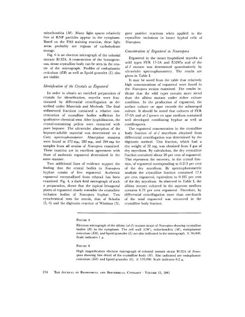
mitochondria (M). Many light spaces relativelyfree of RNP particles appear in the cytoplasm.Based on the PAS staining reaction, these lightareas probably are regions of carbohydratestorage.Fig. 6 is an electron micrograph of the colonialmutant BI32A. A cross-section of the homogeneous,dense crystalline body can be seen in the centerof the micrograph. Profiles of endoplasmicreticulum (ER) as well as lipoid granules (L) alsoare visible.Identification of the Crystals as ErgosterolIn order to obtain an enriched preparation ofcrystals for identification, mycelia were fractionatedby differential centrifugation as describedunder Materials and Methods. The finalsedimented fi'actions contained a relative concentrationof crystalline bodies sufficient forqualitative chemical tests. After lyophilization, thecrystal-containing pellets were extracted withpure heptane. The ultraviolet absorption of theheptane-soluble material was determined on aCary spectrophotometer. Absorption maximawere found at 272 m/~, 282 m#, and 294 m/~ forsamples from all strains of Neurospora examined.These maxima are in excellent agreement withthose of authentic ergosterol determined in thesame manner.Two additional lines of evidence support thefinding that the crystal bodies in Neurosporahyphae consist of free ergosterol. Authenticergosterol recrystallized from ethanol has beenexamined. Fig. 4, a dark field micrograph of sucha preparation, shows that the typical hexagonalplates of ergosterol closely resemble the crystallineinclusion bodies of Neurospora hyphae. Twocytochemical tests for sterols, that of Schultz(5, 6) and the digitonin reaction of Windaus (5),gave positive reactions when applied to thecrystalline inclusions in intact hyphal cells ofNeurospor a.Concentration of Ergosterol in NeurosporaErgosterol in the intact lyophilized mycelia ofwild types SYR 17-3A and E5297a and of theal-2 mutant was determined quantitatively byultraviolet spectrophotornetry. The results aregiven in Table I.It may be noted from the table that relativelyhigh concentrations of ergosterol were found inthe Neurospora strains examined. The results indicatethat the wild types contain more sterolthan the albino mutant under either culturecondition. In the production of ergosterol, thesurface culture on agar exceeds the submergedculture. It should be noted that cultures of SYR17-3A and al-2 grown on agar medium containedwell developed conidiating hyphae as well asconidiospores.The ergosterol concentration in the crystallinebody fraction of al-2 mycelium obtained fromdifferential centrifugation was determined by thedigitonin method. This fraction, which had adry weight of 32 rag, was obtained from 3 gm ofdry mycelium. By calculation, the dry crystallinefraction contained about 20 per cent of ergosterol.This represents the recovery, in the crystal fraction,of ergosterol corresponding to 0.213 per centof the dry mycelium. By spectrophotometricanalysis the crystalline fraction contained 17.4per cent ergosterol, equivalent to 0.185 per centof the dry mycelium. As observed in Table I, thealbino mutant cultured in the aqueous mediumcontains 0.73 per cent ergosterol. Therefore, bydifferential centrifugation more than one-fourthof the total ergosterol was recovered in thecrystalline body fraction.FIGURE 5Electron micrograph of the albino (M-2) mutant strain of Nvurospora showing crystallinebodies (H) in the cytoplasm. The cell wall (CW), mitochondria (M), endoplasmicreticulum (ER), and lipoid granules (L) are also indicated in the micrograph. X 50,000.Scale indicates 1 ~.FIGURE 6High magnification electron micrograph of colonial mutant strain B132A of Neurosporashowing fine detail of the crystalline body (H). Also indicated arc endoplasmiereticulum (ER) and lipoid granules (L). X 135,000. Scale indicates 0.2 ~.174 THE JOURNAL OF BIOPHYSICAL AND BIOCHEMICAL CYTOLOGY - VOLUME 11, 1961