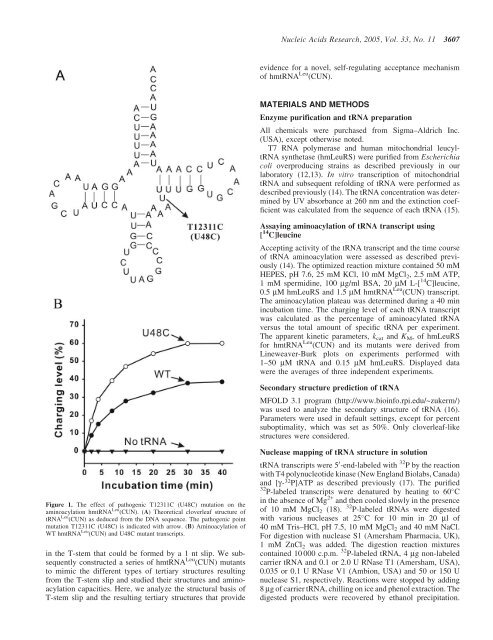
<strong>Nucleic</strong> <strong>Acids</strong> <strong>Research</strong>, 2005, Vol. 33, No. 11 3607evidence for a novel, self-regulating acceptance mechanismof hmtRNA Leu (CUN).MATERIALS AND METHODSEnzyme purification and tRNA preparationAll chemicals were purchased from Sigma–Aldrich Inc.(USA), except otherwise noted.T7 RNA polymerase and human mitochondrial leucyltRNAsynthetase (hmLeuRS) were purified from Escherichiacoli overproducing strains as described previously in ourlaboratory (12,13). In vitro transcription of mitochondrialtRNA and subsequent refolding of tRNA were performed asdescribed previously (14). The tRNA concentration was determinedby UV absorbance at 260 nm and the extinction coefficientwas calculated from the sequence of each tRNA (15).Assaying aminoacylation of tRNA transcript using[ 14 C]leucineAccepting activity of the tRNA transcript and the time courseof tRNA aminoacylation were assessed as described previously(14). The optimized reaction mixture contained 50 mMHEPES, pH 7.6, 25 mM KCl, 10 mM MgCl 2 , 2.5 mM ATP,1 mM spermidine, 100 mg/ml BSA, 20 mM L-[ 14 C]leucine,0.5 mM hmLeuRS and 1.5 mM hmtRNA Leu (CUN) transcript.The aminoacylation plateau was determined during a 40 minincubation time. The charging level of each tRNA transcriptwas calculated as the percentage of aminoacylated tRNAversus the total amount of specific tRNA per experiment.The apparent kinetic parameters, k cat and K M , of hmLeuRSfor hmtRNA Leu (CUN) and its mutants were derived fromLineweaver-Burk plots on experiments performed with1–50 mM tRNA and 0.15 mM hmLeuRS. Displayed datawere the averages of three independent experiments.Secondary structure prediction of tRNAMFOLD 3.1 program (http://www.bioinfo.rpi.edu/~zukerm/)was used to analyze the secondary structure of tRNA (16).Parameters were used in default settings, except for percentsuboptimality, which was set as 50%. Only cloverleaf-likestructures were considered.Figure 1. The effect of pathogenic T12311C (U48C) mutation on theaminoacylation hmtRNA Leu (CUN). (A) Theoretical cloverleaf structure oftRNA Leu (CUN) as deduced from the DNA sequence. The pathogenic pointmutation T12311C (U48C) is indicated with arrow. (B) Aminoacylation ofWT hmtRNA Leu (CUN) and U48C mutant transcripts.in the T-stem that could be formed by a 1 nt slip. We subsequentlyconstructed a series of hmtRNA Leu (CUN) mutantsto mimic the different types of tertiary structures resultingfrom the T-stem slip and studied their structures and aminoacylationcapacities. Here, we analyze the structural basis ofT-stem slip and the resulting tertiary structures that provideNuclease mapping of tRNA structure in solutiontRNA transcripts were 5 0 -end-labeled with 32 P by the reactionwith T4 polynucleotide kinase (New England Biolabs, Canada)and [g- 32 P]ATP as described previously (17). The purified32 P-labeled transcripts were denatured by heating to 60 Cin the absence of Mg 2+ and then cooled slowly in the presenceof 10 mM MgCl 2 (18).32 P-labeled tRNAs were digestedwith various nucleases at 25 C for 10 min in 20 ml of40 mM Tris–HCl, pH 7.5, 10 mM MgCl 2 and 40 mM NaCl.For digestion with nuclease S1 (Amersham Pharmacia, UK),1 mM ZnCl 2 was added. The digestion reaction mixturescontained 10 000 c.p.m. 32 P-labeled tRNA, 4 mg non-labeledcarrier tRNA and 0.1 or 2.0 U RNase T1 (Amersham, USA),0.035 or 0.1 U RNase V1 (Ambion, USA) and 50 or 150 Unuclease S1, respectively. Reactions were stopped by adding8 mg of carrier tRNA, chilling on ice and phenol extraction. Thedigested products were recovered by ethanol precipitation.
3608 <strong>Nucleic</strong> <strong>Acids</strong> <strong>Research</strong>, 2005, Vol. 33, No. 11The pellets were resuspened in 10 ml of dye mix solution[0.0125% xylene cyanol and 0.0125% bromphenol blue in50% formamide]. Samples were run on a 21 cm (width) ·50 cm (height) · 0.4 mm (diameter) 12% polyacrylamidegel containing 8 M urea buffered with 1· TBE, pH 8.3).Alkaline ladders and G-ladders were obtained as describedpreviously (19).RESULTSAminoacylation capacity of tRNA Leu (CUN) wasincreased by the pathogenic T12311C (U48C)mutationIn the hmtRNA Leu (CUN) gene, the pathogenic T12311Cmutation changes the U to C at position 48, which isthe connecting base between the variable loop and theT-stem (Figure 1A). We constructed the wild-type (WT)hmtRNA Leu (CUN) and the U48C mutant in vitro and testedtheir aminoacylation efficiency by the cognate hmLeuRS.Interestingly, the accepting activity of the mutant tRNAwith the U48C substitution was increased compared withthe WT (Figure 1B). Under optimal conditions, 40% of theWT transcript was aminoacylated (compared with the calculatedtheoretical accepting activity of the transcript, seeMaterials and Methods), while the accepting activity ofthe U48C mutant was >60%. Kinetic aminoacylation assayswere further performed with 1–50 mM tRNA and 0.15 mMhmLeuRS. The kinetic parameters k cat and K M were determinedfor both the mutant and WT transcripts. Establishmentof these parameters enables a valuable comparison ofaminoacylation efficiency (k cat /K M ). The increase of chargingcapacity was further evidenced by the finding that the U48Cmutant was leucylated with a 1.31-fold higher efficiency thanthe WT hmtRNA Leu (CUN) (Table 1).Because magnesium ions may affect tRNA folding,we assayed the denaturing/renaturing processes with severalconcentrations of magnesium (from 3 to 20 mM) for eachtranscript. Similar aminoacylation levels of WT and mutanttRNA were obtained under all of these conditions (data notshown), indicating that magnesium ion concentration hadlittle effect on hmtRNA Leu (CUN) folding.Structure analysis of hmtRNA Leu (CUN)In order to understand the relatively lower aminoacylationefficiency of the WT transcript, we examined the secondarystructure of hmtRNA Leu (CUN) using the MFOLD program(16) and enzymatic probing (20).Secondary structure calculation revealed that there were twotypes of T-stem base pair alignments in the hmtRNA Leu (CUN)transcript. In one alignment (Ta alignment), the U48 waslocated between the T-stem and variable loop as an unpairedconnector and the T-loop contained 7 nt. In the other alignment(Tb alignment), the U48 paired with A65 in the T-stem andthe enlarged T-loop contained 8 nt. Thus, the Ta alignmentcould become the Tb alignment by a 1 nt slip, which formeda base pair between 48 and 65 bases (Figure 2A). Becausethe U48C substitution decreased the tendency to form a basepair between C48 and A65, thus prohibiting the T-stem slip,most of the U48C transcript was stabilized in Ta alignment.Considering the limitations of the MFOLD prediction(e.g. its ignorance of RNA 3D structure), we carried out anenzymatic probing experiment to detect the solution structureof the WT and mutant transcripts. The results of the enzymaticprobing of hmtRNA Leu (CUN) were in agreement withthe MFOLD prediction described above. Nuclease S1 andRNase T1 (guanine specific) were used to probe the unpairednucleotides, and RNase V1 was used to monitor base-paired orstacked nucleotides. The 5 0 -end-labeled cleaved fragmentswere separated by PAGE/urea. As the nonspecific cleavage ofthe transcripts at certain weak points could affect the results,each of these enzymes was used with different concentrations.Only dose-dependent enzymatic cleavages were considered.The autoradiograms are presented in Figure 3A and resultsof enzymatic mapping are summarized in Figure 3B.Most of the nucleotides within the theoretical stem regionsof the WT hmtRNA Leu (CUN) were cleaved by RNase V1 andmost of those within the canonical loop regions were recognizedby nuclease S1. The hmtRNA Leu (CUN) transcriptappears to fold into the classical cloverleaf structure despitethe absence of post-transcriptional modification. The generalcleavage patterns of the WT and U48C mutant were similar toeach other except for some minor differences in their T-loops.The WT transcript was cleaved by RNase T1 at G53, which isthought to be paired with C61 in the Ta alignment. This indicatedthat at least some proportion of the WT transcript existedin the Tb alignment, in which G53 remained unpaired in theT-loop. The cleavage at G53 by RNase T1 was dose-dependentin three separate experiments (data not shown), suggesting thatthe result is not related to the quality of the gel. However, inthe case of U48C mutant, no corresponding cleavage wasobserved. The differences in the enzymatic digestion patternsTable 1. Apparent kinetic parameters of in vitro aminoacylation of WT and mutant hmtRNA Leu (CUN) transcripts by human mitochondrial leucyl-tRNA synthetasehmtRNA Leu (CUN) transcripts k cat (10 3 s 1 ) K M (mM) k cat /K M (10 3 s 1 mM 1 ) Relative k cat /K MWild type 52.4 – 1.8 23.9 – 3.9 2.19 1U48C 112.3 – 1.0 39.1 – 5.7 2.87 1.31U48G 115.5 – 3.2 36.3 – 6.9 3.18 1.45U48A 110.3 – 2.2 37.2 – 5.3 2.96 1.35D48 123.1 – 4.7 34.9 – 2.6 3.53 1.61G52A/C62G 53.8 – 3.2 11.5 – 2.5 4.68 2.13U51A/A63U 56.2 – 4.9 8.8 – 1.6 6.39 2.91U51G