Chapter 3: Summary of the Gross Anatomy of the Extraocular Muscles
Chapter 3: Summary of the Gross Anatomy of the Extraocular Muscles
Chapter 3: Summary of the Gross Anatomy of the Extraocular Muscles

Create successful ePaper yourself
Turn your PDF publications into a flip-book with our unique Google optimized e-Paper software.
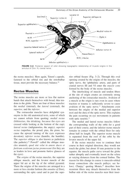
<strong>Summary</strong> <strong>of</strong> <strong>the</strong> <strong>Gross</strong> <strong>Anatomy</strong> <strong>of</strong> <strong>the</strong> <strong>Extraocular</strong> <strong>Muscles</strong> 39FIGURE 3–2. Posterior aspect <strong>of</strong> orbit showing topographic relationship <strong>of</strong> muscle origins in <strong>the</strong>annulus <strong>of</strong> Zinn. N, cranial nerve.<strong>the</strong> rectus muscles). Here again, Tenon’s capsule,fastened to <strong>the</strong> orbital rim and <strong>the</strong> retrobulbartissue, must provide <strong>the</strong> necessary balance. 39Rectus <strong>Muscles</strong>The rectus muscles are more or less flat narrowbands that attach <strong>the</strong>mselves with broad, thin tendonsto <strong>the</strong> globe. There are four <strong>of</strong> <strong>the</strong>se muscles:<strong>the</strong> medial (internal), <strong>the</strong> lateral (external), <strong>the</strong>superior, and <strong>the</strong> inferior.The extraocular muscles have delightful synonymsin <strong>the</strong> old anatomical texts, some <strong>of</strong> whichwe cannot refrain from quoting: medial rectus(bibitorius, <strong>the</strong> drinking, because <strong>the</strong> eyes arecrossed while looking at <strong>the</strong> bottom <strong>of</strong> <strong>the</strong> cup);lateral rectus (indignatorius, <strong>the</strong> angry); superiorrectus (superbus, <strong>the</strong> proud; pius, <strong>the</strong> pious, because<strong>the</strong> upward turning <strong>of</strong> <strong>the</strong> eyes expressesdevotion); inferior rectus (humilis, <strong>the</strong> humble).The superior oblique is also known as pa<strong>the</strong>ticus(<strong>the</strong> pa<strong>the</strong>tic). 26 Powell 31 called <strong>the</strong> oblique musclesamatorii, quod sint velut in amore duces etfurtivum oculorum jactus promoveant (for <strong>the</strong>y areas leaders in love and promote furtive glances <strong>of</strong><strong>the</strong> eyes).The origins <strong>of</strong> <strong>the</strong> rectus muscles, <strong>the</strong> superioroblique muscle, and <strong>the</strong> levator muscle <strong>of</strong> <strong>the</strong>upper lid are at <strong>the</strong> tip <strong>of</strong> <strong>the</strong> orbital pyramid.There <strong>the</strong> origins <strong>of</strong> <strong>the</strong> muscles are arranged in amore or less circular fashion (<strong>the</strong> annulus <strong>of</strong> Zinn),surrounding <strong>the</strong> optic canal and in part <strong>the</strong> superiororbital fissure (Fig. 3–2). Through this ovalopening created by <strong>the</strong> origins <strong>of</strong> <strong>the</strong> muscles, <strong>the</strong>optic nerve, <strong>the</strong> ophthalmic artery, and parts <strong>of</strong>cranial nerves III and VI enter <strong>the</strong> muscle coneformed by <strong>the</strong> body <strong>of</strong> <strong>the</strong> rectus muscles.The interlocking <strong>of</strong> muscle and tendon fibersat <strong>the</strong> site <strong>of</strong> origin creates an extremely stronganchoring <strong>of</strong> <strong>the</strong> extraocular muscles. Avulsion <strong>of</strong>a muscle at <strong>the</strong> origin is rare even in cases wheretraction or trauma is sufficiently severe to causeavulsion <strong>of</strong> <strong>the</strong> optic nerve. 33 Attachments existbetween <strong>the</strong> origins <strong>of</strong> <strong>the</strong> medial and superiorrecti and <strong>the</strong> dura <strong>of</strong> <strong>the</strong> optic nerve. This explains<strong>the</strong> pain occurring on eye movements in patientswith optic neuritis. 33The medial and lateral rectus muscles follow<strong>the</strong> corresponding walls <strong>of</strong> <strong>the</strong> orbit for a goodpart <strong>of</strong> <strong>the</strong>ir course, and <strong>the</strong> inferior rectus muscleremains in contact with <strong>the</strong> orbital floor for onlyabout half its length. The superior rectus muscleis separated from <strong>the</strong> ro<strong>of</strong> <strong>of</strong> <strong>the</strong> orbit by <strong>the</strong>levator muscle <strong>of</strong> <strong>the</strong> upper lid.If <strong>the</strong> rectus muscles were to continue <strong>the</strong>ircourse in <strong>the</strong>ir original direction, <strong>the</strong>y would nottouch <strong>the</strong> globe; but about 10 mm posterior to <strong>the</strong>equator, <strong>the</strong> muscle paths curve toward <strong>the</strong> globera<strong>the</strong>r abruptly and eventually insert on <strong>the</strong> scleraat varying distances from <strong>the</strong> corneal limbus. Thereason for this change in course is musculo-orbitaltissue connections (<strong>the</strong> muscle pulleys; see below).Charpy, 5 quoting Motais, 28 describes howrecurrent fibers may detach <strong>the</strong>mselves from <strong>the</strong>bulbar side <strong>of</strong> <strong>the</strong> rectus muscles near <strong>the</strong>ir inser-