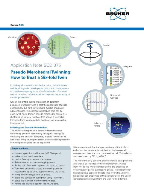
How to Treat a Six-fold Twin - Bruker
How to Treat a Six-fold Twin - Bruker
How to Treat a Six-fold Twin - Bruker

Create successful ePaper yourself
Turn your PDF publications into a flip-book with our unique Google optimized e-Paper software.
<strong>Bruker</strong> AXSVisualizeSelectCreateApplication Note SCD 375Pseudo Merohedral <strong>Twin</strong>ning:<strong>How</strong> <strong>to</strong> <strong>Treat</strong> a <strong>Six</strong>-<strong>fold</strong> <strong>Twin</strong>IntegrateIn dealing with pseudo-merohedral twins, cell refinementand data integration need special care due <strong>to</strong> the presenceof closely overlapping spots. Careful selection of trustedareas in which <strong>to</strong> refine the cell will improve the reliability ofthe cell parameters.One of the pitfalls during integration of data frompseudo-merohedral twins is that the spot shape changescontinuously due <strong>to</strong> the systematic overlap of areas ofadjacent spots. The approach described here can beused for all multi-domain pseudo-merohedral cases. It isillustrated using a six-<strong>fold</strong> twin that shows a reversibletransition from triclinic cells <strong>to</strong> single crystal state with ahexagonal cell.Indexing and Domain OrientationThe initial indexing result is severally biased <strong>to</strong>wardsthe overlap position, resembling hexagonal setting. Byvisualizing the peaks in 3D space, ‘trusted’ areas can beidentified. The pseudo precession pictures will help identifyin which planes spots can be separated.Solve andRefineScale andCorrectSteps and Tools• Harvest spots from all frames (~10.000 spots.)• Index on low resolution data.• Lattice Overlay <strong>to</strong> isolate one domain.• Select area <strong>to</strong> remove overlapping peaks.• Refine cell of domain 1 against the selected peaks.• Transform unit cell <strong>to</strong> create 5 remaining cells byrotating multiples of 60 degrees around the c-axis.• Integrate the images with all 6 cells.• Scale and correct for absorption using TWINABS 1 .• Solve the structure from the HKLF4 data.• Refine the structure against the HKLF5 data.It is also apparent that the spot positions of the tricliniccell at low temperature have inherited the hexagonalarrangement from the room temperature cell. This relationwas confirmed by CELL_NOW 2 .The hk0 plane only contains exactly overlaid peak positionsand should be included in the cell refinement. Planesfrom hk1 <strong>to</strong> hk5 were excluded due <strong>to</strong> the presence ofsystematically partial overlapping peaks. The higher orderhk-planes have separated spots. The reversible (triclinic/hexagonal) cell properties of this sample favors the use ofgenerated cells derived from one well-refined domain.
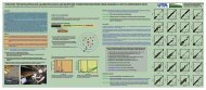
Data CollectionMeasurements were done using Cu radiation. Datacollection with 0.5 degrees images from 10 <strong>to</strong> 30 secondsper image. Detec<strong>to</strong>r distance 50 mm.IntegrationAll six domains were integrated concurrently using SAINT 3 .The cells were not refined <strong>to</strong> avoid refining on peaksaffected by overlap.ResultsThe structure behaves well in unconstrained refinementusing the HKLF5 data. The freely refined batch fac<strong>to</strong>rs foreach domain range from 0.154 <strong>to</strong> 0.191.Refinement results (shelxl-97 4 ):R1=0.0673, 24906 Fo>4sig(Fo)R1=0.0754 for all 29361 datawR2 = 0.2402,Restrained GooF= 1.032 all data = 0.0026Conclusions• Visual representation and interactive <strong>to</strong>ols <strong>to</strong> selectreliable peaks are essential.• Absorption correction and scaling can be successfullyapplied through TWINABS.• Generating idealized classical twin cells by pseudosymmetry operations, provides an excellent descriptionof the spot positions• All operations are possible through the APEX2 graphicaluser interface (version2.2).References1 Sheldrick, G.M. (2002). TWINABS, <strong>Bruker</strong> AXS, Madison.2 Sheldrick, G.M. (2003). CELL_NOW, <strong>Bruker</strong> AXS, Madison.3 <strong>Bruker</strong> (2001). SAINT, <strong>Bruker</strong> AXS, Madison.4 Sheldrick, G.M. (1997). SHELXL-97, Uni. Göttingen.Thanks <strong>to</strong> R. Pietschnig for providing the crystal.AuthorsLeo Straver and Rob Hooft<strong>Bruker</strong> AXS, Oostsingel 209, 2612HL Delft,The NetherlandsFigure 1: Visual representation and selection <strong>to</strong>ols6-<strong>fold</strong> spot 3D lattice view The hk8Lattice overlay"precession" selection <strong>to</strong>olplaneRefinementresult using alldomains<strong>Bruker</strong> AXS Inc.Madison, WI, USAPhone +1 (800) 234-XRAYPhone +1 (608) 276-3000Fax +1 (608) 276-3006info@bruker-axs.comwww.bruker-axs.com<strong>Bruker</strong> AXS GmbHKarlsruhe, GermanyPhone +49 (721) 5 95-28 88Fax +49 (721) 5 95-45 87info@bruker-axs.dewww.bruker-axs.de<strong>Bruker</strong> AXS BVDelft, The NetherlandsPhone +31 (15) 215 2400Fax +31 (15) 215 2599info@bruker-axs.comwww.bruker-axs.comAll configurations and specifications are subject <strong>to</strong> change without notice. Order No. A86-EXS002 © 2007 <strong>Bruker</strong> AXS Inc.Printed in the United States of America. Any trademarks are the property of the respective owners.