Table of Lectures - Society for Mycotoxin Research
Table of Lectures - Society for Mycotoxin Research
Table of Lectures - Society for Mycotoxin Research
Create successful ePaper yourself
Turn your PDF publications into a flip-book with our unique Google optimized e-Paper software.
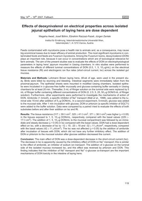
May 14 th –16 th 2007, Fellbach P-63<br />
Effects <strong>of</strong> deoxynivalenol on electrical properties across isolated<br />
jejunal epithelium <strong>of</strong> laying hens are dose dependent<br />
Wageha Awad, Josef Böhm, Ebrahim Razzazi-Fazeli, Jürgen Zentek<br />
Institut für Ernährung, Veterinärmedizinische Universität Wien,<br />
Veterinärplatz 1, A-1210 Vienna, Austria<br />
Feeds contaminated with mycotoxins pose a health risk to animals and, as a consequence, may cause<br />
big econimical losses due to lower efficacy <strong>of</strong> animal production. The most significant mycotoxins in contaminated<br />
foods and feeds are Fusarium mycotoxins. Among the Fusarium toxins, deoxynivalenol (DON)<br />
plays an important role, because it can occur in concentrations which are <strong>of</strong> toxicological relevance <strong>for</strong><br />
farm animals. The aim <strong>of</strong> the present studies was to evaluate the effects <strong>of</strong> DON on electrophysiological<br />
parameters in laying hens’ jejunum mounted in Ussing chambers. In vitro studies were per<strong>for</strong>med to<br />
measure the effects <strong>of</strong> different luminal concentrations <strong>of</strong> DON (0.5, 1, 5, 10 µg/mL) on the electrical<br />
tissue resistance (Rt) and electrogenic ion flux rates (short-circuit current, Isc) across the isolated gut<br />
mucosa.<br />
Materials and Methods: Lohmann Brown laying hens, 48 wk <strong>of</strong> age, were used in the present study.<br />
Birds were killed by stunning and bleeding. Intestinal segments were immediately taken from the<br />
proximal-jejunum. The epithelial sheets were mounted in modified Ussing chambers. Isolated epithelia<br />
were incubated in D-glucose-free buffer mucosally and glucose-containing buffer serosally in Ussing<br />
chambers <strong>for</strong> at least 20 min. Thereafter, 5 mL <strong>of</strong> Ringer solution on the luminal side were replaced by 5<br />
mL <strong>of</strong> Ringer buffer containing different concentrations <strong>of</strong> DON (0, 2.5, 5, 25, 50 µg DON/5 mL <strong>of</strong> Ringer<br />
solution). Furthermore, other experiments were per<strong>for</strong>med to investigate the mechanisms <strong>of</strong> action <strong>of</strong><br />
DON. Amiloride (1 mmol/l), a specific inhibitor <strong>of</strong> Na + transport (Mall et al., 1998), was added to the luminal<br />
side 15 min after addition <strong>of</strong> 5 µg DON/mL. In a second experiment, 5 mmol/L glucose was added<br />
to the mucosal side. After 1 min incubation with glucose, DON or phlorizin (a specific inhibitor <strong>of</strong> SGLT1)<br />
were added to the buffer solution. Data were compared by a paired t-test to evaluate the effects <strong>of</strong> both<br />
substrates be<strong>for</strong>e and after their addition on Isc and Rt.<br />
Results: The tissue resistance (317 ± 39 Ω·cm 2 , 325 ± 41 Ω·cm 2 , 371 ± 38 Ω·cm 2 ) was higher (p < 0.05)<br />
in the tissues exposed to 1, 5, 10 µg DON/mL, respectively, compared with the basal values (229 ±-<br />
17 Ω·cm 2 ). The addition <strong>of</strong> 1, 5, 10 µg DON/mL to the mucosal compartment was followed by an immediate<br />
and steady decrease (p < 0.05) in Isc compared with the basal values. DON had a dose-dependent<br />
effect on Isc, with a decrease <strong>of</strong> Isc to -12 ± 19, -20 ± 18 and -32 ± 11 µA/cm 2 , respectively, compared<br />
with the basal values (42 ± 11 µA/cm 2 ). The Isc was not affected (p > 0.05) by the addition <strong>of</strong> amiloride<br />
after incubation <strong>of</strong> tissues with DON, which did not have any further inhibitory effect. The addition <strong>of</strong><br />
DON or phlorizin to the mucosal solution after glucose addition decreased the current.<br />
Conclusion: The main effect <strong>of</strong> DON was a dose-dependent decrease in the short-circuit current (Isc).<br />
The decrease in Isc is apparently caused by the inhibitory effect <strong>of</strong> DON on Na + -transport; this is similar<br />
to the effect <strong>of</strong> amiloride, an inhibitor <strong>of</strong> sodium ion transport. The addition <strong>of</strong> D-glucose on the luminal<br />
side <strong>of</strong> the isolated mucosa increased Isc, and this effect was reversed by phlorizin and DON. This<br />
finding indicates that the inhibition <strong>of</strong> Na + -transport and Na + -D-glucose co-transport are the important<br />
mechanisms <strong>of</strong> DON toxicity in the intestine <strong>of</strong> laying hens.<br />
119


