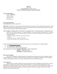
And finally, the utero-placental tissues play a major role in producting <strong>and</strong> transportingvarious hormones. Some steroid precursors, such as cholesterol, are transported frommaternal to fetal circulation <strong>and</strong> metabolized within the fetal <strong>and</strong> placental tissues. Themajor products of these chemical reactions are progestagens <strong>and</strong> estrogens, which aresecreted back to the maternal circulation (10). The feto-placental unit is essentiall insupporting pregnancy by production of progestagens, such as 5αDHP (5α-pregnan-3,20-dione). This hormon circulate in maternal plasma during the second half of thepregnancy, but it is also ruturned to the fetus, especially near term. The total progestagenconcentration in maternal plasma increases rapidly during the last two weeks ofpregnancy thanks to the increased level of delivery of the substrates, doubling of the fetaladrenal gl<strong>and</strong>s, <strong>and</strong> increased expression of the progestagenic enzymes in the fetaladrenals <strong>and</strong> placenta.The final decline in progestagen concentration occurs usually soonbefore parturition. Other hormons produced by the equine placenta are: estrogens <strong>and</strong>relaxin. The precursor for estrogens, DHEA (dehydroepi<strong>and</strong>rostenone), is produced byenlarging fetal gonads, regardles of fetal sex. DHEA is excreted to the umbilical artery,transported into the placenta <strong>and</strong> aromatased into estrogens <strong>–</strong> estrone, estradiol 17-α <strong>and</strong>estradiol 17-β. Relaxin is produced mainly by the placenta. This hormon reaches its highconcentration in maternal circulation by day 80 <strong>and</strong> remains high until it increases againduring parurition, as a response to rising concentrations of oxytocin.Summarizing, the equine placenta is a very complex, marvelous organ which acts in asynchrony with fetal development <strong>and</strong> its needs, as well as with a dam <strong>and</strong> her ability tosupport her pregnancy. Unfortunately, things not always go, as planned, <strong>and</strong> equineplacenta may become a deadly trap for the fetus <strong>and</strong>/or a silent killer for the mare.Evaluation of equine placenta post partumThe fetal part of the equine placenta (fetal membranes) is usually expelled within 30minutes after delivery of the foal. A delay in expulsion of the fetal membranes beyond 3hours is considered abnormal in the mare <strong>and</strong> may have serious consequences, such asmetritis/laminitis/septicemia complex. It is important to remember that even a very smallpiece of chorioallantois retained in mare’s uterus is just as lethal as fetal membranesretained as a whole. Therefore, a thorough examination of the fetal membranes that werepassed post partum is so crucial for planning both foal’s <strong>and</strong> mare’s care. <strong>Placenta</strong>consists of maternal <strong>and</strong> fetal part, <strong>and</strong> only fetal part is expelled in the horse. However,many horsemen, farmers, animal scientists, as well as veterinarians, refer to this tissues asjust the placenta. Therefore, we will use this term from now on in this paper as theequivalent to the fetal membranes.The best time to evaluate equine placenta is immidiately after foaling, <strong>and</strong> placentaexpulsion. <strong>Placenta</strong> should be protected from tearing <strong>and</strong> excessive contamination withfeces, shavings, straw or s<strong>and</strong>. Therefore, any freely hanging from the mare’s vulvafragment of the placenta should be tied immediately after foaling. Farm crew should betrained to collect expelled placenta <strong>and</strong> to place it in a clean backet or trash bag forevaluation. Preparing an outline of the examination <strong>and</strong> a form, which has to be filled outis very helpful in collecting <strong>and</strong> saving good records.<strong>Equine</strong> placenta is usually expelled intact, inside out <strong>–</strong> with the chorionic surface inside<strong>and</strong> the allantoic surface facing outside. The exception to this rule is a prematureplacental separation, when the entire chorioallantoic sac comes out with the chorionic2
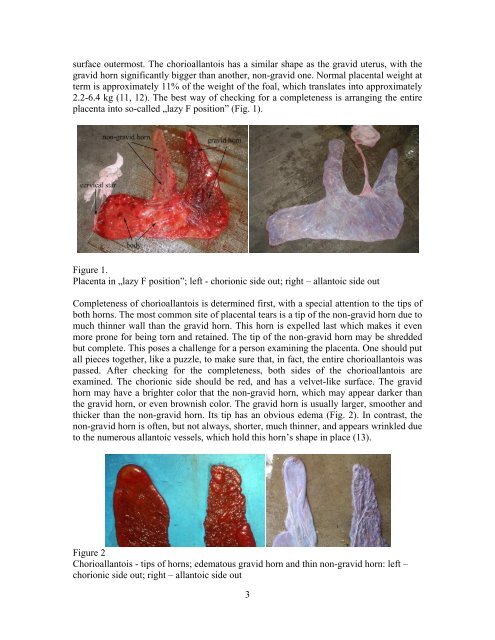
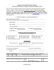
surface outermost. The chorioallantois has a similar shape as the gravid uterus, with thegravid horn significantly bigger than another, non-gravid one. Normal placental weight atterm is approximately 11% of the weight of the foal, which translates into approximately2.2-6.4 kg (11, 12). The best way of checking for a completeness is arranging the entireplacenta into so-called „lazy F position” (Fig. 1).Figure 1.<strong>Placenta</strong> in „lazy F position”; left - chorionic side out; right <strong>–</strong> allantoic side outCompleteness of chorioallantois is determined first, with a special attention to the tips ofboth horns. The most common site of placental tears is a tip of the non-gravid horn due tomuch thinner wall than the gravid horn. This horn is expelled last which makes it evenmore prone for being torn <strong>and</strong> retained. The tip of the non-gravid horn may be shreddedbut complete. This poses a challenge for a person examining the placenta. One should putall pieces together, like a puzzle, to make sure that, in fact, the entire chorioallantois waspassed. After checking for the completeness, both sides of the chorioallantois areexamined. The chorionic side should be red, <strong>and</strong> has a velvet-like surface. The gravidhorn may have a brighter color that the non-gravid horn, which may appear darker thanthe gravid horn, or even brownish color. The gravid horn is usually larger, smoother <strong>and</strong>thicker than the non-gravid horn. Its tip has an obvious edema (Fig. 2). In contrast, thenon-gravid horn is often, but not always, shorter, much thinner, <strong>and</strong> appears wrinkled dueto the numerous allantoic vessels, which hold this horn’s shape in place (13).Figure 2Chorioallantois - tips of horns; edematous gravid horn <strong>and</strong> thin non-gravid horn: left <strong>–</strong>chorionic side out; right <strong>–</strong> allantoic side out3