Equine Placenta – Marvelous Organ and a Lethal Weapon
Equine Placenta – Marvelous Organ and a Lethal Weapon
Equine Placenta – Marvelous Organ and a Lethal Weapon

You also want an ePaper? Increase the reach of your titles
YUMPU automatically turns print PDFs into web optimized ePapers that Google loves.
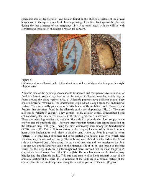
(placental area of degeneration) can be also found on the chorionic surface of the gravidhorn, close to the tip, as a result of chronic pressing of the fetal foot against the placentaduring the last trimester of the pregnancy (14). Any other areas with no villi or withsignificant discoloration should be a reason for concern.Figure 5Chorioallantois <strong>–</strong> allantoic side: left <strong>–</strong> allantoic vesicles; middle <strong>–</strong> allantoic pouches; right- hippomaneAllantoic side of the equine placenta should be smooth <strong>and</strong> transparent. Accumulation offluid in allantoic stroma may lead to the formation of allantoic vesicles, which may befound around the blood vessels. (Fig. 5) Allantoic pouches have different origin. Theycontain necrotic remains of the endometrial cups which slough from the endometrialsurface. They are usually present near the attachment of the umbilical cord. Characteristicfeatures that are often found in the allantoic cavity are hippomanes (Fig. 5). There arealso called “allantoic calculi”. They contain lipids, cellular debris, degenerated bloodcells <strong>and</strong> irregular mineralized material (11). Their significance is unknown.There are many big arteries <strong>and</strong> veins on this side that provide the blood supply to thechorion <strong>and</strong> the chorionic villi. There are three vascular patterns that can be identified onthe allantoic side, with type I being the most commonly seen among the St<strong>and</strong>ardbred(STD) mares (16). Pattern II is consistent with changing location of the fetus from onehorn where implantation took place to another one, where the fetus is present at term.Pattern III is considered abnormal <strong>and</strong> is associated with having a co-twin, which diedspontaneously or was reduced early. The umbilical cord should be attached on the dorsalside at the base of one of the horns. It consists of one vein <strong>and</strong> two arteries on the fetalside <strong>and</strong> two arteries <strong>and</strong> two veins on the maternal side (Fig. 6). The length of the cordvaries, but the large study on 143 Thoroughbred mares showed that the mean length is 55cm, with a broad range from 32 - 90 cm (14). The urachus connects the fetal urinarybladder <strong>and</strong> the allantoic cavity. This structure runs within loose stromal tissue of theamniotic section of the cord (14). A remnant of the yolk sac is a normal feature of theequine placenta <strong>and</strong> is often present along the allantoic portion of the cord (Fig. 6).5