Assessment of gonad staging systems and other ... - CiteSeerX
Assessment of gonad staging systems and other ... - CiteSeerX
Assessment of gonad staging systems and other ... - CiteSeerX

You also want an ePaper? Increase the reach of your titles
YUMPU automatically turns print PDFs into web optimized ePapers that Google loves.
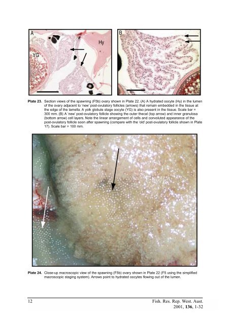
AYGHyBPlate 23. Section views <strong>of</strong> the spawning (F5b) ovary shown in Plate 22. (A) A hydrated oocyte (Hy) in the lumen<strong>of</strong> the ovary adjacent to ‘new’ post-ovulatory follicles (arrows) that remain embedded in the tissue atthe edge <strong>of</strong> the lamella. A yolk globule stage oocyte (YG) is also present in the tissue. Scale bar =300 mm. (B) A ‘new’ post-ovulatory follicle showing the outer thecal (top arrow) <strong>and</strong> inner granulosa(bottom arrow) cell layers. Note the linear arrangement <strong>of</strong> cells <strong>and</strong> convoluted appearance <strong>of</strong> thepost-ovulatory follicle soon after spawning (compare with the ‘old’ post-ovulatory follicle shown in Plate17). Scale bar = 100 mm.Plate 24. Close-up macroscopic view <strong>of</strong> the spawning (F5b) ovary shown in Plate 22 (F5 using the simplifiedmacroscopic <strong>staging</strong> system). Arrows point to hydrated oocytes flowing out <strong>of</strong> the lumen.12 Fish. Res. Rep. West. Aust.2001, 136, 1-32