Standard Anatomical and Visual Space for the Mouse Retina ...
Standard Anatomical and Visual Space for the Mouse Retina ...
Standard Anatomical and Visual Space for the Mouse Retina ...

You also want an ePaper? Increase the reach of your titles
YUMPU automatically turns print PDFs into web optimized ePapers that Google loves.
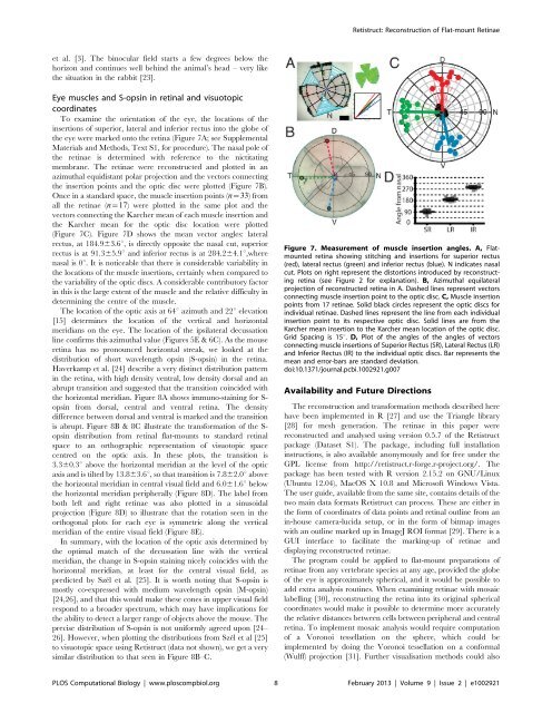
Retistruct: Reconstruction of Flat-mount <strong>Retina</strong>eet al. [3]. The binocular field starts a few degrees below <strong>the</strong>horizon <strong>and</strong> continues well behind <strong>the</strong> animal’s head – very like<strong>the</strong> situation in <strong>the</strong> rabbit [23].Eye muscles <strong>and</strong> S-opsin in retinal <strong>and</strong> visuotopiccoordinatesTo examine <strong>the</strong> orientation of <strong>the</strong> eye, <strong>the</strong> locations of <strong>the</strong>insertions of superior, lateral <strong>and</strong> inferior rectus into <strong>the</strong> globe of<strong>the</strong> eye were marked onto <strong>the</strong> retina (Figure 7A; see SupplementalMaterials <strong>and</strong> Methods, Text S1, <strong>for</strong> procedure). The nasal pole of<strong>the</strong> retinae is determined with reference to <strong>the</strong> nictitatingmembrane. The retinae were reconstructed <strong>and</strong> plotted in anazimuthal equidistant polar projection <strong>and</strong> <strong>the</strong> vectors connecting<strong>the</strong> insertion points <strong>and</strong> <strong>the</strong> optic disc were plotted (Figure 7B).Once in a st<strong>and</strong>ard space, <strong>the</strong> muscle insertion points (n~33) fromall <strong>the</strong> retinae (n~17) were plotted in <strong>the</strong> same plot <strong>and</strong> <strong>the</strong>vectors connecting <strong>the</strong> Karcher mean of each muscle insertion <strong>and</strong><strong>the</strong> Karcher mean <strong>for</strong> <strong>the</strong> optic disc location were plotted(Figure 7C). Figure 7D shows <strong>the</strong> mean vector angles: lateralrectus, at 184.963.6u, is directly opposite <strong>the</strong> nasal cut, superiorrectus is at 91.365.9u <strong>and</strong> inferior rectus is at 284.264.1u,wherenasal is 0u. It is noticeable that <strong>the</strong>re is considerable variability in<strong>the</strong> locations of <strong>the</strong> muscle insertions, certainly when compared to<strong>the</strong> variability of <strong>the</strong> optic discs. A considerable contributory factorin this is <strong>the</strong> large extent of <strong>the</strong> muscle <strong>and</strong> <strong>the</strong> relative difficulty indetermining <strong>the</strong> centre of <strong>the</strong> muscle.The location of <strong>the</strong> optic axis at 64u azimuth <strong>and</strong> 22u elevation[15] determines <strong>the</strong> location of <strong>the</strong> vertical <strong>and</strong> horizontalmeridians on <strong>the</strong> eye. The location of <strong>the</strong> ipsilateral decussationline confirms this azimuthal value (Figures 5E & 6C). As <strong>the</strong> mouseretina has no pronounced horizontal streak, we looked at <strong>the</strong>distribution of short wavelength opsin (S-opsin) in <strong>the</strong> retina.Haverkamp et al. [24] describe a very distinct distribution patternin <strong>the</strong> retina, with high density ventral, low density dorsal <strong>and</strong> anabrupt transition <strong>and</strong> suggested that <strong>the</strong> transition coincided with<strong>the</strong> horizontal meridian. Figure 8A shows immuno-staining <strong>for</strong> S-opsin from dorsal, central <strong>and</strong> ventral retina. The densitydifference between dorsal <strong>and</strong> ventral is marked <strong>and</strong> <strong>the</strong> transitionis abrupt. Figure 8B & 8C illustrate <strong>the</strong> trans<strong>for</strong>mation of <strong>the</strong> S-opsin distribution from retinal flat-mounts to st<strong>and</strong>ard retinalspace to an orthographic representation of visuotopic spacecentred on <strong>the</strong> optic axis. In <strong>the</strong>se plots, <strong>the</strong> transition is3.360.3u above <strong>the</strong> horizontal meridian at <strong>the</strong> level of <strong>the</strong> opticaxis <strong>and</strong> is tilted by 13.863.6u, so that transition is 7.862.0u above<strong>the</strong> horizontal meridian in central visual field <strong>and</strong> 6.061.6u below<strong>the</strong> horizontal meridian peripherally (Figure 8D). The label fromboth left <strong>and</strong> right retinae was also plotted in a sinusoidalprojection (Figure 8D) to illustrate that <strong>the</strong> rotation seen in <strong>the</strong>orthogonal plots <strong>for</strong> each eye is symmetric along <strong>the</strong> verticalmeridian of <strong>the</strong> entire visual field (Figure 8E).In summary, with <strong>the</strong> location of <strong>the</strong> optic axis determined by<strong>the</strong> optimal match of <strong>the</strong> decussation line with <strong>the</strong> verticalmeridian, <strong>the</strong> change in S-opsin staining nicely coincides with <strong>the</strong>horizontal meridian, at least <strong>for</strong> <strong>the</strong> central visual field, aspredicted by Szél et al. [25]. It is worth noting that S-opsin ismostly co-expressed with medium wavelength opsin (M-opsin)[24,26], <strong>and</strong> that this would make <strong>the</strong>se cones in upper visual fieldrespond to a broader spectrum, which may have implications <strong>for</strong><strong>the</strong> ability to detect a larger range of objects above <strong>the</strong> mouse. Theprecise distribution of S-opsin is not uni<strong>for</strong>mly agreed upon [24–26]. However, when plotting <strong>the</strong> distributions from Szél et al [25]to visuotopic space using Retistruct (data not shown), we get a verysimilar distribution to that seen in Figure 8B–C.Figure 7. Measurement of muscle insertion angles. A, Flatmountedretina showing stitching <strong>and</strong> insertions <strong>for</strong> superior rectus(red), lateral rectus (green) <strong>and</strong> inferior rectus (blue). N indicates nasalcut. Plots on right represent <strong>the</strong> distortions introduced by reconstructingretina (see Figure 2 <strong>for</strong> explanation). B, Azimuthal equilateralprojection of reconstructed retina in A. Dashed lines represent vectorsconnecting muscle insertion point to <strong>the</strong> optic disc. C, Muscle insertionpoints from 17 retinae. Solid black circles represent <strong>the</strong> optic discs <strong>for</strong>individual retinae. Dashed lines represent <strong>the</strong> line from each individualinsertion point to its respective optic disc. Solid lines are from <strong>the</strong>Karcher mean insertion to <strong>the</strong> Karcher mean location of <strong>the</strong> optic disc.Grid Spacing is 15u. D, Plot of <strong>the</strong> angles of <strong>the</strong> angles of vectorsconnecting muscle insertions of Superior Rectus (SR), Lateral Rectus (LR)<strong>and</strong> Inferior Rectus (IR) to <strong>the</strong> individual optic discs. Bar represents <strong>the</strong>mean <strong>and</strong> error-bars are st<strong>and</strong>ard deviation.doi:10.1371/journal.pcbi.1002921.g007Availability <strong>and</strong> Future DirectionsThe reconstruction <strong>and</strong> trans<strong>for</strong>mation methods described herehave been implemented in R [27] <strong>and</strong> use <strong>the</strong> Triangle library[28] <strong>for</strong> mesh generation. The retinae in this paper werereconstructed <strong>and</strong> analysed using version 0.5.7 of <strong>the</strong> Retistructpackage (Dataset S1). The package, including full installationinstructions, is also available anonymously <strong>and</strong> <strong>for</strong> free under <strong>the</strong>GPL license from http://retistruct.r-<strong>for</strong>ge.r-project.org/. Thepackage has been tested with R version 2.15.2 on GNU/Linux(Ubuntu 12.04), MacOS X 10.8 <strong>and</strong> Microsoft Windows Vista.The user guide, available from <strong>the</strong> same site, contains details of <strong>the</strong>two main data <strong>for</strong>mats Retistruct can process. These are ei<strong>the</strong>r in<strong>the</strong> <strong>for</strong>m of coordinates of data points <strong>and</strong> retinal outline from anin-house camera-lucida setup, or in <strong>the</strong> <strong>for</strong>m of bitmap imageswith an outline marked up in ImageJ ROI <strong>for</strong>mat [29]. There is aGUI interface to facilitate <strong>the</strong> marking-up of retinae <strong>and</strong>displaying reconstructed retinae.The program could be applied to flat-mount preparations ofretinae from any vertebrate species at any age, provided <strong>the</strong> globeof <strong>the</strong> eye is approximately spherical, <strong>and</strong> it would be possible toadd extra analysis routines. When examining retinae with mosaiclabelling [30], reconstructing <strong>the</strong> retina into its original sphericalcoordinates would make it possible to determine more accurately<strong>the</strong> relative distances between cells between peripheral <strong>and</strong> centralretina. To implement mosaic analysis would require computationof a Voronoi tessellation on <strong>the</strong> sphere, which could beimplemented by doing <strong>the</strong> Voronoi tessellation on a con<strong>for</strong>mal(Wulff) projection [31]. Fur<strong>the</strong>r visualisation methods could alsoPLOS Computational Biology | www.ploscompbiol.org 8 February 2013 | Volume 9 | Issue 2 | e1002921