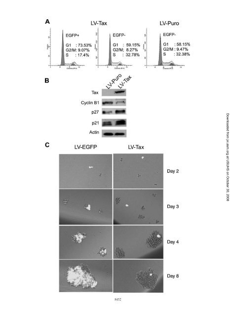
VOL. 82, 2008 HTLV-1 INFECTION LEADS TO SENESCENCE-LIKE G 1 ARREST 8451FIG. 7. HTLV-1-infected Sup-T1 cells ceased proliferation. (A) Half a million 18X21-EGFP SupT1 reporter T cells were cocultured with <strong>the</strong>same number <strong>of</strong> MT2 cells in RPMI medium supplemented with 10% fetal bovine serum. After 48 h, cells were collected, counted, dispersed assingle cells, and cultured in two 12-well plates at a density <strong>of</strong> approximately 2,000 cells/well in <strong>the</strong> same medium. Cells were visualized at 1, 3, or5 days after limiting dilutions. (B) HTLV-1-infected Sup-T1 cells (EGFP positive) are enlarged and expressed hair-like surface protrusions. EGFPfluorescence and bright-field images and a merged image <strong>of</strong> <strong>the</strong> two (merge) <strong>of</strong> <strong>the</strong> same visual field are shown. Two randomly selected visual fieldsfrom two separate wells are shown.days. As shown in Fig. 7A, approximately 0.5% <strong>of</strong> SupT1/18x21-EGFP cells became EGFP positive after coculture. Asexpected, <strong>the</strong> EGFP-positive SupT1 cells remained as singlecells 5 days after coculture, while <strong>the</strong> surrounding EGFP-negativecells proliferated continuously. Because <strong>of</strong> <strong>the</strong> tendency<strong>of</strong> T cells to form clumps, after prolonged culture, <strong>the</strong> Taxtransducedsingle cells were <strong>of</strong>ten found attached to largeclusters <strong>of</strong> EGFP-negative cells (Fig. 7A, day 5). Flow cytometryanalysis fur<strong>the</strong>r indicated that <strong>the</strong> EGFP-positive cells aremostly in <strong>the</strong> G 1 phase <strong>of</strong> <strong>the</strong> cell cycle (not shown). Interestingly,most EGFP-positive cells were enlarged and expressedhair-like protrusions on <strong>the</strong>ir cell surface (Fig. 7B).To demonstrate that <strong>the</strong> cell cycle arrest <strong>of</strong> HTLV-1-infectedEGFP-positive SupT1 is mediated by Tax, we transduced<strong>the</strong> SupT1/18x21-EGFP cells with LV-Tax or LV-Puroat an MOI <strong>of</strong> 1 (titered on HeLa/18x21-EGFP cells) and analyzed<strong>the</strong>m by flow cytometry (Fig. 8A). As expected, <strong>the</strong>fraction <strong>of</strong> EGFP-positive SupT1 cells (transduced with tax)that were in S phase was significantly smaller (17%) than <strong>the</strong>fraction <strong>of</strong> untransduced EGFP-negative counterparts (33%).Indeed, most <strong>of</strong> <strong>the</strong> EGFP-positive SupT1 cells became accumulatedat G 1 in contrast to <strong>the</strong> untransduced cells (Fig. 8A,74% versus 59%). As anticipated, <strong>the</strong> levels <strong>of</strong> p21 CIP1/WAF1and p27 KIP in <strong>the</strong> Tax-transduced cells increased significantly,while that <strong>of</strong> cyclin B1 decreased (Fig. 8B). In a separateexperiment, SupT1/18x21-EGFP cells were transduced withLV-Tax or LV-EGFP. The lentiviral vector-transduced cellswere <strong>the</strong>n dispersed and seeded at a density <strong>of</strong> 5 cells/well in a96-well plate for 8 days. Here again, <strong>the</strong> LV-Tax-transduced(EGFP-positive) SupT1 cells failed to divide and remained assingle cells over <strong>the</strong> entire time course <strong>of</strong> <strong>the</strong> experiment, while<strong>the</strong> untransduced (EGFP-negative) cells continued to divide.As in Fig. 7A, after prolonged culture, single EGFP-positivecells were <strong>of</strong>ten found attached to large clusters <strong>of</strong> EGFPnegativecells (Fig. 8C, LV-Tax day 4 and day 8). In contrastand as expected, <strong>the</strong> LV-EGFP-transduced SupT1 cells expressedEGFP and grew to form large clusters <strong>of</strong> EGFP-positivecells. In aggregate, <strong>the</strong>se results strongly suggest thatHTLV-1-infected T cells, like HeLa cells, are likely to be in astate <strong>of</strong> Tax-induced senescence/G 1 arrest, contrary to <strong>the</strong> prevailingnotion that HTLV-1 infection leads to T-cell proliferation.DISCUSSIONIt is generally thought that after HTLV-1 infection, viralproteins, such as Tax, promote cell proliferation. The ensuingoligoclonal expansion <strong>of</strong> infected T cells in turn leads to <strong>the</strong>onset <strong>of</strong> ATL. This line <strong>of</strong> thinking, however, cannot satisfactorilyexplain <strong>the</strong> low penetrance and long clinical latency <strong>of</strong>Downloaded from jvi.asm.org at USUHS on October 30, 2008
8452Downloaded from jvi.asm.org at USUHS on October 30, 2008