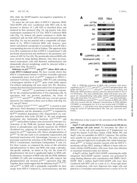
VOL. 82, 2008 HTLV-1 INFECTION LEADS TO SENESCENCE-LIKE G 1 ARREST 8449<strong>of</strong> AZT. As expected, AZT treatment drastically inhibited viralantigen expression, albeit it did not affect syncytium formation.While extensive washing did remove most, if not all, <strong>of</strong> <strong>the</strong>MT2 cells from MT2-HeLa or MT2-HOS cocultures, someHTLV-1 particles apparently remained attached to <strong>the</strong> surfaces<strong>of</strong> HeLa and HOS cells or had entered <strong>the</strong> cells but didnot reverse transcribe and integrate. This is reflected by <strong>the</strong>presence <strong>of</strong> gp46 and p24 in <strong>the</strong> AZT-treated samples (Fig.2B); however, no Tax expression was detected in <strong>the</strong>se samples,consistent with <strong>the</strong> inhibition <strong>of</strong> productive infection.These results indicate that cell-mediated infection <strong>of</strong> HeLaand HOS cells by HTLV-1 readily occurred and <strong>the</strong> amplification<strong>of</strong> HTLV-1 DNA template after reverse transcriptionand integration was principally responsible for <strong>the</strong> increase in<strong>the</strong> production <strong>of</strong> viral antigens.HeLa cells infected by HTLV-1 become arrested in G 1 ina senescence-like state. We have shown recently that Taxdirectly interacts with <strong>the</strong> APC and activates it in an unscheduledmanner (20, 21). Most recently, we have found thatAPC activation by Tax leads to premature polyubiquitination/degradation <strong>of</strong> Skp2, inactivation <strong>of</strong> SCF Skp2 , and dramaticstabilization <strong>of</strong> p27 KIP1 . Concurrent with <strong>the</strong>se cell cyclechanges, <strong>the</strong> levels <strong>of</strong> p21 CIP1/WAF1 mRNA and protein alsobecame greatly activated by Tax. The buildup <strong>of</strong> p21 CIP1/WAF1and especially p27 KIP1 <strong>the</strong>n commits <strong>the</strong> cells to senescence(16).To determine whe<strong>the</strong>r HTLV-1 infection also leads to similarcell cycle abnormalities, HeLa/18x21-EGFP cells wereagain cocultured with MT2 cells at a cell ratio <strong>of</strong> 1 to 1 in <strong>the</strong>presence or absence <strong>of</strong> 1 M AZT as described above. Sixteenhours after coculture, MT2 cells were removed, and <strong>the</strong> infectedHeLa cells were washed extensively to remove any residualMT2 cells. Two days after infection, <strong>the</strong> cells were collected,fixed, stained with propidium iodide, and analyzed for<strong>the</strong>ir cell cycle pr<strong>of</strong>iles by flow cytometry. As shown in Fig. 3A,<strong>the</strong> majority <strong>of</strong> HTLV-1-infected/EGFP-positive cells in <strong>the</strong>AZT-free coculture were arrested at G 1 (84% G 1 , 6.38% S,and 9.47% G 2 /M). This contrasted with <strong>the</strong> uninfected EGFPnegativeHeLa cells in <strong>the</strong> same cell population (58.61% G 1 ,28.38% S, and 13.01% G 2 /M). AZT treatment effectivelyblocked HTLV-1 infection (as indicated by <strong>the</strong> absence <strong>of</strong>EGFP-positive cells from <strong>the</strong> coculture), and as expected, <strong>the</strong>cell cycle pr<strong>of</strong>ile <strong>of</strong> HeLa cells from <strong>the</strong> AZT-treated coculture(Fig. 3A, 62.14% G 1 , 26.04% S, and 11.55% G 2 /M) was similarto <strong>the</strong> pr<strong>of</strong>ile <strong>of</strong> <strong>the</strong> uninfected EGFP-negative cells in <strong>the</strong>AZT-free coculture (Fig. 3A, compare <strong>the</strong> bottom middle andbottom right panels).In a separate approach, 16 h after coculture with MT2, HeLacells were trypsinized and plated on 10-cm plates as single cells.They were <strong>the</strong>n monitored by fluorescence microscopy over<strong>the</strong> course <strong>of</strong> 1 week. As shown in Fig. 3B, HTLV-1-infectedHeLa cells (EGFP positive) quickly ceased cell division afterinfection; 72% remained as single cells, and 23% underwentonly one or two cell division cycles (Fig. 3B, bar diagram) 7days postinfection. The uninfected cells, in contrast, proliferatedand formed large EGFP-negative colonies that were easilyobserved (Fig. 3B, compare <strong>the</strong> EGFP and Phase panels).We next examined whe<strong>the</strong>r HTLV-1 infection also led toaccumulation <strong>of</strong> p21 CIP1/WAF1 and p27 KIP1 . To this end, HeLa/18x21-EGFP cells were plated at 30% confluence and cocultivatedwith MT2 cells for 16 h. Under this condition, <strong>the</strong> celldensity reached 70% confluence 2 days postinfection, and approximately5 to 6% <strong>of</strong> <strong>the</strong> cell population became infectedwith HTLV-1. Even though <strong>the</strong> efficiency <strong>of</strong> HTLV-1 infectionis modest, <strong>the</strong> increase in p21 CIP1/WAF1 and p27 KIP1 in cellscocultivated with MT2 cells was readily apparent in <strong>the</strong> infectedculture versus <strong>the</strong> AZT-treated control (Fig. 3C). Consistentwith <strong>the</strong>ir being in a state <strong>of</strong> G 1 arrest, <strong>the</strong> infectedEGFP-positive cells expressed significant p27 KIP1 in <strong>the</strong>ir nucleicompared to <strong>the</strong> neighboring uninfected EGFP-negativecells (Fig. 3D). Similar nuclear accumulation <strong>of</strong> p27 KIP1 wasalso observed for HeLa cells transduced with LV-Tax (notshown). Finally, HTLV-1-infected HeLa/18x21-EGFP cells becameenlarged, flattened, highly granulated, and expressed <strong>the</strong>senescence-associated -galactosidase (Fig. 3E), much likewhat we have previously reported for LV-Tax-transducedHeLa cells (16). On <strong>the</strong> basis <strong>of</strong> <strong>the</strong>se results, we conclude thatHTLV-1-infected HeLa cells are in a senescence-like state.HTLV-1-infected HOS cells escaped G 1 arrest but developedmitotic abnormalities. Efficient and persistent HTLV-1 infection<strong>of</strong> a human osteosarcoma cell line, HOS, following coculturewith <strong>the</strong> C91PL cell line had been previously reported (6).This suggests that HOS cells may circumvent <strong>the</strong> senescencelikeG 1 arrest after HTLV-1 infection. To investigate <strong>the</strong> cellcycle effect <strong>of</strong> Tax expression on HOS cells, HOS/18x21-EGFPreporter cells were transduced with LV-Tax as previously described(16). As indicated in Fig. 4A, LV-Tax-transducedHOS/18x21-EGFP cells (scored as EGFP positive) progressedthrough cell division cycle without arrest (Fig. 4A), and amajority (around 60%) formed large colonies on plates (Fig.4B). Consistent with <strong>the</strong> absence <strong>of</strong> G 1 arrest, little increase inp21 CIP1/WAF1 and p27 KIP1 levels was observed in <strong>the</strong>se cellsover a course <strong>of</strong> 14 days compared to <strong>the</strong> untransduced control(Fig. 4C). While Tax-expressing HOS cells continued to divide,Tax-induced mitotic abnormalities persisted, as evidenced by<strong>the</strong> appearance <strong>of</strong> large multinucleated cells (Fig. 4B; also seeFig. 5B). Finally, <strong>the</strong> dampening effect <strong>of</strong> Tax on cell cycleprogression was also readily apparent from <strong>the</strong> decrease in <strong>the</strong>EGFP-positive cell population during passage. Within 14 daysafter LV-Tax transduction, <strong>the</strong> population <strong>of</strong> EGFP-positive(tax-transduced) HOS cells declined from 80% to 60% (Fig.Downloaded from jvi.asm.org at USUHS on October 30, 2008FIG. 5. HTLV-1-infected-HOS cells escape G 1 arrest. (A) HOS/18x21-EGFP cells were cocultivated with MT2 cells with or without (w/o) <strong>the</strong>addition <strong>of</strong> AZT as described in <strong>the</strong> legend to Fig. 3. Three days postinfection, infected and uninfected HOS cells were analyzed by flow cytometryas described in <strong>the</strong> legend to Fig. 3A for cell cycle pr<strong>of</strong>ile. EGFP , EGFP positive; EGFP , EGFP negative. (B) HOS/18x21-EGFP cells wereinfected with HTLV-1 as described above for panel A. Cells were trypsinized and plated, grown for 6 days, photographed, and analyzed asdescribed in <strong>the</strong> legend to Fig. 3B and 4B. The colonies/cell clusters <strong>of</strong> different sizes were counted as described in <strong>the</strong> legend to Fig. 4B. (C) HOScells persistently infected by HTLV-1 were cloned by using limiting dilutions. A representative clone is shown. Cells with mitotic abnormalities aremarked by arrows.
8450 LIU ET AL. J. VIROL.4D), while <strong>the</strong> EGFP-negative (tax-negative) population increasedin number.To assess <strong>the</strong> cell cycle effect <strong>of</strong> HTLV-1 infection, HOS/18x21-EGFP cells were cocultivated with MT2 cells in <strong>the</strong>presence or absence <strong>of</strong> 1 M AZT as described above andanalyzed by flow cytometry (Fig. 5A, top panels). Like <strong>the</strong>ircounterparts transduced by LV-Tax, HTLV-1-infected HOScells (Fig. 5A, bottom left panel) continued to divide likeuninfected cells (in both AZT-treated and untreated populations[Fig. 5A, top two panels]) with a comparable cell populationin G 1 . HTLV-1-infected HOS cells, however, grewslower and showed a propensity to accumulate in G 2 /M with acorresponding decrease <strong>of</strong> cells in S phase. This apparent delayin G 2 /M is reminiscent <strong>of</strong> that <strong>of</strong> HTLV-1-transformed T cellspreviously characterized and attributed to <strong>the</strong> premature activation<strong>of</strong> APC by Tax (20). The persistently infected HOS cellswere cloned by using limiting dilutions. Like <strong>the</strong>ir tax-transducedcounterparts, cells with flattened, multinucleated, anddramatically altered morphology could be detected within agiven clone (Fig. 5B and C).Deficiency in p21 CIP1/WAF1 and p27 KIP1 allows HOS cells toevade HTLV-1-induced arrest. We have recently shown thatHTLV-1-transformed human T-cell lines invariably expresseda dramatically lower level <strong>of</strong> p27 KIP1 compared to HTLV-1-unrelated T-cell lines. Fur<strong>the</strong>rmore, NIH 3T3 cells containinga homozygous deletion <strong>of</strong> p27 KIP1 gene could stably expressTax without undergoing cell cycle arrest (16). We, <strong>the</strong>refore,surmise that functional inactivation and/or loss <strong>of</strong> expression <strong>of</strong>p21 CIP1/WAF1 and p27 KIP1 in particular is most likely responsiblefor <strong>the</strong> continued proliferation <strong>of</strong> Tax-expressing cells. Inagreement with our previous prediction, <strong>the</strong> levels <strong>of</strong>p21 CIP1/WAF1 and p27 KIP1 in HOS cells were very low and notaltered by Tax compared to those seen in HeLa cells (Fig. 6A,p21 and p27 blots).The control <strong>of</strong> p21 CIP1/WAF1 and p27 KIP1 is exerted at multiplelevels. Their abundance is regulated by both transcriptionand translation, and <strong>the</strong> abundance <strong>of</strong> p27 KIP1 is regulated bySCF Skp2 -mediated degradation (30). Sequestration via complexformation with cyclin D/Cdk4 (4, 38) and cytoplasmiclocalization also functionally inactivate p21 CIP1/WAF1 andp27 KIP1 and facilitate S-phase entry (reviewed in reference 41).Activation <strong>of</strong> <strong>the</strong> PI3K-Akt (protein kinase B) pathway isknown to phosphorylate and <strong>the</strong>reby inactivate <strong>the</strong> FOXO3(Forkhead box, subgroup O) transcription factor that is requiredfor p27 KIP1 expression (28). Direct phosphorylation <strong>of</strong>p27 KIP1 by Akt also leads to its cytoplasmic sequestration andfunctional inactivation (18). Interestingly, Cereseto et al. hadpreviously reported <strong>the</strong> activation <strong>of</strong> <strong>the</strong> PI3K-Akt pathway inHTLV-1-transformed, interleukin-2-independent T cells (3).For <strong>the</strong>se reasons, we compared <strong>the</strong> status <strong>of</strong> Akt activation(phosphorylation) in HeLa and HOS cells using a phospho-Akt-specific antibody. As shown in Fig. 6A, <strong>the</strong> serine-473phosphorylated Akt level is significantly higher in HOS cells.Fur<strong>the</strong>rmore, treatment <strong>of</strong> HOS cells with <strong>the</strong> PI3K inhibitorsLy294004 (25 M) and wortmannin (100 M) reduced Aktphosphorylation and elevated p27 KIP1 expression (Fig. 6B).Toge<strong>the</strong>r, <strong>the</strong>se results suggest that <strong>the</strong> deficiency in p21 CIP1/WAF1and p27 KIP1 in HOS cells is principally responsible for <strong>the</strong>irescape from HTLV-1-induced G 1 arrest/senescence and thatFIG. 6. PI3K/Akt activation in HOS cells correlates with downregulation<strong>of</strong> p21 CIP1/WAF1 and p27 KIP1 . (A) S473-phosphorylated Akt[pAkt (S473)], p21 CIP1/WAF1 , and p27 KIP1 protein levels in HeLa andHOS cells. HOS and HeLa reporter cells were infected with LV-Tax orLV-SV40-puro. Two days postinfection, whole-cell lysates were preparedfor immunoblotting with pAkt (S473) (S473-phosphorylatedAkt), Akt (total Akt), Tax, p21 CIP1/WAF1 , p27 KIP1 , and actin antibodies.(B) PI3K inhibitors caused p27 KIP1 level to increase in HOS cells. HOScells were treated with <strong>the</strong> indicated concentration <strong>of</strong> PI3K inhibitorLY294002 or wortmannin for 36 h or not treated with LY294002 orwortmannin (). Cell lysates were <strong>the</strong>n prepared for immunoblottingusing <strong>the</strong> serine-473-phosphorylated Akt, Akt, p27, and actin antibodies.this deficiency is due in part to <strong>the</strong> activation <strong>of</strong> <strong>the</strong> PI3K-Aktpathway.SupT1 T cells infected by HTLV-1 or transduced with <strong>the</strong> taxgene undergo cell cycle arrest. To determine <strong>the</strong> effect <strong>of</strong>HTLV-1 on <strong>the</strong> cell cycle progression <strong>of</strong> T lymphocytes, wetested five human T-cell lines, Jurkat, CEM, Molt4, H9, andSupT1 cells, for suitability for HTLV-1 infection. The cell linemost suitable for such analysis is SupT1. The o<strong>the</strong>r cell linesei<strong>the</strong>r expressed high levels <strong>of</strong> phospho-Akt, which downregulatedp27 KIP1 or formed extensive syncytia with <strong>the</strong> HTLV-1-producing cell line MT2, making it difficult to know with certaintywhe<strong>the</strong>r EGFP-positive cells arose from virus infectionor syncytium formation (data not shown).A SupT1 cell line, SupT1/18x21-EGFP, containing <strong>the</strong> stablyintegrated 18x21-EGFP reporter cassette was generated byusing limiting dilutions after transduction with SMPU-18x21-EGFP. Half a million SupT1/18x21-EGFP cells were <strong>the</strong>ncocultured with MT2 cells at a ratio <strong>of</strong> 1:1. Forty-eight hoursafter cocultivation, cells were dispersed into single cells andplated in a 12-well plate and monitored over a course <strong>of</strong> 5 to 10Downloaded from jvi.asm.org at USUHS on October 30, 2008