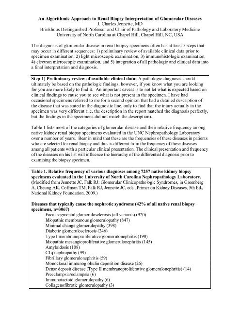
An Algorithmic Approach to Renal Biopsy Interpretation of ...
An Algorithmic Approach to Renal Biopsy Interpretation of ...
An Algorithmic Approach to Renal Biopsy Interpretation of ...

Create successful ePaper yourself
Turn your PDF publications into a flip-book with our unique Google optimized e-Paper software.
<strong>An</strong> <strong>Algorithmic</strong> <strong>Approach</strong> <strong>to</strong> <strong>Renal</strong> <strong>Biopsy</strong> <strong>Interpretation</strong> <strong>of</strong> Glomerular DiseasesJ. Charles Jennette, MDBrinkhous Distinguished Pr<strong>of</strong>essor and Chair <strong>of</strong> Pathology and Labora<strong>to</strong>ry MedicineUniversity <strong>of</strong> North Carolina at Chapel Hill, Chapel Hill, NC, USAThe diagnosis <strong>of</strong> glomerular disease in renal biopsy specimens <strong>of</strong>ten has at least 5 steps thatmay occur in different sequences: 1) preliminary review <strong>of</strong> available clinical data prior <strong>to</strong>specimen examination, 2) light microscopic examination, 3) immunohis<strong>to</strong>logic examination,4) electron microscopic examination, and 5) integration <strong>of</strong> all pathologic and clinical data in<strong>to</strong>a final interpretation and diagnosis.Step 1) Preliminary review <strong>of</strong> available clinical data: A pathologic diagnosis shouldultimately be based on the pathologic findings; however, if you know what you are lookingfor you are more likely <strong>to</strong> find it. <strong>An</strong> important caveat is <strong>to</strong> not let what is expected based onclinical findings <strong>to</strong> cause you <strong>to</strong> see what is not present in the specimen. I have hadoccasional specimens referred <strong>to</strong> me for a second opinion that had a detailed description <strong>of</strong>the disease that was stated in the diagnostic line, only <strong>to</strong> find that the injury actually in thespecimen was very different (i.e. the description in the report matched the diagnosis perfectly,but the findings in the specimens did not match the description).Table 1 lists most <strong>of</strong> the categories <strong>of</strong> glomerular disease and their relative frequency amongnative kidney renal biopsy specimens evaluated in the UNC Nephropathology Labora<strong>to</strong>ryover a number <strong>of</strong> years. Bear in mind that these are the frequencies <strong>of</strong> these diseases in patientswho are selected for renal biopsy and thus is different from the frequency <strong>of</strong> these diseasesamong all patients with a particular clinical presentation. The clinical presentation and frequency<strong>of</strong> the diseases on his list will influence the hierarchy <strong>of</strong> the differential diagnosis prior <strong>to</strong>examining the biopsy specimen.Table 1. Relative frequency <strong>of</strong> various diagnoses among 7257 native kidney biopsyspecimens evaluated in the University <strong>of</strong> North Carolina Nephropathology Labora<strong>to</strong>ry.(Modified from Jennette JC, Falk RJ: Glomerular Clinicopathologic Syndromes, in GreenbergA, Cheung AK, C<strong>of</strong>fman TM, Falk RJ, Jennette JC, eds., Primer on Kidney Diseases, 5th Ed.,National Kidney Foundation, 2009.)Diseases that typically cause the nephrotic syndrome (42% <strong>of</strong> all native renal biopsyspecimens, n=3067)Focal segmental glomerulosclerosis (all variants) (920)Idiopathic membranous glomerulopathy (847)Minimal change glomerulopathy (398)Diabetic glomerulosclerosis (246)Type I membranoproliferative glomerulonephritis (190)Idiopathic mesangioproliferative glomerulonephritis (145)Amyloidosis (108)C1q nephropathy (99)Fibrillary glomerulonephritis (59)Monoclonal immunoglobulin deposition disease (26)Dense deposit disease (Type II membranoproliferative glomerulonephritis) (14)Preeclampsia/eclampsia (6)Immunotac<strong>to</strong>id glomerulopathy (6)Collagen<strong>of</strong>ibrotic glomerulopathy (3)
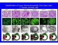
Table 1 continued.Diseases that typically cause hematuria and nephritis (29% <strong>of</strong> biopsies, n=2109)Lupus nephritis (all classes) (636)IgA nephropathy (538)Idiopathic immune complex proliferative glomerulonephritis (375)Pauci-immune/antineutrophil cy<strong>to</strong>plasm antibody glomerulonephritis (301)Postinfectious acute diffuse proliferative glomerulonephritis (86)Thin basement membrane lesion (82)<strong>An</strong>ti-glomerular basement membrane antibody glomerulonephritis (56)Alport syndrome (35)Diseases other than glomerulonephritis that typically cause acute renal failure (5% <strong>of</strong>biopsies, n=371)Thrombotic microangiopathy (all types) (126)Acute tubulointerstitial nephritis (101)Acute tubular necrosis (69)Atheroembolization (34)Light chain cast nephropathy (31)Cortical necrosis (10)Diseases other than glomerulonephritis that typically manifest as chronic renal failure(8%, n=583)Arterionephrosclerosis (229)Chronic sclerosing glomerulonephritis (166)End-stage renal disease not otherwise specified (114)Chronic tubulointerstitial nephritis (74)Miscellaneous other diseases (3%, n=199)Adequate tissue with nonspecific abnormalities (5%, n=370)No pathologic lesion identified (2%, n=141)Inadequate tissue for definitive diagnosis (6%, n=417)Step 2) Light microscopic examination: Thin (~3 micrometer) paraffin sections should beexamined with appropriate stains (e.g. H&E, PAS, Jones, Masson) at multiple levels <strong>of</strong>section. The different tissue compartments should be evaluated systematically, i.e. glomeruli,tubules, interstitium, and arteries and arterioles. Terms commonly used <strong>to</strong> describealterations in glomeruli are defined in Table 2.Table 2. Glossary <strong>of</strong> terms used <strong>to</strong> describe his<strong>to</strong>logic lesions in glomeruli (Modified fromfrom Jennette JC, Olson JL, Schwartz MM, Silva FG: Primer on the Pathologic Diagnosis <strong>of</strong><strong>Renal</strong> Disease in Heptinstall's Pathology <strong>of</strong> the Kidney, 6th Edition, Jennette JC, Olson JL,Schwartz MM, Silva FG (eds), LWW, 2007, Chapter 3.FocalDiffuseSegmentalGlobalMesangial hypercellularityEndocapillary hypercellularityExtracapillary hypercellularityInvolving onelayer <strong>of</strong> parietal or visceral epithelial cells, ormonocytes/macrophages
Table 2 continued.CrescentFibrinoid necrosisSclerosisHyalineMembranoproliferativeLobular (hypersegmented)MesangiolysisExtracapillary hypercellularity other than the epithelialhyperplasia <strong>of</strong> collapsing variant <strong>of</strong> FSGSLytic destruction <strong>of</strong> cells and matrix with deposition <strong>of</strong>acidophilic fibrin-rich materialIncreased collagenous extracellular matrix that isexpanding the mesangium, obliterating capillary lumensor forming adhesions <strong>to</strong> Bowmans capsuleGlassy acidophilic extracellular materialCombined capillary wall thickening and mesangial orendocapillary hypercellularityConsolidated expansion <strong>of</strong> segments that aredemarcated by intervening urinary spaceDetachment <strong>of</strong> the paramesangial GBM from themesangial matrix or lysis <strong>of</strong> mesangial matrixTable 3, lists the major patterns <strong>of</strong> glomerular injury that can be observed by lightmicroscopy. Note that a specific category <strong>of</strong> disease (e.g. lupus nephritis, IgA nephropathy,post-infectious glomerulonephritis) can cause more than one pattern <strong>of</strong> injury.Table 3: Patterns <strong>of</strong> glomerular injury observed by light microscopy and some but not all<strong>of</strong> the diseases that can cause each patterns <strong>of</strong> injury.No abnormality by light microscopy:1. No glomerular disease2. Glomerular disease with no light microscopic changes (e.g. minimal change glomerulopathy,thin basement membrane nephropathy)3. Mild or early glomerular disease (e.g. ISN/RPS Class I lupus nephritis, IgA nephropathy, C1qnephropathy, membranous glomerulopathy, amyloidosis, Alport syndrome, etc.)Thick capillary walls without hypercellularity or mesangial expansion:1. Membranous glomerulopathy (primary or secondary) (>Stage I)2. Thrombotic microangiopathy with expanded subendothelial zone3. Preeclampsia/eclampsia with endothelial swelling3. Fibrillary glomerulonephritis with predominance <strong>of</strong> capillary wall depositsThick walls with mesangial expansion but little or no hypercellularity:1. Diabetic glomerulosclerosis with diffuse rather than nodular sclerosis2. Secondary membranous glomerulopathy with mesangial immune deposits3. Amyloidosis4. Monoclonal immunoglobulin deposition disease5. Fibrillary glomerulonephritis6. Dense deposit disease (type II membranoproliferative glomerulonephritis)Focal segmental glomerular sclerosis without hypercellularity:1. Focal segmental glomerulosclerosis (primary or secondary)2. Chronic sclerotic phase <strong>of</strong> a focal glomerulonephritis3. Hereditary nephritis (Alport syndrome)
Table 3 continued.Mesangial or endocapillary hypercellularity:1. Focal or diffuse mesangioproliferative glomerulonephritis*2. Focal or diffuse (endocapillary) proliferative glomerulonephritis*3. Acute (exudative) diffuse proliferative postinfectious glomerulonephritis4. Membranoproliferative glomerulonephritis (type I, II or III)Extracapillary hypercellularity:1. ANCA crescentic glomerulonephritis (paucity <strong>of</strong> immunoglobulin by IFM)2. Immune complex crescentic glomerulonephritis ((granular immunoglobulin by IFM)3. <strong>An</strong>ti-GBM crescentic glomerulonephritis (linear immunoglobulin by IFM)4. Collapsing variant <strong>of</strong> focal segmental glomerulosclerosis (including HIV nephropathy)Membranoproliferative, lobular or nodular pattern:1. Membranoproliferative glomerulonephritis (type I, II/DDD, or IIIB/IIIS)2. Diabetic glomerulosclerosis with nodular mesangial expansion (KW nodules)3. Monoclonal immunoglobulin deposition disease with nodular sclerosis4. Idiopathic (smoking associated) nodular glomerulosclerosis5. Thrombotic microangiopathy6. Fibrillary glomerulonephritis7. Immunotac<strong>to</strong>id glomerulopathyAdvanced diffuse global glomerular sclerosis1. End stage glomerular disease2. End stage vascular disease3. End stage tubulointerstitial diseaseFigure 1 on the following page illustrates some <strong>of</strong> the patterns <strong>of</strong> glomerular injury observedby light microscopy (all stained with PAS). Panel A shows membranous glomerulopathywith thick capillary walls without hypercellularity or mesangial expansion. Panel B shows verymild segmental mesangial hypercellularity (e.g. ~5 nuclei in mesangial matrix at ~8:00).Panel C shows global endocapillary hypercellularity with numerous neutrophils in a patientwith acute diffuse proliferative GN caused by strep<strong>to</strong>coccal infection. Panel D showsperihilar segmental sclerosis and hyalinosis in a patient with FSGS. Panel E shows globalendocapillary hypercellularity and two small foci <strong>of</strong> extracapillary hypercellularity (e.g.arrow) in a patient with Class IV-G lupus GN. Panel F shows a cellular crescent (arrow) and afocus <strong>of</strong> segmental fibrinoid necrosis (~6:00) in a patient with ANCA disease. Panel G showscapillary wall thickening, mild endocapillary hypercellularity and hyaline thrombi in apatient with cryoglobulinemic glomerulonephritis. Panel H shows nodular glomerulosclerosisin a patient with diabetic glomerulosclerosis (arrow points <strong>to</strong> a small KW nodule).
Figure 1. Patterns <strong>of</strong> glomerular injury observed by light microscopy (all stained with PAS).See text for explanation. (Reproduced from Heptinstall's Pathology <strong>of</strong> the Kidney, 6thEdition, Jennette JC, Olson JL, Schwartz MM, Silva FG (eds), LWW, 2007, Chapter 3, 100-126)
Step 2) Immunohis<strong>to</strong>logic microscopic examination: Immun<strong>of</strong>luorescence orimmunohis<strong>to</strong>chemistry are required for diagnosis <strong>of</strong> glomerular disease.Figure 2: Some <strong>of</strong> the staining patterns that must be discerned when evaluatingglomerular diseases by direct immun<strong>of</strong>luorescence microscopy. <strong>An</strong>tibody specificity isshown in parentheses. The patterns <strong>of</strong> staining are mesangial in IgA nephropathy, mesangialand capillary wall in proliferative lupus GN, peripheral capillary wall granular in type IMPGN, band-like capillary wall and coarsely granular mesangial in DDD (type II MPGN),coarsely granular capillary wall in acute postinfectious GN, finely granular capillary wall inmembranous glomerulopathy, linear GBM staining in anti-GBM glomerulonephritis, andcoarsely granular <strong>to</strong> chunky in fibrillary GN. (From Heptinstall's Pathology <strong>of</strong> the Kidney).
Step 4: Electron microscopic examination. Electron microscopy is not essential for thediagnosis <strong>of</strong> most forms <strong>of</strong> glomerular disease, but EM is helpful in many diseases for furtherconfirmation and nuanced observations, EM is required for some diagnoses (e.g. fibrillaryGN, immunotac<strong>to</strong>id glomerulopathy, thin basement membrane nephropathy), and EM ishelpful in recognize unsuspected categories <strong>of</strong> disease that were not recognized by LM or IM(e.g. hereditary nephritis, collagen<strong>of</strong>ibrotic glomerulopathy, Fabry disease).Figure 3. Diagrams and electron micrographs <strong>of</strong> some <strong>of</strong> the glomerular capillary changesthat must be discerned when evaluating glomerulonephritis by electron microscopy. A)mesangial electron dense deposits, B) subendothelial electron dense deposits, C) subepithelialhump-like electron dense deposits (postinfectious GN), D) subendothelial dense deposits withmesangial interposition (type I MPGN).