Acuity Cone Beam CT - Behestan Darman
Acuity Cone Beam CT - Behestan Darman
Acuity Cone Beam CT - Behestan Darman

You also want an ePaper? Increase the reach of your titles
YUMPU automatically turns print PDFs into web optimized ePapers that Google loves.
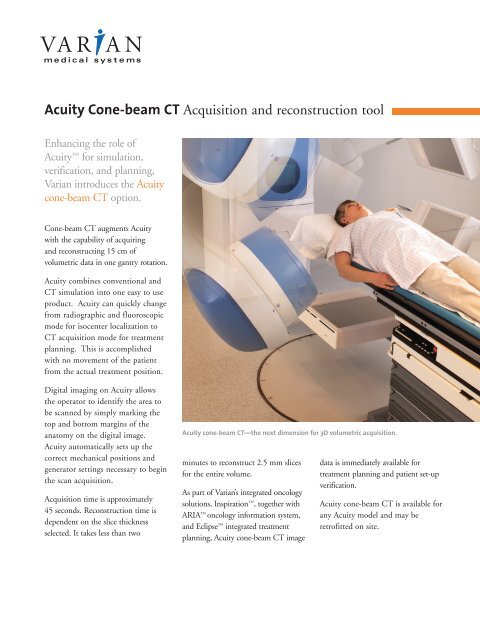
<strong>Acuity</strong> <strong>Cone</strong>-beam <strong>CT</strong> Acquisition and reconstruction toolEnhancing the role of<strong>Acuity</strong> for simulation,verification, and planning,Varian introduces the <strong>Acuity</strong>cone-beam <strong>CT</strong> option.<strong>Cone</strong>-beam <strong>CT</strong> augments <strong>Acuity</strong>with the capability of acquiringand reconstructing 15 cm ofvolumetric data in one gantry rotation.<strong>Acuity</strong> combines conventional and<strong>CT</strong> simulation into one easy to useproduct. <strong>Acuity</strong> can quickly changefrom radiographic and fluoroscopicmode for isocenter localization to<strong>CT</strong> acquisition mode for treatmentplanning. This is accomplishedwith no movement of the patientfrom the actual treatment position.Digital imaging on <strong>Acuity</strong> allowsthe operator to identify the area tobe scanned by simply marking thetop and bottom margins of theanatomy on the digital image.<strong>Acuity</strong> automatically sets up thecorrect mechanical positions andgenerator settings necessary to beginthe scan acquisition.Acquisition time is approximately45 seconds. Reconstruction time isdependent on the slice thicknessselected. It takes less than two<strong>Acuity</strong> cone-beam <strong>CT</strong>—the next dimension for 3D volumetric acquisition.minutes to reconstruct 2.5 mm slicesfor the entire volume.As part of Varian’s integrated oncologysolutions, Inspiration , together withARIA oncology information system,and Eclipse integrated treatmentplanning, <strong>Acuity</strong> cone-beam <strong>CT</strong> imagedata is immediately available fortreatment planning and patient set-upverification.<strong>Acuity</strong> cone-beam <strong>CT</strong> is available forany <strong>Acuity</strong> model and may beretrofitted on site.
<strong>Acuity</strong> <strong>Cone</strong>-beam <strong>CT</strong> Acquisition and reconstruction toolSpecifications1.0 Software Features• Window/level• Pan/zoom• Multiple image display• HU Calibration• Measurement tool• Patient file management2.0 Hardware2.1 Acquisition workstation• Pentium ® -class computer• 3.2 GHz single processor• Minimum 4 GB RAM• Minimum 40 GB hard disk• GB Ethernet card2.2 Reconstruction computer• Dell Precision 670 dualCPU Workstation orequivalent• Dual 3.2 GHz Intel ®Xeon CPUs; 800 MHzFSB• 2 GB DDR2 RAM• 160 GB Serial ATA 7200RPM hard drive• Windows ® XPProfessional operatingsystem SP1 or later2.3 CB<strong>CT</strong> Phantoms• Body normalizationphantom: 45 cm diameter• Head normalizationphantom: 25 cm diameter• Geometry calibrationphantom• <strong>CT</strong> image qualityphantom (e.g., Catphan504 or equivalent)2.4 Image performancespecificationsSee Table 1.Table 1: Performance SpecificationsCharacteristicPerformance<strong>CT</strong> Number Range<strong>CT</strong> Number AccuracySpatial ResolutionLow Contrast ResolutionNoise<strong>CT</strong> Number Uniformity (cupping)<strong>CT</strong> Number LinearityAperture SizeReconstruction Field of ViewLength of Image Volume-1024 to +3072 or greater± 40 HU for 20 cm diameter (water equivalent) phantomAxial = 7 lp/cm (measured using a full-fan acquisition with Catphan 504 phantom)Axial = 6 lp/cm (measured using a half-fan acquisition with Catphan 504 phantom)1.0%; 15 mm diameter object visible; using a dose of 90 mGy (<strong>CT</strong>DIw) in 20 cmCatphan 504 phantom using a 2.5 mm slice thickness1.0% using a dose of 50 mGy (<strong>CT</strong>DIw) in a 20 cm H2O phantom using a 10 mmslice thickness±40 HU from center to edge in a 20 cm diameter uniform phantom (uniformitysection of a Catphan 504 phantom)<strong>CT</strong> # versus true attenuation (mu) has a regression co-efficient of 0.95 for a fourpoint calibrationApproximately 95 cm25 cm dia. x 15 cm axial length (head scan)45 cm dia. x 15 cm axial length (body scan)Three contiguous volumes of 15 cm can be merged via software to provide avolume length of approximately 45 cm.Reconstruction Matrix 128 x 128, 256 x 256, 512 x 512Image Display1280 x 1024 x 8 bitsSlice Thickness0.5 – 10 mmAcquisition and Reconstruction Time Acquisition time approximately 45 secReconstruction time approximately < 2 min for 2.5 mm slice widthTypical DosesBody: 38 mGy (<strong>CT</strong>DIw) [@125 kVp and using bow-tie filter]Head: 90 mGy (<strong>CT</strong>DIw) [@125 kVp and using bow-tie filter]Specifications subject to change without notice.Varian and Varian Medical Systems are registered trademarks, and <strong>Acuity</strong>, ARIA, Eclipse, and Inspiration are trademarks of Varian Medical Systems, Inc.The names of other companies and products mentioned herein are used for identification purposes only and may be trademarks or registered trademarks oftheir respective owners.The <strong>Acuity</strong> <strong>Cone</strong>-beam <strong>CT</strong> is part of theInspiration integrated oncology environment.Inspiration, the Varian advantageUSA Headquarters, CaliforniaVarian Medical SystemsPalo Alto, CATel: 650.424.5700800.544.4636Fax: 650.493.5637www.varian.comHeadquarters Europe, Eastern Europe, Africa,Middle & Near EastVarian Medical Systems International AGZug, SwitzerlandTel: 41.41.749.8844Fax: 41.41.740.3340info.europe@varian.comRAD 2259B © 2005 Varian Medical Systems, Inc. Printed in USA 10/05 (5K)