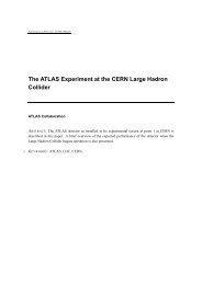
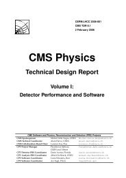
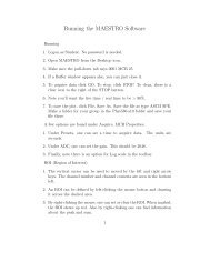
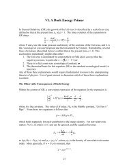
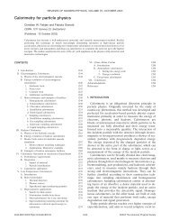
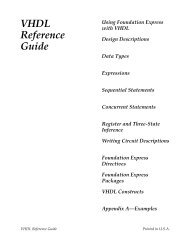
DA Wilkinson. Biomed <strong>Imaging</strong> Interv J 2006; 2(2):e34 2This page number is notfor citation purposeThe Clevel<strong>and</strong> Clinic FoundationMANCHESTER TEMPLATEThe Clevel<strong>and</strong> Clinic FoundationThe Clevel<strong>and</strong> Clinic Foundation<strong>HDR</strong> Fletcher-suitGYN TEMPLATEFigure 1 Examples of <strong>HDR</strong> applicators used for lung, rectal, <strong>and</strong> gynecologic diseases.(AAPM). The latter are intended to provide the medicalphysicist in the USA (<strong>and</strong> it is hoped other nations aswell) the proper guidance to ensure that <strong>brachytherapy</strong>procedures are carried out safely <strong>and</strong> with due attentionto these rules. There are two AAPM task group (TG)reports that are particularly relevant to <strong>HDR</strong> QA. Theyare TG 56: Code of practice for <strong>brachytherapy</strong> physics[3], <strong>and</strong> especially TG 59: <strong>HDR</strong> <strong>brachytherapy</strong> treatmentdelivery [4]. Another useful reference for <strong>brachytherapy</strong><strong>quality</strong> <strong>assurance</strong> has been published by ESTRO <strong>and</strong> isavailable on their website [5]. This paper will providedetails about <strong>HDR</strong> QA as it is performed in our radiationtherapy department (on a Nucletron Microselectronsystem) <strong>and</strong> how the QA program is related to the NRCregulations <strong>and</strong> the task group recommendations.APPLICATOR QAPrior to the initial use of a new (or replacement)applicator, it is necessary to verify that the source dwellpositions correspond to the radiographic markerpositions used in simulation <strong>and</strong> treatment planning. TG-56 recommends that coincidence of dummy <strong>and</strong>radioactive sources be checked annually as well. Thereare many st<strong>and</strong>ard applicators; photographs of several ofthose used in our institution are shown in Figure 1.The method we employ to verify coincidence ofdwell position <strong>and</strong> radiographic marker isautoradiography. An applicator is taped securely to asealed film envelope (Figure 2) <strong>and</strong> the <strong>HDR</strong> after loaderis programmed to send the source to a few appropriatelychosen dwell positions for less than 1 second (e.g. 0.3 sfor 0.31 GBq source). Next, the film plus applicator istransferred to a diagnostic X-ray source such as asimulator, the dummy source markers are placed in theapplicator <strong>and</strong> the film exposed <strong>and</strong> developed (e.g. 125kVp, 125 mAs for Kodak XV film). An example of thisis shown in Figure 3. TG-56 recommends that the
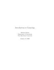
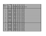
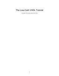
DA Wilkinson. Biomed <strong>Imaging</strong> Interv J 2006; 2(2):e34 3This page number is notfor citation purpose1713915Figure 2 Photograph of a ring applicator secured to a base platewith film taped securely in place.Figure 3 Autoradiograph of a 3 cm ring showing 5 dwellpositions (1,5,9,13,17) as well as the interveningdummy markers (unnumbered arrows). Dwell positionsare for the 0.5 cm step-size.coincidence of dummy <strong>and</strong> active sources be within 2mm; the NRC regulations call for ± 1 mm.PERIODIC SPOT-CHECKThe new NRC regulations require a periodic spotcheckof each <strong>HDR</strong> unit prior to the first use on anygiven day that the after loader is in operation <strong>and</strong> aftereach new source installation. These spot-checks need notbe done by the authorized medical physicist, but thelatter must review the results <strong>and</strong> notify the licensee inwriting of his findings. Table 1 lists the checks that mustbe performed at a minimum to assure proper operation ofthe unit according to NRC Regulations 10 CFR Part 35.A convenient way of implementing <strong>and</strong> recordingthe above <strong>quality</strong> <strong>assurance</strong> is by using a checklist suchas the one our clinic uses as shown in figure 4.Certain tests require only a simple inspection toensure that materials are present, viz. User manual,Removal kit, Emergency instructions, Bailout pig,Radiation alarm setting, <strong>and</strong> Printer paper. Switching onthe system allows the tests in item 7 to be performed.The source activity comparison can be made using atable generated by the medical physicist (Figure 5). Thiswill also satisfy the requirement (see Full Calibrationbelow) for performing decay correction which must bedone by the authorized medical physicist. Agreementshould easily be within 1 percent tolerances.For the remaining tests, the active source will needto be deployed. For this, the system can be programmedmanually each time or a st<strong>and</strong>ard program recalled fromthe system memory. A single dwell time of 20 to 30 ssuffices to test the door interlock, the interrupt button,<strong>and</strong> the emergency off button as well as to verify that theappropriate exposure indicators <strong>and</strong> radiation monitorsare functioning properly. The spot check form requirestesting of the functioning of the meter in the treatmentroom (Figure 6) under battery power alone. Its alarmsetting of 4 mR/hr was established so as to be aboveexposure levels in the room due to an adjacent linactherapy suite. As an additional safety measure, we have acalibrated GM meter that is carried by h<strong>and</strong> by personnelupon entering the treatment room. It is checked using a 1mCi Cs-137 source that yields a 10 mR/hr contact value.The remaining item is an estimate of timer accuracy. Forthis, a stopwatch is used to time a 30 s dwell. Typicalerror estimates are well below 1 second.FULL CALIBRATIONA “full calibration” is m<strong>and</strong>ated for several differentcircumstances, e.g. before first medical use, following asource change or any major repair, etc. Since the sourcein most, if not all, modern <strong>HDR</strong> after loaders is Ir-192with a half-life of approximately 74 days, therequirement for quarterly calibration [2] does notformally apply. However, it is usual to replace an iridiumsource four times a year so as to maintain reasonabledose rates <strong>and</strong> treatment times. Quarterly QA testing of<strong>HDR</strong> after loaders was recommended in the report of TG56. The components of a full calibration as defined inNRC Regulations 10 CFR Part 35 are listed in Table 2.