May 2008 - bcps
May 2008 - bcps
May 2008 - bcps

You also want an ePaper? Increase the reach of your titles
YUMPU automatically turns print PDFs into web optimized ePapers that Google loves.
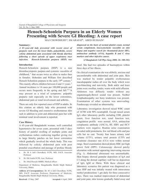
Journal of Bangladesh College of Physicians and SurgeonsVol. 26, No. 2, <strong>May</strong> <strong>2008</strong>Henoch-Schonlein Purpura in an Elderly WomenPresenting with Severe GI Bleeding: A case reportMAJ CHOWDHURY a , SM ARAFAT b , ABED HUSSAIN cSummary:A 65–year-old lady presented with recent onset ofpurpuric rash over the lower limbs, polyarthritis, severecolicky abdominal pain associated with bloody diarrheafollowing a short episode of upper respiratory tractinfection. Henoch-Schonlein purpura (HSP) wasdiagnosed on the basis of normal platelet count, normalserum complement, leucocytoclastic vasculitis on skinbiopsy and negative search for rheumatoid factor (RF),antinuclear antibody (ANA), hepatitis B and C virusmarkers and other infective causes.(J Bangladesh Coll Phys Surg <strong>2008</strong>; 26: 100-102)IntroductionHenoch-Schonlein purpura (HSP) is a nonthrombocytopenic purpura and systemic vasculitis ofchildhood, 1 that occurs twice as often in males thenin females. Heberden and William first describedHenoch-Schonlein purpura in the early 19 th century. 2This mainly affects children between 4 and 11 years. 3Annual incidence is 14 cases per 100,000 people andoccurs more frequently in the spring and fall. 3’4 Itmay present as a triad of symptoms: palpablepurpuric rash especially on the lower extremities,abdominal pain or renal involvement and arthritis.There are only few reported cases of HSP in adults. Inthis citation an elderly lady who presented withsevere GI bleeding and extensive erythematous skinrash in addition to joint and abdominal pain but withminimal renal involvement is reported.Case HistoryA 65-year-old Bangladeshi woman, well controlledhypertensive for seven years, presented with 5 dayshistory of painful swelling of multiple joints anderythematous rashes coalescing together giving riseto large blotchy patches on her lower extremitieswhich gradually involved the whole body. This wasfollowed by colicky abdominal pain with postprandial exacerbation and passage of profuse bloodya. Dr. M A Jalil Chowdhury FCPS, MD, FACP, AssociateProfessorb. Dr. SM Arafat FCPS, Asst. Professorc. Dr. Abed Hussain MBBS, Medical officerDepartment of Medicine, Bangabandhu Sheikh Mujib MedicalUniversity, Dhaka.Address of Correspondence: Dr. M A Jalil Chowdhury FCPS,MD, FACP, Associate Professor, Department of Medicine,Bangabandhu Sheikh Mujib Medical University, Dhaka.Received: 15 <strong>May</strong>, 2007 Accepted: 6 April, <strong>2008</strong>stool. She had two episodes of haemoptysis withinthree days of her illness.On clinical examination she was afebrile, anxious anduncomfortable with abdominal and joint pain. Skinwas marked by tender palpable erythematousmaculopapular rashes all over the body which werenon-blanching and non-itchy. Both ankle and kneejoints were swollen, tender, warm with mild effusion.Abdomen was diffusely tender without anyorganomegaly.Bowel sound was present. Neitherlymphadenopathy nor bony tenderness was present.Examination of other systems was unrevealing.Funduscopy revealed no abnormality.Laboratory investigation showed raised WBC countof 16700 with 90% neutrophill. Except raised serumIgA other laboratory profile including ESR, plateletcount, liver function test, renal function test,coagulation profile were normal. ANA, antinuclearcytoplasmic antibody (ANCA), RF, HBV and HCVmarkers were negative. Routine urine examinationrevealed mild proteinuria, few red blood cells and puscells but no cast. Twenty four hours urinary totalvolume (UTV), urinary total protein (UTP) andcreatinine clearance rate (CCr) were within normalrange. Stool examination showed plenty RBC per highpower field (HPF). Colonoscopy showed patchyulceration with normal appearing intervening mucosa(Fig. 1). Abdominal ultrasound was unremarkable.Skin biopsy showed granular deposition of IgA andC3 along the dermal capillary wall but no depositionof IgG, IgM or fibrin. With these clinical andlaboratory scenarios this patient was diagnosed as acase HSP and was initially treated with intravenousmethyl prednisolone 1 gm daily for three consecutivedays. There was marked improvement of abdominaland joint pain; skin lesions gradually faded away and