September 2004 - bcps
September 2004 - bcps
September 2004 - bcps

You also want an ePaper? Increase the reach of your titles
YUMPU automatically turns print PDFs into web optimized ePapers that Google loves.
ISSN 1015-0870<strong>September</strong> <strong>2004</strong>Vol. 22, No. 3Journal ofBangladesh College ofPhysicians and SurgeonsOfficial Journal ofThe Bangladesh College of Physicians and Surgeons
Journal of Bangladesh College ofPhysicians and SurgeonsVol. 22, No. 3, <strong>September</strong> <strong>2004</strong>Official Journal of the Bangladesh College of Physicians and SurgeonsBCPS Bhaban, 67 Shaheed Tajuddin Ahmed SaraniMohakhali, Dhaka-1212, BangladeshJOURNAL COMMITTEEChairpersonProfessor M. A. MajedEditor-in-ChiefProf. T.I.M. Abdullah-Al-FaruqMembersProfessor M. A. MajidProfessor Tofayel AhmedProfessor Mahmud HasanProfessor Choudhury Ali KawserProfessor Sayeba AkhterProfessor Salim Md. JahanagirProfessor U.H. Shahera KhatunDr. Syed Kamaluddin AhmedDr. Projesh Kumar RoyDr. A.K.M. Anwarul IslamProf. Shafquat Hussain KhundkerDr. Emran Bin YunusDr. Barendra ChakrabortyDr. A.K.M. Fazlul HaqueDr. Md. Rajibul AlamDr. Syed Azizul HaqueDr. Nooruddin AhmedDr. Abid Hossain MollahDr. Md. Zulfiquar Rahman KhanDr. Md. Mazibur Rahman BhuiyanDr. Dewan Saifuddin AhmedDr. Abdul Wadud ChowdhuryDr. A.K.M. Aminul HoqueDr. Hasina AfrozDr. Mohammad Monir HossainEDITORIAL BOARDChairpersonProfessor M. A. MajedEditor-in-ChiefProf. T.I.M. Abdullah-Al-FaruqMembersProf. Md. Abdul HadiProf. Md. Abdul Mobin KhanProf. MA MajidProf. Tofayel AhmedProf. AHM TA ChowdhuryProf. AHM AhsanullahProf. AZM Zahid HossainProf. Quazi Deen MohammadProf. Md. TahirProf. Nazmun NaharProf. Md. Sanawar HossainProf. Choudhury Ali KawserProf. MA Hadi FaquirProf. Syed Atiqul HaqProf. Mahmud HasanProf. SAM Golam KibriaProf. Md. Ruhul AminProf. Abdul Bayes BhuiyanProf. Shafiqul HaqueProf. Shafquat Hussain KhundkarDr. Syed Kamaluddin AhmedPUBLISHED BYProf T.I.M. Abdullah-Al-Faruqon behalf of the Bangladesh Collegeof Physicians and SurgeonsPRINTED ATAsian Colour Printing130 DIT Extension Road, FakirerpoolDhaka-1000, Phone : 9357726, 8362258ANNUAL SUBSCRIPTIONTk. 300/- for local and US$ 30for overseas subscribersThe Journal of BangladeshCollege of Physicians andSurgeons is a peer reviewedJournal. It is published threetimes in a year, (January,May and <strong>September</strong>). Itaccepts original articles,review articles, and casereports. Complimentary copiesof the journal are sent tolibraries of all medical andother relevant academic institutionsin the country and selectedinstitutions abroad.While every effort is alwaysmade by the Editorial Boardand the members of the JournalCommittee to avoid inaccurateor misleading informationappearing in the Journal ofBangladesh College of Physiciansand Surgeons, informationwithin the individual article arethe responsibility of its author(s).The Journal of BangladeshCollege of Physicians andSurgeons, its Editorial Boardand Journal Committee acceptno liability whatsoever for theconsequences of any suchinaccurate and misleadinginformation, opinion or statement.ADDRESS OF CORRESPONDENCEEditor-in-Chief, Journal of Bangladesh College of Physicians and Surgeons, BCPS Bhaban, 67, Shaheed Tajuddin Ahmed Sarani, Mohakhali,Dhaka-1212, Tel : 8825005-6, 8856616-7, Fax : 880-2-8828928, E-mail : <strong>bcps</strong>@bdonline.com
INFORMATION FOR AUTHORSThe Journal of Bangladesh College of Physicians andSurgeons agrees to accept manuscript prepared inaccordance with the 'Uniform Requirements Submitted tothe Biomedical Journals' published in the New EnglandJournal of Medicine 1991; 324 : 424-8.Aims and scope:The Journal of Bangladesh College of Physicians andSurgeons is one of the premier clinical and laboratory basedresearch journals in Bangladesh. Its International readershipis increasing rapidly. It features the best clinical andlaboratory based research on various disciplines of medicalscience to provide a place for all medical scientists to relateexperiences which will help others to render better patientcare.Conditions for submission of menuscript: All manuscripts are subject to peer-review.Manuscripts are received with the explicit understandingthat they are not under simultaneous consideration by anyother publication.Submission of a manuscript for publication implies thetransfer of the copyright from the author to the publisherupon acceptance. Accepted manuscripts become thepermanent property of the Journal of Bangladesh Collegeof Physicians and Surgeons and may not be reproducedby any means in whole or in part without the writtenconsent of the publisher.It is the author's responsibility to obtain permission toreproduce illustrations, tables etc. from otherpublications.Ethical aspects: Ethical aspect of the study will be very carefullyconsidered at the time of assessment of the manuscript.Any menuscript that includes any table, illustration orphotographs that have been published earlier shouldaccompany a letter of permission for re-publication fromthe author(s) of the publication and editor/publisher ofthe Journal where it was published earlier. Permission of the patients and/or their families toreproduce photographs of the patients where identity isnot disguised should be sent with the manuscript.Otherwise the identity will be blackened out.Preparation of manuscript:Criteria:Informations provided in the menuscript are important andlikely to be of interest to an international readership.Preparation:a) Menuscript should be written in English and typed onone side of A4 (290 x 210cm) size white paper.b) Double spacing should be used throughout.c) Margin should be 5 cm for the header and 2.5 cm for theremainder.d) Style should be that of modified Vancouver.e) Each of the following section should begin on separatepage :Title pageSummary/abstractTextAcknowledgementReferencesTables and legends.f) Pages should be numbered consecutively at the upperright hand corner of each page beginning with the titlepage.Title Page :The title page should contain: Title of the article (should be concise, informative andself-explanatory).Name of each author with highest academic degreeName of the department and institute where the work wascarried outName and address of the author to whom correspondenceregarding manuscript to be madeName and address of the author to whom request forreprint should be addressedSummary/Abstract :The summary/abstract of the menuscript : Should be informative Should be limited to less than 200 words Should be suitable for use by abstracting journals andinclude data on the problem, materials and method, resultsand conclusion. Should emphasize mainly on new and important aspectsof the study Should contain only approved abbreviations
Introduction:The introduction will acquaint the readers with the problemand it should include:Nature and purpose of the studyRationale of the study/observationStrictly pertinent referencesBrief review of the subject excepting data and conclusionMaterials and method :This section of the study should be very clear and describe: The selection criteria of the study population includingcontrols (if any).The methods and the apparatus used in the research.The procedure of the study in such a detail so that otherworker can reproduce the results.Previously published methods (if applicable) withappropriate citations.Results:The findings of the research study should be described hereand it should be:Presented in logical sequence in the text, tables andillustrations.Described without comment.Supplemented by concise textual description of the datapresented in the tables, charts and figures where it isnecessaery.Tables:During preparation of tables following principles should befollowed Tables should be simple, self-explanatory and shouldsupplement, not duplicate the text. Each table should have a tittle and typed in double spacein separate sheet. They should be numbered consecutively with romannumerical in order of text. Page number should be in theupper right corner.If abbreviations are to be used, they should be explainedin footnotes.Illustrations:Only those illustrations that clarify and increaseunderstanding of the text should be used and:All illustrations must be numbered and cited in the text.Print photograph of each illustration should be submitted.Figure number, tittle of manuscript, name ofcorresponding author and arrow indicating the top shouldbe typed on a sticky label and affixed on the back of eachillustration.Original drawings, graphs, charts and lettering should beprepared on an illustration board or high-grade whitedrawing paper by an experienced medical illustrator.Figures and photographs:The figures and photographs :Should be used only where data can not be expressed inany other formShould be unmounted glossy print in sharp focus, 12.7 x17.3 cms in size. Should bear number, tittle of manuscript, name ofcorresponding author and arrow indicating the top on asticky label and affixed on the back of each illustration.Legend:The legend: Must be typed in a separate sheet of paper. Photomicrographs, should indicate the magnification,internal scale and the method of staining.Units: All scientific units should be expressed in SystemInternational (SI) units. All drugs should be mentioned in their generic form. Thecommercial name may however be used within brackets.Discussion:The discussion section should reflect:The authors' comment on the results and to relate them tothose of other authors.The relevance to experimental research or clinicalpractice.Well founded arguments.References:This section of the menuscript : Should be numbered consecutively in the order in whichthey are mentioned in the text. Should be identified in the text by superscript in Arabicnumerical. Should use the form of references adopted by USNational Library of Medicine and used in Index Medicus.Acknowledgements :Individuals, organizations or bodies may be acknowledgedin the article and may include:Name (or a list) of funding bodies. Name of the organization(s) and individual(s) with theirconsent.Manuscript submission:Manuscript should be submitted to the Editor-in-Chief andmust be accompanied by a covering letter and followinginclusions:
a) A statement regarding the type of article beingsubmitted.b) A statement that the work has not been published orsubmitted for publication elsewhere.c) A statement of financial or other relationships that mightlead to a conflict of interests.d) A statement that the manuscript has been read, approvedand signed by all authors.e) A letter from the head of the institution where the workhas been carried out stating that the work has beencarried out in that institute and there is no objection toits publication in this journal.f) If the article is a whole or part of the dissertation or thesissubmitted for diploma/degree, it should be mentioned indetail and in this case the name of the investigator andguide must be specifically mentioned.Submissions must be in triplicates with three sets ofillustrations. Text must be additionally submitted in a floppydiskette.Editing and peer review:All submitted manuscripts are subject to scrutiny by theEditor in-chief or any member of the Editorial Board.Manuscripts containing materials without sufficientscientific value and of a priority issue, or not fulfilling therequirement for publication may be rejected or it may besent back to the author(s) for resubmission with necessarymodifications to suit one of the submission categories.Manuscripts fulfilling the requirements and found suitablefor consideration are sent for peer review. Submissions,found suitable for publication by the reviewer, may needrevision/ modifaications before being finally accepted.Editorial Board finally decides upon the publishability of thereviewed and revised/modified submission. Proof ofaccepted manuscript may be sent to the authors, and shouldbe corrected and returned to the editorial office within oneweek. No addition to the manuscript at this stage will beaccepted. All accepted manuscript are edited according tothe Journal's style.Reprints for the author(s):Ten copies of each published article will be provided to thecorresponding author free of cost. Additional reprints maybe obtained by prior request and only on necessary payment.Subscription information:Journal of Bangladesh College of Physicians and SurgeonsISSN 1015-0870Published by the Editor-in-Chief three times a year inJanuary, May and <strong>September</strong>Annual SubscriptionLocal BDT = 300.00Overseas $ = 30.00Subscription request should be sent to:Editor-in-ChiefJournal of Bangladesh College of Physicians and Surgeons67, Shaheed Tajuddin Ahmed SaraniMohakhali, Dhaka-1212.Any change in address of the subscriber should be notifiedat least 6-8 weeks before the subsequent issue is publishedmentioning both old and new addresses.Communication for menuscript submission:Communication information for all correspondence isalways printed in the title page of the journal. Anyadditional information or any other inquiry relating tosubmission of the article the Editor-in-Chief or the Journaloffice may be contacted.Copy right :No part of the materials published in this journal may bereproduced, stored in a retrieval system or transmitted in anyform or by any means electronic, mechanical, photocopying,recording or otherwise without the prior written permissionof the publisher.Reprints of any article in the Journal will be available fromthe publisher.
JOURNAL OF BANGLADESH COLLEGE OFPHYSICIANS AND SURGEONSVol. 22, No. 3, Page 79 - 126 <strong>September</strong> <strong>2004</strong>CONTENTSEDITORIALMedical Ethics : Our duty now 79ORIGINAL ARTICLESStudy to Document Pre Admission Risk Factors for Development of Severe Malaria and the Spectrum 83of It and Outcome in Different Categories of Hospitals in Malaria Endemic Zone of BangladeshEB YunusEvaluation of Menstrual Pattern in Young College Girls 89S N Begum, MH Khan, MS BasherStudy of Protein Level of Cerebrospinal Fluid (CSF) in Motor Neuron Disease 93M A Hayee, A Haque, QD MohammadBenign Lesions Causing Facial Disfiguration 97M Abdullah, M A Chowdhury, Y HaiderHbA1c in diagnosis of diabetes mellitus without ketonuria in young adult 100K NazimUddin, T Ahmed, MS haque, AKM MUSA, RSC SarkerREVIEW ARTICLEAngiotensin Converting Enzyme (ACE) inhibitors and Anogiotensin II 104Receptor Blockers (ARBs) in renal diseaseS AhmedPulmonary Valvuloplasty: Analysis of Fifteen Cases 111NN FatemaCASE REPORTRhino Cerebral Mucormycosis - A Case Report 115MAJ Chowdhury, TAK Mahmud, ZM Sarker, S Huda, S Ahmed, F Jahan, AKM RafiquddinPost Traumatic Female Urethral Stricture : A Case Report with Review of Current Literature 119MZH Bhuiyan, M Siraj, MF Islam, KMH TawhidCOLLEGE NEWS122
EDITORIALMedical Ethics : Our duty nowThe word ethics is from Greek word ethos, themanner and habits of man or of animals. It indicatesthe rules or principles which govern right conduct. Incase of medical ethics, for a long time till date, itapplies about the values and guidelines that shouldgovern decisions in medicine. From timeimmemorial, several opinions had come in differentcountries all centered around it (ethics), as an issue ofright and wrong. There are several definations. Somewants to relate it with professionalism. These areclose to one another and there is need of relationship.History of Medical Ethics e.g. Hippocrates (460-377BC), code of Hammurabi (about, 2200 BC) Thomaspercival (1803, Geneva Declaration 1948) etc. arethere. Even one can see the changes of Hippocraticoath as the old Hippocratic Oath –425 BC and a newHippocratic Oath –1998 AD 1 . Many of us readdifferent books of western ethics. But our socialenvironment, educational systems, religious beliefsand feelings, cultural heritage, available resourcesetc, are different.As such the two could not be same in several aspectsand should be so.The codes or systems of western countries are notmade applicable or accepted by the people of easternparts.We have less formal teaching programme aboutmedical ethics in undergraduate education. It isencouraging to see that BMDC has published thecode of Medical Ethics and is distributing it to thedoctors 2 . The oath is taken by the members andfellows of BCPS during convocation. Another step isits inclusion in the text books as chapters 3,4 .Ethics in medicine was complementary to theteaching of symptoms and signs in Islamic Universityof Medina, Kufa, Bagdad, Bokhara, Samarkand from7 th century onwards. Between 8 th century and the endof the 15 th century Universities of Cordova, Toledo,Almeria, Eadiz etc. taught ethics along with medicineto their students.From the relegions points of view ethics has got veryhigh value. ‘On the day of Judgement the weightiestitem is one’s balance shall be one’s good ethics, andnothing shall weight more than that’- asserts theProphet (P,BHU) in one hadiath 5 .The Quranic verse wa mun uhyaahaa fakurramauhannaasa jamee 'aa' (And he who saveth one life isas if he saveth the whole mankind –5 : 32) affirmsthat medical ethics occupy a position among thenoblest of human morals 5 .The old Indian Medical Ethics are the CarakaSamhita and the Susruta Samhita. In C. Samhita thephysicians were told to lead a life a celibate, speakonly the truth, eat no meat, be free from envy andcarry no arms. Confucian ethics had differentunderstanding. The centre of each person’s life is nothimself or herself but the family 6,7 .Going through the different books of different timesand of different countries, the truth is ‘doctors belongto fraternity of noblest of professions’.Because of different factors between east and west, asmentioned earlier, any attempt for adaptation ofwestern ideas of medical ethics in Asia will faceproblems. Transferring an idea is like transplantation.Rejection is expected. We above two very importantpoints in our civilisation- ‘benevolence’ andcompassion. Previously we had the impression thatbioethics was a western product. But these sort ofideas are now rapidly changing. Several countries likeJapan, China etc are preparing the ethics as per theirown need. American Academy of OrthopaedicSurgeons have their ethics 8 .We the Asians should interpret the principles ofbiomedical ethics from the perspective of Asianbelief. We should remember the following pointswhile discussing the ethics - a) moral bindings, b)legal bindings, c) conventional rules and d) codes ofconduct.
Moral BindingsFor example, we should take the history verycordially and examine the patients very gentlykeeping in mind that he has reached me as he is indistress. In this doctor patient relationship, thepatients should always feal that he is in the hand of arelation. We should have sufficient time to listen tothe patients. Humane touch is mandatory.Legal BindingsPhysician should introduce himself to the patient andalso record the details of patient with date, time andsignature. Maintenance of privacy is a must .Confidentiality to be maintained and informedconsent for examination and any procedure to betaken.Conventional RulesIn an era with society of progressively loosing humanvalues altention is to be paid in all respects inparticular to confidentiality. One should honour thecomment of other doctor (colleagues). Patients'findings, clinical datas, diagnosis and prognosisshould not be discussed in front of patients (if consentwas not taken) 8 . Higher education or study on a topicneeds due permission from Ethical Committee whichhas again rules of its own.Codes of ConductThe crafts of medical science is as old as mankind.We should believe the changes of codes over theyears but always for the welfare of the patients. Aglobal or universal code of medical ethics also seemsparadoxical in the era of pluralism andpostmodernism 9 . In ‘the moral foundation of medicalleadership the professional virtues of the physician asfiduciary of the patients- the detail description hasbeen made 10 .Violation of Ethics (Unacceptable Parts)There are certain topics which embarrass a physician.Debates about public health ethics, equity and justicewere discussed in “International debate about theright to health” 11 . Here few of those are mentioned.They need discussion and each may be a chapter forsolution and decision :Diagnostic procedures, private practice, gratificationfrom pharmaceutical industries, self-promotion,medical certificate, public hospital vs privateconsultation, decision about right or wrong treatment,chair-related post holding with special motives,accepting gifts, lunch and dinner, ethics in medicaljournalism (code of Ethics for Medical Press) 5 etc.OathOaths are commitments. This is true for otherprofessionals also. Several aspects are included in it.For us the following are few of those aspects1. Confidence,2. Physical, Mental, Social and spiritual help,3. Help for healing4. Obligations (Positive and negative)Examples of positive obligations area) Always to promote health and happiness,b) To inform dangers / risk of illness to a near oneof patients (fatality, incurability, disabilities,complications etc),c) Informed consent, record maintaining,postmorten, confidentiallty.d) Social and legal obligationsExamples of negative aspect are :a) Not to indulge in malpractice, any type of social,illegal professional practice.b) Never to have unusual personal relationship,black mailing, distortion of wittiness /medicolegal records,c) Never to cause personal, social embarrassment..Health is a vital concern of every human being.Technology being its entrée into human activitieslong ago. The future sends ethical signals in twoways-man’s hope to do and to be something; andsignal of consequence. “There is no profession”, SaysPoul Ramsey, “that comes close to medicine in itsconcern to inculcate, transmit and keep in constantrepair its standards governing the conduct of itsmembers” 12 .Physicians were thought to be man of wisdom – assuch they were called, Hakeem derives from Hikmator wisdom. Now also knowledgeable persons aredoing this job. We should have relink our past andrecent past, do self analysis, disuss among us andhave necessary alterations and additions. The80
editors/authors of ‘Medical Ethics in theContemporary Era’ have righty mentioned in thepreface of the book’. The effort may prove to be acandle attempting to burn on a windy night todissipate darkness to identify a path for the lonelywonderer.AKM Mahbubur RahmanFCPSProfessor of Surgery (Retd.)(J Bangladesh Coll Phys Surg <strong>2004</strong>; 22 : 79-81)References :1. Hope RA, Longmore JM, Mc Mamus SK, Wood-Allum CA :Hippocratic Oaths : old and new in Oxford Handbook of clinicalMedicine (4th Ed) 1998 : Oxford University Press : XIV=XV.2. Bangladesh Medical and Dental Council : Code of MedicalEthics : Printed by Quality Printing Ltd. 6 Rajani Bose Lane,Dhaka : 1-8.3. Russel RCG, Williams NS, Bulstrode CJK (Editors) : SurgicalEthics in Baily and Love's Short Practice of Surgery (24th Ed)<strong>2004</strong> : Arnold, A member of the Holder Headline Group,London : 251-256.4. Way LW, Doherth GM: Legal Medicine for the Surgery inCurrent Surgical Diagnosis and Treatment (11th Ed) 1994 :Int. Ed. 2003 : 72-85.5. Khalid JA: Intraprofessional and Socio-economic Ethics ofMedical Practice in Jaida HS, Jafary MH, Niaz-Anwar IL,Jawaid SA (Editors) : Medical Ethics in the ContemporaryEra, 1995 : Royal Book Co. Korachi : 125-146 and Ethics inMedical Journalism SA Jawaid : 171-179.6. Tai MC, Lin CS : Developing a culturally relevant bioethicsfor Asian people: J Med Ethics 2001; 27 : 51-54.7. Bishop WJ: The Early History of Surgery: Hale London 1960in Burnand KG, Young AE: The new Air's Companion inSurgical Studies (2nd ed) 1998: Churchill Livingstone;London: 1.8. Code of Medical Ethics and Professionalism for OrthopaedicSurgeons by American Academy of Orthopaedic Surgeons,revised in May, 2002.9. Neitzke G: Global aspects of Medical ethics: conditions andpossibilities: Wein Med. Wochenschr, 2001; 151(9-10) : 108-12.10. Chervenak FA, McCullough LB: the moral foundation ofMedical leadership: the professional virtues of the physician asfiduciary of the patient : Am J Obstet. Gynecol, 2001 Apr :184(5) : 875-9.11. Ehglish V, Romano-Critchley G, Sommeriville A and GardnerL : Ethics Briiefings : J Med Ethics 2000; 26 : 473-4.12. Voux K, Biomedical Ethics Mortality for the New Medicine1976 : Harper and Row, Publishers, London : 3-9 and 37-45.81
Study to Document Pre Admission Risk Factors for Development of Severe Malaria and the SpectrumEB YunusORIGINAL ARTICLESStudy to Document Pre Admission Risk Factors forDevelopment of Severe Malaria and the Spectrum of Itand Outcome in Different Categories of Hospitals inMalaria Endemic Zone of BangladeshEB YUNUSSummary :As per inclusion and exclusion criteria based on World HealthOrganization formulated case definitions 1303 cases werescreened. Out of these 909 severe malaria cases were selected at3 different categories of hospitals of Chittagong zone. Hospitalswere one Tertiary Health Care, 2 Secondary Health Care and 4Primary Health Care providers which were, Chittagong MedicalCollege Hospital, District General Hospitals and Thana HealthComplex Hospitals respectively. The study area is the mostmalaria endemic zone of Bangladesh at the south-eastern part ofthe country. In this zone a National Malaria Control Programbased on WHO sponsored country specific `Early DiagnosisPrompt Treatment' strategy has been in operation since 1994. Inview of the high mortality and morbidity in presence of good careproviding network and availability of effective drugs, this studyenvisaged documenting some preadmission factors in order todescribe and if possible relate their influence on the outcome ofSM cases. Moreover it tried to document the pattern and outcomeof severe malaria cases at different tiers of hospitals. It was doneover a period of 6 months in 1996 covering peak and off-peakseasons of malaria transmission. Data collector, who weredesignated Medical Officers of respective centers, were trainedthrough workshop on the study, protocol, definitions, diagnosis,management and documentation. It was found that males in their3rd decades were most frequent, significant numbers of pregnantcases, delays in initiating appropriate treatment, failure torecognize and prior use of first line antimalarials and significantmortality. The clinical patterns were different amongst differentcategories of hospitals, with severe ones were more at tertiaryhealth care center with highest fatality, but as a whole all patternsare more frequent at secondary health care facility.We conclude that more awareness and orientation training areneeded both for the community and the professionals working inthe endemic zone to ensure early diagnosis and prompt treatment.Secondary Health Care Hospitals should be more equipped forhandling severe malaria cases and Chittagong Medical CollegeHospital should be made as a center of excellence for the same asit is the only tertiary health care provider at the perimeter of themalaria endemic zone of Bangladesh. Malaria issue should beincorporated in national MCH program. Further studies areneeded including those directed to KAP.(J Bangladesh Coll Phys Surg <strong>2004</strong>; 22 : 83-88)IntroductionMortality from malaria is often due to severe andcomplicated form. The World Health Organisation(WHO) had a target to reduce the malaria specificmortality by at least 25% by the year 2000 and 50%by 2010 by application of new strategy for control ofmalaria. Mortality due to malaria 1 remained high(around 25%) among severe malaria (SM) cases inChittagong Medical College Hospital (CMCH),which is the only tertiary health care setup at theperimeter of most malarious zone of Bangladesh,Chittagong 2 . Till recently quinine resistance has notbeen documented in the malaria-affected areas of thecountry. Even then the mortality is found to be quitehigh in comparison to other malaria-affectedcountries. It is conceivable that there are some riskAddress of Correspondence : Dr. Emram Bin Yunus, AssociateProfessor of Nephrology, Chittagong Medical College, Chittagong.factors operating at the community and the sojourntill attendance at the appropriate facilities. SM is amedical emergency demanding immediatemanagement and, appropriate care can only beprovided at hospital setting, the lowest tier is theprimary health care (PHC) one. Most of the patientsof cerebral malaria admitted in CMCH have beenfound to have fever for about a week beforehospitalisation and about 85% patients after a periodof unconsciousness for more than 24 hours 3 . On theother hand there are some subtle SM criteria whichcan easily be overlooked like 'prostration' if one is notused to these. Obviously these factors influence theoutcome even after appropriate treatment. Thereforeit is imperative to describe the similar risk factors asfar as possible to identify the modifiable aspects,which can be picked up by the national controlprogram for prudent intervention.
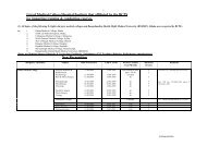
Journal of Bangladesh College of Physicians and Surgeons Vol. 22, No. 3, <strong>September</strong> <strong>2004</strong>Aims of the studyThis study has been done to describe various riskfactors for the development of SM prior to admissionin hospital and, to document the clinical patterns ofSM at different categories of hospitals in Bangladesh.Experimental Design and Methods :All cases of SM admitted were documented in a TertiaryReferral Hospital, Chittagong Medical College Hospital(Tertiary Health Center - THC); two Secondary HealthCare Facilities, Khagrachari and Bandarban DistrictHospitals (Secondary Health Center - SHC); and, 4Primary Health Centers: Ramu, Teknaf, Matiranga andFatikchari Thana Health Complex Hospitals (PrimaryHealth Center - PHC); in high risk endemic area ofBangladesh, the southern and eastern part of the country.The period of study was six months from May toOctober 1998, which included pre-monsoon and postmonsoon.Diagnosis and management of SM were basedon WHO guidelines customized in the 'NationalGuidelines for Clinical Management of Malaria 1 . Anintensive care with minimum facilities required fornursing care, clinical monitoring, laboratory service andfollow-up was established within the existing setups ofCMCH and other study site hospitals. Ethical clearancewas taken from the Ethical Review committee ofCMC&H. Written informed consent was taken from theresponsible person of each patient after elaborating thepurpose of the study, the procedures and theimplications. Attending doctors recorded base line datafrom tile patients on admission to hospital and ensured12 hourly follow-up subsequently. For diagnosis ofmalaria thick and thin blood films were done for everypatient. Depending on availability of blood glucosemonitoring, renal and hepatic function assessment, chestx-ray, ECG and CSF study (in cerebral malaria casesonly) were also done as indicated. The story precedinghospital admission was noted from tile accompanyingperson or patient (when able to communicate reliably) ina pre-designed proforma. A five day-long trainingcourse was arranged for twenty Medical Officersselected from all the participating hospitals to documentthe cases and manage them uniformly. All patients ofSM were treated with parenteral quinine to be followedby oral formulation of the same as depicted by thenational guidelines. Data were double entered andanalysed utilizing EPINFO 6 software to determine therisk factors related to the development of SM. Thedifferent parameters of SM were compared in respect ofdifferent levels of care.ResultsOut of total screened 1308 cases, 909 fulfilled theselection criteria and enrolled for the study of which58 patients died. Of these 339 (37.4%) cases admittedat PHCs, 382 (42.0%) at SHCs and 188 (20.6%) atTHC. The male female ratio was 1.9 and mean agewas 21.4 (± 14.4 SD, 95% CI 20.5 - 22.3) andmaximum was 95, minimum 0.2 years with medianvalue of 20 and mode 25. Of the female patients 39(12.7.5.) cases were pregnant. 47.7% of cases hadsome form of antimalarials prior to admission withinprevious 2 weeks with a mean period of 2.4 (± 2.7)days. The case fatality rate was 6.4% and recoverywithout sequele 82.2%. But 1.4% cases recoveredwith some form of sequele. The outcomes of the othercases (N=91, 10%,) were not known because they lefthospital before attaining the outcome.Table – IDescription: Patient Profile of SM cases N=909Particulars Total 909 (100%), PHCs 339 (37.4%),SHCs 382 (42.0%), THCs 188 (20.6%)Sex: Male N (%) 601(66%)Male female ratio 1.9Pregnancy N.('%) 39(12.7%)Mean Age (SD, 95% CI), Maximum, Minimum, Median and Mode 21.4 (14.4, 20.5-22.3) 95 0.2 20.0 25.0Within 2wks AM usage N (%) 434(47.7%)Period of prior AM usage Days (SD) 2.4 (2.7)Case fatality 58(6.4%)Recovery without sequele 747(82.2%)Recovery with sequele 13(1.4%)AM=Antimalarials, PHC=Primary Health Center, SHC=Secondary Health Center, THC=Tertiary Health Center84
Study to Document Pre Admission Risk Factors for Development of Severe Malaria and the SpectrumEB YunusOn admission-duration of illness, severe symptoms,inability to eat/drink (NPO: Non Per Os) and prostrationwere longest at the THC, and most of them were lowestat SHCs. The travel time was lowest at SHCs followedby THC and longest at the PHCs. The hospital stay waslongest at THC followed by SHCs. These durationssaving NPO were significantly different betweendifferent categories of hospitals.All patterns of presentation of SM were found incases at district hospitals (SHCs). Amongst the typesas predominant presentation severe prostration rankedthe top followed by hypoglycemia, unarousable coma.convulsion, severe anemia, impaired consciousness,hyperpyrexia and hyperparasitemia. Some cases ofhemoglobinuria, jaundice, abnormal behavior,pulmonary edema, acute renal failure, shock,DIC/bleedings and acidosis were also encountered.Many of the cases had more than one type (7S.4%).Severe types of SM predomiently unarousable comawere more in tertiary care hospital (THC).Table-IIDescription: Different parameters with respect to different durations at different categories of hospitalsParticulars Total PHC SHC THC PN=909 N=339 N=382 N=188Duration: Illness Days (SD) 6.2 (5.5) 5.7(3.4) 5.0 (3.9) 9.7 (8.9) < 0.001Duration: Severe symptoms Hours (SD) 41.3 37(48) 37(33) 59(78) < 0.001(51.3)Duration: NPO Hours (SD) N 35(60) 26(21)49 31 (53) 41 (72) > 0.1293 102 142Duration: Impaired 30.7 14 (14) 35 24(27) 42(78) < 0.01Consciousness hours (SD) N (54.8) 317 142 126Duration: Prostration hours (SD) N 37(39) 34(39) 32(28) 70(57)81 < 0.001448 309 58Travel time: To reach hospital 2(3.2) 3.2 (4.4) 1.0 (1.9) 1.9 (2.1) < 0.00 lhours (SD) NDuration: Hospital stay Hours (SD) 107(76) 81 (43) 110(54) 147 (12G) < 0.001PHC=Primary Health Center, SHC=Secondary Health Center, THC=Tertiary Health CenterTable-IIIPredominant Presentations of patients of SM by categories of hospitalsPresentations Total = 909 PHC=339* SHC=382* THC=188* P*Severe prostration N(%) 379 (41.7) 248 (73.2) 101 (26.4) 30(16.0) < 0.001Hypoglycemia N(%) 135 (14.9) 9(2.7) 123 (32.2) 3 (1.6) < 0.001Unarousable coma N (%) 115 (12.7) 7 (2.1) 17(4.5) 91 (48.4) < 0.001Convulsion N(%) 99 (10.9) 23 (6.8) 54 (14.1) 22(11.7) < 0.05Severe anemia N(%) 53 (5.8) 10(2.9) 27(7.1) 16(8.5) < 0.05Impaired consciousness N(%) 44(4.8) 20(5.9) 16(4.2) 8(4.3) NSHype pyrexia N(%) 39(4.3) 15 (4.4) 18(4.7) 6(3.2) < 0.05Hyperarasitemia N(%) 14(1.5) 4(1.2) 8(2.1) 2(1.1) > 0.5Hemoglobinuria N(%) 8(0.9) 0 6(1.6) 2(1.1) NSJaundice N(%) 8 (0.9) 0 2(0.5) 6(3.2) NSAbnormal behavior N(%) 7(0.8) I (0.3) 6(1.6) 0 NSPulmonaryedema N (%) 3 (0.3) 1 (0.3) 2(0.5) 0 NSAcute renal failure N (%) 2(0.2) 0 2(0.5) 0 NSShock N(%) 2(0.2) 0 0 2(1.1) NSDIC/Bleedings N(%) 1 (0.1) 1 (0.3) 0 0 NSMore than one 678 (78.4%) 233 (8.7%) 353 (92.4%) 92(48.9%) < 0.001PHC=Primary Health Center, SHC=Secondary Health Center, THC=Tertiary Health Center85
Journal of Bangladesh College of Physicians and Surgeons Vol. 22, No. 3, <strong>September</strong> <strong>2004</strong>Table-IVOutcome pattern of SM categories of hospitals N=909Outcome Total = 909 PHC =339 SHC=382 THC N=188N (%) N(%) N(%) N(%)Full Recovery 747 (82.2) 286 (84.4) 321 (84.0) 140 (74.5) P > 0.05Recovery with sequele 13(1.4) 9(2.7) 2(0.5) 2(1.1) P < 0.05Death 58(6.4) 5 (1.5) 19(5.0) 34 (18.1) P < 0.01Not known 91 (10.0) 39 (11.5) 40 (10.4) 12(6.4) P < 0.05PHC=Primary Health Center, SHC=Secondary Health Center, THC=Tertiary Health Center747 (82.20%) cases recovered fully. But 13 cases(1.4%) had some sequele after recovery. The outcomeof 91 (10%) cases was not known as they left thehospital without advice and or referred to otherfacilities. Case fatality was highest at THCfollowed by SHCs. But there was also differencesof various outcome patterns among variouscategories of hospitals both significant and nonsignificant as well.DiscussionsWe have described the patient profile and time linesand presentations of patients admitted with SM indefferent categories of hospitals in high risk areas inBangladesh for malaria. There is in operation the'National Malaria Control Program' to control themalaria as per the WHO guidelines, whichemphasizes on early diagnosis on clinical criteria andtreatment with affordable and readily available'chloroquine' as first line agent for uncomplicatedmalaria and parental quinine for severe category. Fora successful campaign the need is to describe relevantinfluencing factors for appropriate addressing.Patients were documented in three differentcategories of hospitals. Most cases were male of theage group of third decade. This signifies the sectionsof population who were economically active and aremost exposed to the vector were affected. Thereforemalaria is not only a health problem rather adevelopmental economic issue 4 . A significantnumber of patients were pregnant. Pregnancy is asusceptible state for SM, and carries more risk forcase fatality. Studies in Thailand revealed that SM is3 times more common amongst pregnant women 5 .The morbidity is also high in pregnant state as well.The 'National MCH Program' should developguidelines for malaria with pregnancy andincorporate them in field operation.The National Guidelines for Malaria emphasizes useof chloroquine as first line antimalarials (AMs) for allfever cases without focal signs for any other febrileillness in endemic zone. But it was found that morethan half of the enrolled cases didn't get any AM priorto development of SM. This issue call for moreawareness of the people and professional in thisregard. The over all case fatality was 6.4%. In ahospital-based series in Papua-New-Guinea it wasfound that the mortality was 18% 6 . On the other handthe same was found in an ICU in Singapore to be12.5% 7 . Compared to these, the mortality in ourseries is low. One of the reasons for this is the nonhomogeneity of study stations plus less severepatterns in most cases. About 1.4% of casesdeveloped some form of sequele. Variousneurological sequele, mostly psychiatric weredescribed in cases of cerebral malaria but theassociation with malaria was doubtful 8 . So SM is notonly regrettable for high mortality but also for lastingmorbidity and or infirmity with related social andeconomic consequences. A study dedicated todescribe the sequele is warranted in this behalf.86
Study to Document Pre Admission Risk Factors for Development of Severe Malaria and the SpectrumEB YunusSM is a medical emergency, which needs immediatemanagement in a hospital setup. The differentdurations, which were documented by this study fromthe onset to hospital admission, will provide someimportant insight. These durations: onset of illness,severe symptoms, impaired consciousness,prostration, travel time, and hospital stays are allfound to be in appropriately prolonged leading todelay in starting specific management. The delay maybe important contributing factor to influence theoutcome. The travel time to hospital was found to becomparatively less if the commuter mechanism of thecountry is considered. Therefore the delays arepossibly due to lack of awareness about the conditionand consequences. Other authors in this region ofAsia also observed this 9 . Even one can speculate thislack of awareness is also prevailing amongst thepracticing and operational health care personnel. AKAP study is needed to describe the situationprevailing among people and professionals.Detail examination of different durations by differentcategories of hospitals provided that these are moreprolong in THC. This is possibly because of the factthat patients developing complications over time andmore serious cases are used to seek admission at THCand or being referred from PHCs and SHCs. Butsurprisingly the travel time was lowest at the SHCsthat should be supposed to be in between PHCs andTHCs. This needs further study. But all the durationswere found to be significantly different betweendifferent categories of hospitals saving duration ofprostration.Most of the patients had more than one types orpresentation of SM, types as described by WHO. Butout of these in descending order the most frequenttypes were: Severe prostration, Hypoglycemia,Unarousable coma, Convulsion, Severe anemia,Impaired consciousness, Hyperpyrexia andHyperparasitemia. But in a small number of casesjaundice, abnormal behavior, pulmonary edema,shock and DIC/bleedings were also present. Therewas a striking difference between the THC and PHCsfor different types with SHCs in betweenencountering most of the types that the other twolacked in this series. But it was found that moresevere types like unarousable coma and impairedconsciousness were more frequent at THC. Thisfeature emphasizes that the center of attention for SMmanagement should be more focused to SHCsthrough making them more efficient in all respect.The outcome data revealed that the case fatality washighest at THC followed by SHCs. This is lowest atthe PHCs. As more severe cases with more serioustypes were conglomerated naturally and or by referralto THC and SHCs obviously the fatality rate washigher. So these centers should be upgraded andrefurnished so that critical cases can be appropriatelyand comprehensively managed. A good number ofcases, with insignificant difference between allcategories of hospitals, left the hospital before theattainment of outcome. So outcome of these caseswere unknown and therefore acting as an artefact forgeneral implication of outcome data. Theinterpretation of those therefore needs somereservation for this reason.Conclusion :It can be concluded that more awareness andorientation training are needed both for thecommunity and the professionals working in theendemic zone to ensure early diagnosis and prompttreatment. Secondary Health Care Hospitals shouldbe more equipped for handling severe malaria casesand Chittagong Medical College Hospital should memade as a center of excellence for the same as it is theonly Tertiary Health Care Provider at the perimeter ofthe malaria endemic zone of Bangladesh. Malariaissue should be incorporated in National MCHprogram. Further studies are needed to explore thesituation critically.Acknowledgement & Conflicts of InterestThis study was supported by Research GroupStrengthening Grant of Tropical Disease ResearchWing of World Health Organization and wascollaborated by Malaria and Parasitic DiseasesControl Unit of Directorate General of HealthServices of Bangladesh. The authorities and staffs ofthe study site hospitals deserve special mentioning forthe continued help. There was no conflict of interest.87
Journal of Bangladesh College of Physicians and Surgeons Vol. 22, No. 3, <strong>September</strong> <strong>2004</strong>References1. M&PDC Unit of DGHS and WHO. (1994). Recommendationsof the workshop on "Malaria Clinical Case Definition - Theiruse for Early Diagnosis and Prompt Treatment (EDPT) andEpidemiological surveillance".2. Rhaman MR, Faiz MA, Das JC, Yunus EB: A prospectivedocumentation of prognostic factors of severe malaria amongadult patients in Chittagong Medical College Hospital,Bangladesh. JCMCTA 1996;7(S3):32-45.3. Faiz MA, Rhaman MR, Hussain MA, Rashid HU: Factorscontributing to outcome in cerebral malaria. Bang Med ResCourt Bull (in press).4. Brundfand GH: Roll Back Malaria answering the call. 1998http://rbm.who.int/docs/rbm brochure.htm5. Luxemburg C, Riccf F, Nostcn F, Raimond D, Bathed S, WhiteNJ:The epidemiology of severe malaria in an area of lowtransmission in Thailand. Trans R See Trop Med Hyg91 : 256-626. Brown N: Severe malaria in children at Port Moresby GeneralHospital, Papua New Guinea. Trop Geogr Med 1995; 47 : 107-10.7. Kool KL, Tan WL, Eng P, Ong YY: Malaria requiring intensivecare. Ann Acad Med Singapore 1995 May 27 : 353-7.8. Grag RK, Karak B, Misra S: Neurological manifestations ofmalaria : an update. Neurol India 1999 Jun 47 : 85-919. Kochar D, Kumawat BL, Karan S, Kochar SK, Karan AgarwalRP: Severe and complicated malaria in Bikaner, WesternIndia. Southeast Asian J Trop Med Public Health 1997; 28(2): 259-267.88
Journal of Bangladesh College of Physicians and SurgeonsVol. 22, No. 3, <strong>September</strong> <strong>2004</strong>Evaluation of Menstrual Patternin Young College GirlsS N BEGUM a , MH KHAN b , MS BASHER cSummaryWith a view to cast a glance on menstrual pattern, a descriptivecross-sectional study was carried out among 158 purposelyselected young college girls of Sylhet MAG Osmani MedicalCollege and Sylhet Nursing Institute through self-administeredstructured questionnaire. Age of the respondents was between 18- 26 years with a mean of 22.4 years. Minimum age at menarchewas 10 years, while maximum age was 16 years with mean 12.8years and median 13 years. It was observed that as many as 125(79.11 %) respondents had regular menstrual cycle, whereas 33(20.89%) had irregular cycle. Menstrual flow was average in 129(81.65%), scanty in 8 (5.06%) and heavy in 21 (13.29%)respondents. At least 116 (73.42%) respondents conceded thatthey had painful menstruation (dysmenorrhoea) with a varyingdegree of severity. Of them, as many as 60 (51.72%) neededmedical intervention either by analgesic and / or antispasmodic.About 57 respondents has family history of dysmenorrhoea.To establish relationship between dysmenorrhoea and its familyhistory, conduction of a large scale study has been suggested.(J Bangladesh Coll Phys Surg <strong>2004</strong>; 22 : 89-92)Introduction :Menstruation is a periodic and cyclical shedding ofprogestational endometrium accompanied by loss ofblood. This peculiar function only present in womenand in higher apes 1 . Menstruation is the visiblemanifestation at the conclusion of one cycle ofhormonal activity, marks the beginning of the next 2 .It needs coordinated interplay of hypothalamopituitary-ovarian axis, functioning ovary, responsiveendometrium and presence of patent utero-vaginalcanal for the onset of menstruation. The first 4 - 5days of menstrual cycle is menstrual phase withshedding of two third to four fifth of endometrium.The remaining days consists basically of aproliferative and secretary phases. Menarche, the firstmenstrual period of life, usually occurs between theages of 10 - 16 years, the average being 13.5 years.The age at menarche varies to some extent withfamily, race, social class, family size, birth order,environment, diet and general health but not withclimate. 3 Menstruation tends to occur earlier in thehigher social classes and in urban surroundingsprobably reflecting general health. It is more closelyrelated to bone age than to chronological age. For thepast couple of decades, the age of menarche isgradually declining with improvements of nutritionand environmental condition 3 .a. Dr. Shamsun Nahar Begum, FCPS, Associate Professor, Gyaneand Obs, Sylhet MAG Osmani Medical College, Sylhet.b. Dr. Mohafizul Hoque Khan, Director, Centre for NeucularMedicine & Ultrasound, Sylhetc. Dr. Md. Shahidul Basher, Lecturer of Community Medicine,Sylhet MAG Osmani Medical College, SylhetAddress for Correspondence : Dr. Shamsun Nahar Begum,FCPS, Associate Professor Gyane and Obs, Sylhet MAG OsmaniMedical College, Sylhet.During active reproductive life menstruation occursat approximately 28 days interval, but 21 - 35 days isaccepted as normal. Duration of menstrual periodbetween 2 - 7 days is accepted as normal, but it mustbe individualized. Nonetheless, there is widevariation in duration and amount of blood loss.Menstrual flow varies from 50 - 80 ml with anaverage of 45 ml. In practice, however, menseslasting more than seven days, or occur at an intervalof 21 days or less, or which are subjectively thoughtto be heavy are considered as excessive 3 .In teenage or in nullipara, menstruation may beassociated with tolerable colicky pain at thebeginning of mense due to uterine contraction. Painof sufficient magnitude incapacitating the day-to-dayactivities is called dysmenorrhoea. Nearly 50 per centwomen experience some discomfort in relation tomenstruation. But in 5-10% individuals severe painincapacitate them for several hours in each month. 1First period of life is usually anovular, followed byirregular ovulation. Moreover, it takes about 2 yearsfor regular ovulation to occur. Anovulatory cycles canresult in excessive bleeding. This is typically found inthe post-pubertal teenager with an immaturehypothalamopituitary- ovarian axis. 1This study was conducted among 158 young collegegirls to throw light on the menstrual pattern andproblem related to menstruation.
Journal of Bangladesh College of Physicians and Surgeons Vol. 22, No. 3, <strong>September</strong> <strong>2004</strong>Materials and Methods :This descriptive cross-sectional study was conductedamong 158 purposively selected young college girls ofNursing Institute, Sylhet, and undergraduate students ofdifferent session of Sylhet MAG Osmani MedicalCollege, Sylhet. The study period was four monthsranging from March 2002 to June 2002. Age atmenarche, menstrual cycle with duration, menstrualflow including associated complaint such asdysmenorrhoea were the study variables. Data werecollected through self-administered structuredquestionnaire.Menstrual cycle was considered as regular one whenit was within 21 - 35 days with a mean of 28 ± 2 days,while it was considered irregular when it was lessthan 21 days or more than 35 days. Menstrual flowwas considered as scanty, average and heavy onebased on number of sanitary towels used per day asmentioned by the respondents (1- 2, 3 -5 and > 5sanitary towel per day as scanty, average and heavymenstrual flow respectively). Painful menstruationwith pain ranging from dull ache to spasmodic ofvarying intensity was categorized as dysmenorrhoea.Data were analyzed manually and with the help ofscientific calculator.Results :A total of 158 college girls were selected purposivelyand interviewed by self-administered structuredquestionnaire. Of them, 128 was from Sylhet MAGOsmani Medical College and 30 from NursingInstitute, Sylhet. Age of the respondents was between18 - 26 years with a mean of 22.4 years. Of them, 9(5.70 %) were married and 149 (94.30%) were single.Shortest age at menarche was 10 years, while highestage was 16 years with a mean of 12.8 years andmedian 13 years. Onset of menarche in 64 (40.51%)respondents was at the age of 13 years. Menstrualcycle was regular in 125 (79.11%) respondents,whereas 33 (20.89%) had irregular cycle (Table-I).Concerning menstrual period, it was found that agood number of respondents (148) had period within2 to 7 days (Table- II).Table- IMenstrual cycle N=158Menstrual cycle Frequency PercentageIrregular 33 20.89Regular 125 79.11Total 158 100Table IIDuration of Menstrual period N=158Menstrual cycle Duration in daysType 1 Day 2-7 Days >8 DaysRegular 1 120 4Irregular 2 28 3Total 3 148 7It was revealed that in 129 (81.65%) respondentsmenstrual flow was average, while in 8 (5.06%) itwas scanty and was heavy in 21 (13.29%)respondents (Table-III). As discussed in materials andmethods; menstrual flow was considered as scanty,average and heavy one based on number of sanitarytowels used per day as mentioned by the respondents(1- 2, 3 -5 and > 5 sanitary towel per day as scanty,average and heavy menstrual flow respectively). Atleast 116 (73.42%) respondents disclosed that theyhad painful menstruation (Table- IV). Regardingseverity, it was found that in 56 (48.27%) respondentspain was mild, whereas in 47 (40.51%) respondents itwas moderate and in 13 (11.20%) pain was severe inintensity (Fig.-1). Those who had dysmenorrhoea, asmany as 60 (51.72%) needed medical interventioneither by analgesic and/or antispasmodic (Fig.-2).Moreover, at least 57 (49.14 %) respondents withdysmenorrhoea had family history of dysmenorrhoea(Table-V). However, the relationship either betweentype of menstrual cycle and dysmenorrhoea (x 2 ,df - -1,1.5; P > 0.05) or between dysmenorrhoea and familyhistory was statistically insignificant. (x 2 , df=1, 2.69;P > 0.05).Table IIIMenstrual bleeding by type of cycle N=158Menstrual cycle Menstrual bleedingType Average Scanty HeavyRegular 106 6 13Irregular 23 2 8Total 129(81.65%) 8(5.06%) 21(13.39%)90
Evaluation of Menstrual Pattern in Young College GirlsSN Begum et al50403020100Table- IVTypes of menstrual cycle with dysmenorrhoea N=158Menstrual cycleDysmenorrhoeaType Present AbsentRegular 89 36Irregular 27 6Total 116(73.42%) 42(26.58%)x 2 , df=l, 1.5; P > 0.05FrequencyFrequency50454035302520151050441235Mild 1 Moderate 2 Severe 3Treatment needed12Treatment no neededRegularIrregular10Intensity ofdysmenorrhoea with types ofmenstrual cycleFig.-1 : Multiple bar diagram showing severity ofdysmenorrhoea with type of menstrual cycle45 44RegularIrregularRequirement of treatment for dysmenorrhoeawith type of cycleFig.-2 : Multiple bar diagram showing requirement oftreatment for dysm enorrhoea with type of cycle15123Table-VDysmenorrhoea with family history N=116Dysmenorrhoea Family history of dysmenorrhoeawith type of cycle Present AbsentRegular 40 49Irregular 17 10Total 57(49.14%) 59(50.86%)x 2 , df=l, 2.69; P > 0.05Discussion :In this study the age at menarche was 10 - 16 yearswith a mean of 12.8 years. This is in line withstatement made by Datta. 3 In a significant number ofrespondents, age of menarche was 13 years which isindicated by findings of study conducted by Chowdhuryet. al. 4 and corroborated by Datta, 3 and Jeffcoate 1 aswell. Median age at menarche was 13 years which isvery much related to findings of study conducted byGrover et. al., Singh et. al. and Hedge et. al. 5In respect of regularity of menstrual cycle, it wasrevealed that it was regular in 125 (79.11%)respondents, whereas 33 (20.89%) had irregular cycle(Table-III). As many as three respondents hadmenstrual cycle over ninety days. Regardingmenstrual flow, it was found that it was average in129 (81.65%) respondents, while it was scanty in 8(85.06%) and heavy in 21 (13.29%) respondents. Thisis more or less similar to the findings of studyconducted by Chowdhury et al. 5A substantial number of respondents (116, 73.41%)disclosed that they had dysmenorrhoea with variousdegree of severity. Furthermore, majority of them (60,37.97%) required medical intervention either withanalgesic and/ or antispasmodic. This can becompared with the study findings of Chowdhury etal. 5 where only 21.3% needed medical intervention.Moreover, at least 57 (49.14%) respondents withdysmenorrhoea had positive family history.Conclusion :This descriptive, cross-sectional study with samplesize 158 was conducted to shed light on menstrualpattern and its related problem. To explore factsregarding menstruation however needs large groupstudy. As incidence of dysmenorrhoea is affected bysocial status, age, occupation and family history 1 . It91
Exomphalos Major: Conservative TreatmentMU Alam et alJournal of Bangladesh College of Physicians and Surgeons Vol. 22, No. 3, <strong>September</strong> <strong>2004</strong>has been suggested to test relationship between typeof menstruation and dysmenorrhoea, anddysmenorrhoea with family history through largegroup study.AknowledgementThe authors like to express their thanks to Dr. NyemaAkhter Khan M O CNMU Sylhet and Ms IshratTahsin for their unflagging help in this study.References :1. Neerja Bhatta (Revised and updated). Menstruation and othercyclical phenomena, In Jeffcoates Principles of Gynaecology,6th international ed. Arnold 2001: 81-86 : 88-99.2. I.D. Cooke Menstrual cycle and ovulation, In DewhurtsTextbook of Obstetrics and Gynaecology for postgraduates,6th edition, Blackwell Science Ltd 1999 : 28.3. D.C. Datta. Menstruation, In Text Book of Gynecology, 3rdedition, New Central Book Agency, Calcutta 2001 : 74-88.4. Chowdhury T.A., Akhter S. Survey of dysmenorrhoea in agroup of college girls at Dhaka city. Journal of BCPS.1985;3(l) : 12 - 16.5. Usha R, Krishna and Vinita Salvi. Adolescent and pediatricgynecological problems, In Ratnam SS, Bhasker Rao K,Arulkumaran S, eds. Obstetrics and Gynaecology forpostgraduates, Vol-2, 1st edition, Orient longman ltd. 1994 :293-301.92
Study of Protein Level of Cerebrospinal Fluid (CSF) in Motor Neuron DiseaseM A HayeeJournal of Bangladesh College of Physicians and SurgeonsVol. 22, No. 3, <strong>September</strong> <strong>2004</strong>Study of Protein Level of Cerebrospinal Fluid (CSF) inMotor Neuron DiseaseM A HAYEE a , A HAQUE b , QD MOHAMMAD cSummary:A prospective study of cerebrospinal fluid (CSF) protein level ofmotor neuron disease (MND) patients was carried out inMedicine Department of Sir Salimullah Medical College, Dhakaand Sher-e-Bangla Medical College, Barisal. The duration ofstudy was from July 1, 2000 to December 31, 2002. One hundredforty MND patients between 25 to 82 years were studied. Amongthem 89 were male and 51 were female. The mean age (with SD)of the patients was 53.12(±13.94) years (male 54.90(±12.32) andfemale 53.40(±14.52)). Clinical typing showed 67 (47.85%) hadamyotrophic lateral sclerosis (ALS), 58 (41.42%) hadIntroduction:Motor neuron disease is one of the importantdebilitating diseases of mankind. It is aneurodegenerative disease. Motor neuron disease canbriefly be described as a complex condition resultingin muscular weakness and wasting without sensorychanges 1 . Motor neurons, grouped as upper and lowermotor neurons, are nerve cells that transmit signalsfor movement from the brain and spinal cord tomuscle fibres. Motor neuron disease is characterizedby progressive deterioration of and loss of theseneurons. The loss of nerve stimulus to specific muscleresults in atrophy and progressive weakness leadingto paralysis. Progressive muscular wasting diseasewas first described in 1850 by Aran and in 1860 byDuchenne. In 1869, the French neurologist Jean-Martin Charcot described a unique conditioncharacterized by deterioration of both upper andlower motor neurons and this condition was termedamyotrophic lateral sclerosis 2 . If only lower motorneuron involvement is evident, the condition istermed progressive muscular atrophy 3 . Clinicala. Dr. Md Abdul Hayee, FCPS, MD, PhD, Associate Professor andHead of the Department of Neuromedicine, SSMC, Dhaka.b. Dr. Md. Anisul Haque, FCPS, MD, PhD, Professor andChairman of Neurology, BSMMU, Dhaka.c. Dr. Quazi Deen Mohammad, FCPS, MD, Professor and Headof the Department of Neuromedicine, DMC, DhakaAddress of Correspondence : Dr. Md A Hayee FCPS MD, PhD,Associate Professor and Head of the Department ofNeuromedicine, SSMC, Dhaka.amyotrophic lateral sclerosis with probable upper motor neuronsigns (ALS-PUMNS), 11 (7.85%) had progressive muscularatrophy (PMA) and four (2.88%) had progressive bulbar palsy(PBP). Maximum patients were between 36 and 60 years andonly 14% patients presented before 35 years of age.Cerebrospinal fluid was collected from each subject and proteinwas estimated. Ninety- eight patients (70%) had cerebrospinalprotein level less than 50 mg/dl, 35 (25%) had protein between 50to 74 mg/dl and seven (5%) had more than 75 mg/dl.(J Bangladesh Coll Phys Surg <strong>2004</strong>; 22 : 93-96)manifestations of amyotrophic lateral sclerosis arelargely dependent on the degree to which the upper orlower motor neurons are affected 4 .Amyotrophic lateral sclerosis is nearly alwaysprogressive and eventually leads to death. Commoncause of death is respiratory failure or cardiacarrythmias. There are some rare reports of patients inwhom the disease is stable 5 . There are three types ofALS – (i) Sporadic, (ii) Familial and (iii) WesternPacific variant. Individuals who have no familymember with this condition are said to have sporadicor classic ALS. It constitutes 90% of all ALS cases.The age of onset of sporadic ALS commonly between55 and 75 years 6,7 and more in females than males 8(M:F = 1:1.6), but recent studies have suggested thatthe sex difference is decreasing 9 . Familial ALS canbe defined as two or more cases occurring in the samefamily. About 5% to 10% of ALS cases are familial 10and related to mutation in Cu/Zn superoxidedismutase gene 11 . There is little information abouttotal CSF protein content in several important generalreviews of CSF of MND 12-15 .Many fields of MND had been explored by manyresearchers. Till now we do not know it’s etiology.Several simple facts are not yet known. How often isthe total CSF protein content abnormally increased?This simple question is not satisfactorily answered tillthis moment. No work had been done in this field inour country. This has stimulated to do this studyamong the Bangladeshi MND patients.
Journal of Bangladesh College of Physicians and Surgeons Vol. 22, No. 3, <strong>September</strong> <strong>2004</strong>The terminologies used in this study needsclarification1. Nomenclature:i. Motor neuron diseases (pleural): any disease inwhich the primary pathology is believed to affectthe perikaryon of motor neurons and themanifestations are usually paretic but someconditions are ascribed to neural over activity(cramps, twitching of muscles, spasm orpersistent abnormal posture of limbs or trunk).ii. Pure motor neuropathy: the term is used when it isbelieved that the primary disorder affects axons ofperipheral nerve which is defined by non-clinicalcriteria, such as electrophysiology, histopathologyor other laboratory tests.iii. Neuronopathy: the term is used to describe thedisease of the neurones when, after all tests, it isstill not possible to assign the primary pathologyeither to perikaryon, peripheral nerve or both 16 .2. Obligatory criteria for motor neuron disease:i. Symptoms must include focal weakness of uppermotor neuron type i.e. spastic gait or limbclumsiness of corticospinal tract disorder.ii. There must be signs of lower motor neuron lesioni.e. evidence of focal weakness and wasting ofmuscles. Focal wasting is needed to differentiateneurogenic atrophy of muscle from generalizedwasting or gaunt appearance after weight loss ordietary restriction. Other lower motor neuronlesion signs, not mandatory but helpful, includingloss of tendon reflexes and fasciculation.“Diminished tendon reflexes” do not countbecause “diminished” is difficult to define. “Lost”means that the patellar reflexes are absent withreinforcement and ankle jerks are absent in thekneeling position.iii. Unequivocal upper motor neuron signs includepositive Hoffmann signs with flexor plantarresponse. Though hypertonicity is a feature ofupper motor neuron lesion, yet it is difficult todefine objectively on clinical ground and is notunder consideration.3. Definitions of some clinical syndromes:i. Progressive muscular atrophy (PMA): lowermotor neuron signs only.ii. Amyotrophic lateral sclerosis (ALS): lower motorneuron signs and unequivocal upper motor neuronsigns.iii. Amyotrophic lateral sclerosis with probable uppermotor neuron signs (ALS-PUMNS): over activetendon reflexes in limbs with weak, wasted andtwitching muscles but no clonus or plantarextensor.iv. Progressive bulbar palsy (PBP): a syndrome inwhich dysarthria and dysphagia are predominantsymptoms and in which there is weakness,wasting and faciculation of tongue. There may ormay not be concomitant upper motor neuronsigns.v. Primary lateral sclerosis (PLS): a syndromecharacterized by upper motor neuron signs and nolower motor neuron signs. It is uncertain whetherPLS is a form of MND 16 . Rarely this syndromemay evolve to full ALS 17 .Materials and methods:Selection of patients: The daily admission register ofMedicine Department of Sir Salimullah MedicalCollege and Mitford Hospital, Dhaka and Sher-e-Bangla Medical College Hospital, Barisal werescreened continuously for patients who were thoughtto have disease related to ALS or PMA. The studyperiod was from July 1, 2000 to December 31, 2002.In the present study all patients had lower motorneuron signs. Patients with PLS were excluded.Patients were divided into four groups: ALS, ALS-PUMNS, PMA and PBP.Exclusion criteria: A total of 260 patients weresuspected to have MND according to admissionregister. They were examined thoroughly to judgewhether inclusion criteria were met. Of the 260patients, so selected, 70 were rejected because theydid not meet the clinical diagnostic criteria. Theseincluded 40 patients with spastic paraparesis orparaplegia without evidence of lower motor neuronsigns and eight patients who met established criteriafor the diagnosis of PLS. Eleven patients haddisorders unrelated to MND. Charcot-Marie-Toothdisease, lumbosacral plexopathy, post-polio muscleatrophy, progressive supranuclear palsy, benignfasciculation, pseudobulbar palsy related to multiple94
Study of Protein Level of Cerebrospinal Fluid (CSF) in Motor Neuron DiseaseM A Hayee et alstrokes were also excluded. In six patients thediagnosis of MND was indeterminate. Five patientswith MND who had coexistent sensory motorneuropathy were excluded.Inclusion criteria: Out of 190 patients who met thecriteria, 50 were excluded because they did not havefull laboratory studies. Cerebrospinal fluid proteinestimation was done in all the remaining 140 patientsfor the study purpose. Following were the inclusioncriteria:Age between 21 and 80 years of any sex; patientswith upper motor neuron lesion signs (hypertonia,hyperreflexia and extensor plantar response); patientswith lower motor neuron lesion signs (flaccidparalysis, muscle wasting and fasciculation) andpatients with both upper motor neuron lesion signsand lower motor neuron lesion signs.Results:Clinical syndromes: Out of 140 patients who metinclusion criteria, there were 89 men with mean age(±SD) 54.9 (±12.32) years with age ranging from 25to 82 years. There were 51 women with mean age(±SD) 53.4 (±14.52) in years with age ranging from15 to 85 years. Sixty seven patients had ALS, 58 hadALM-PUMNS, 11 had PMA and four had PBP.Eleven (12.36%) of the 89 male patients and eight(15.68%) of the 51 female had symptoms before ofage 39 years. Fasciculation was the major symptomwhich was clinically evident in 126 (90%) of the 140patients. The remaining 14 patients without clinicalfasciculation were of PMA six, ALS four and ALS-PUMNS four.Cerebrospinal fluid protein content: Out of 140patients, CSF protein value was greater than 50 mg/dlin 35 (25%). In seven (5%), the CSF protein valuewas greater than 75mg/dl. Rest 98 (70%) had CSFprotein less than 50 mg/dl (ranging from 15 to50mg/dl).Table – IDistribution of patients according to sex (n=140).Number % Mean age (years)Male 89 63.57 54.90Female 51 36.43 53.40Male : Female 1.74:1Table – IIDistribution of MND patients according to clinicaltypes (n=140).Clinical types Number %ALS 67 47.85ALS-PUMNS 58 41.42PMA 11 7.85PBP 04 2.88Table – IIIPresentation of the patients before 35 years.Symptoms before 35 Number %Male 11 12.36Female 08 15.68Table – IVDistribution of the patients according to CSFprotein value (n=140)CSF protein value Number %75mg/dl 07 05Discussion:This study was carried out to know the CSF protein levelof Bangladeshi MND patients. The study subjects weretaken from the medicine department of Sir SalimullahMedical College and Mitford Hospital, Dhaka and Shere-BanglaMedical College Hospital, Barisal. During thestudy period, 140 patients who fulfilled the diagnosticcriteria of MND were evaluated.This study revealed that majority of the patients werebetween 36 to 60 years. This finding correlates withthe finding of a study in Bangladesh 18 but does notcorrelates with the European studies 6,7 . However, thefinding is also similar to an Indian study by K Soodin 1990 19 .The male female ratio of the study subjects was1.74:1 which is similar to different studies 9,10,20 .This study revealed that the mean age (with SD) ofthe male was 54.9 (±12.32) years and that of femalewas 53.4 (±14.52) years. This finding correlates withthat of an Indian 19 and a Mexican study 21 . Motor95
Journal of Bangladesh College of Physicians and Surgeons Vol. 22, No. 3, <strong>September</strong> <strong>2004</strong>neuron disease patients were divided into ALS, ALS-PUMNS, PMA, and PBP. It was found that patientshaving ALS was 47.85%, ALS-PUMNS was 41.42%,PMA was 7.85% and PBP was 2.88%. This is similarto the finding of an American study 22 . It alsocorrelates with that of Indian studies 19,23 but differsfrom other studies 24,25 . This dissimilarity is probablydue to geographical variation, which could also besupported by similarity with the Indian study 19 .The study subjects of this series presented before 35years in 14% and the rest 86% presented after thatage. This picture corroborates with that ofYounger’s 22 and Ashraf’s studies 18 .Seventy percent patients of this study had CSFprotein value less than 50 mg/dl, 25% had between 50to 74 mg/dl and the rest 5% had above 75mg/dl. Thisfinding is similar to that of study in Columbia wherethey showed that CSF protein content was greaterthan 50mg/dl in 25% cases and 5% patients had CSFprotein more than 75gm/dl 22 . Finding of current studyis also similar to the finding of a very old study byGuiloff in 1953 26 . In another study of living MNDpatients, CSF value over 75gm/dl occurred in 5% ofthe patients 27 which is similar to this finding but doesnot corroborate with other studies 28 .In conclusion it may be said that CSF protein ofMND patients usually do not rise but 30% of patientsmay show raised CSF protein. Sometimes the valuemay be more than 100mg/dl. This finding is similar tothe other studies but the age of the patients areyounger in Bangladesh which is similar to that inmany Indian studies.References:1. Brown RH Jr. Motor neuron disease and the progressive ataxia.In: Isselbacher KJ, Martin JB, Braunwald E, Wilson JD, FausiAS, Kasper DL, (editors). Harrison’s Principles of InternalMedicine. New York: McGraw-Hill, 1994 pp 2280-86.2. Bobowick AR, Brody JA. Epidemiology of motor neurondisease. New Eng J Med 1973; 288 : 1047-55.3. Rowland LP, ShneiderNA. Amyotropic lateral sclerosis. NewEng J Med 2001; 344 : 1788-700.4. Swash M. ALS 2000; the past points to the future.Amyotrophic Lateral Sclerosis and Other Motor NeuronDisorders 2001; 2 (Suppl) 1:S 3-9.5. Mulder DW, Kurland LT. Motor neuron disease: epidemiologicstudies. Adv Exp Med & Biol 1987; 209 : 325-32.6. Eisen A. Amyotropic lateral sclerosis is a multifactorialdisease. Muscle and Nerve 1995; 18 : 741-52.7. Chio A. Risk factors in the early diagnosis of ALS: Europianepidemiological studies. Amyotrophic Lateral Sclerosis andOther Motor Neuron Disorders 2000; 1 (Suppl) 1: S 13-8.8. Kondo K. Motor neuron disease: changing populationpatterns and clues for etiology. In: Schoenberg B, (editor).Advances in Neurology. New York: Raven Press, 1978 pp509-43.9. Maasilt P, Jokelainen M, Loytonen M, Sabel CE, Gatrell AC.Mortality from amyotropic lateral sclerosis in Finland, 1986-1995. Acta Neurol Scand 2001; 104: 232-35.10. Rowland LP. Ten central themes in decade of ALS research.Adv Neurol 1991; 56 : 3-23.11. Cudkowicz ME, Mc Kenna Yasek D, Sapp PE, Loytonen M,Sabel CE. Epidemiology of mutations in superoxidedismutase in amyotropic lateral sclerosis. Ann of Neurol1997; 41 : 210 –21.12. Friedman A, Freedman D. Amyotropic lateral sclerosis. JNerve Ment Dis 1950; 3 : 1-18.13. Castaigne P, Lhermitte R, Schuller E, Rouques C. Less Proteindu liquide cephalo-rachidien au cours de la sclerose lateraleamyotrophique. Rev Neurol (Paris) 1971; 125 : 393-400.14. Brownell B, Oppenheimer D R, Trevor Hughes J. The centralnervous system in motor neuron disease. J Neurol NeurosurgPsychiatry 1970; 33 : 338-57.14. Lawyer T, Netsky MG. Amyotrophic lateral sclerosis – aclinico-anatomic study of fifty-three cases. Arc NeurolPsychiat 1953; 69 : 171-92.16. Rowland LP. Peripheral Neuropathy, motor neuron disease orneuropathy? In: Battistin L, Hashim G, Lajtha A (editors).Clinical and Biological Aspects of Peripheral Nerve Disease.New York: Alan R Liss, 1983 pp 27-41.17. Younger DS, Chou S, Oppenheimer DR, Hays AP, SchullerE, Castaigne P. Primary lateral sclerosis. Arch Neurol 1988;45: 1304-07.18. Ashraf Ali. Study of trace elements in MND patients. Thesisfor MD in Neurology, IPGMR, Dhaka.19. K Sood, D Nag, SV Chandra. Role of aluminium in sporadicmotor neuron disease. Indian J Med Res (B) 1990; 36 : 9-12.20. Olivares L, Esteban ES, Alter M. Mexican resistance toamyotrophic lateral sclerosis. Arch Neurol 1972; 27: 397-02.21. E Otero Siliceo, Mencoa NA, Vazquel TC. Frequencies ofmotor neuron disease in a Mexico city referral center. LaRevista de Investigacion Clinica 1997; 49(6) : 445-48.22. DS Younger, LP Lowland, N Latov, Schuller E, W Sherman,M Pesce, D Nag. Motor neuron disease and amyotrophiclateral sclerosis: relation of high CSF protein content toparaproteinemia and clinical syndrome. Neurology 1990; 40: 595-99.23. Caroscio JT, Mulvihil MN, Sterling R, Abra B. Amyotrophiclateral sclerosis- it’s natural history. Neurol Clin 1987; 5 : 11-17.24. Mitsumoto H, Hanson MR, Chand DA. Amyotrophic lateralsclerosis-recent advances in pathogenic and therapeutic trials.Arch Neurol 1988; 45 : 489-99.25. Swash M, Schwatz MS. What do we really know aboutamyotrophic lateral sclerosis. J Neurol Sci 1992; 113 : 4-16.26. Guilloff RJ, McGregor B, Blackwood W, Parre E. Motorneuron disease with elevated cerebrospinal fluid Protein. JNeurol Neurosurg Psychiatry 1980; 43 : 390-96.27. Shy ME, Rowland LP, Smith T, Abra B, McGregor B,Loytonen M. Motor neuron disease and plasma celldyscrasia. Neurology 1986; 36: 1429-36.28. Swank RL, Putnam TJ. Amyotrophic lateral sclerosis. ArcNeurol Psychiat 1943; 49 : 151-77.96
Benign Lesions Causing Facial DisfigurationM Abdullah et alJournal of Bangladesh College of Physicians and SurgeonsVol. 22, No. 3, <strong>September</strong> <strong>2004</strong>Benign Lesions Causing Facial DisfigurationM ABDULLAH a , M A CHOWDHURY b , Y HAIDER cSummary:This retrospective study was aimed to find out diseases althoughbenign but causing facial disfiguration. Lesions affecting themid-face region were more (44.72%) in the study. Among 123patients, the commonest disease was mandibular osteomylitiswith discharging sinus due to dental infection. The diseaseprofile included congenital anomalies, inflammatory as well asneoplastic lesions.The treatment was surgical excision & repair with or withoutreconstruction. The outcome of the treatment was normal toIntroduction :During the course of evolution from the pre-human tomodern humans, the face became smaller in relation tothe overall size of the head 1 . While brain and braincasetripled in volume, the jaws became shorter and the teethsimpler in from and smaller in size. In consequence, theface receded beneath the forehead. Thus the modernhuman face exhibits an essentially vertical profile, inmarked contrast to the protruding facial muzzle of thegorilla, the chimpanzee, and to a lesser extent, extincthominids. The recession of the tooth-bearing portion ofthe jaws beneath the forehead left two distinctivelyhuman features: a prominent, projecting nose and aclearly defined chin 1 .The face grows more slowly than the nasal passagesand the tooth eruption. Viewed in profile, the face atbirth is less than one-fifth of the braincase; byadulthood it has increased to nearly half. Facialdimensions increase most in depth, next in height(length), and least in width; and in general to a greaterextent in males than in females' .In aesthetic point of view, human being do not wantany scar in their face. Everyone is concerned with hisa. Prof. Md. Abdullah FCPS, FICS, Principal & Professor ofENT, SSMC & Mitford Hospital, Dhakab. Dr. Md. Alamgir Chowdhury DLO, MS, Assistant Professor,ENT, SSMC & Mitford Hospital Dhakac. Dr. Yusuf Haider DLO, Assistant Registrar. ENT, SSMC &Mitford Hospital DhakaAddress of Correspondence : Prof. Md. Abdullah FCPS, FICS,Principal & Professor of ENT, Sir Salimulla Medical College &Mitford Hospital Dhakaaccepted facial configuration in 92 (75%) patients. Cases withlate presentation, extensive disease and fibrosis with repeatedinfection had residual deformity. The causes of late presentationare ignorance, poverty, lack of education, fear of surgery andoverall poor facilities. In rural Bangladesh health education,poverty alleviation, adequate orientation, proper referral systemand skilled manpower development will improve the situation.(J Bangladesh Coll Phys Surg <strong>2004</strong>; 22 :97-99 )or her impressive facial outlook. In this retrospectivestudy, we found some diseases although benigncausing facial disfiguration.Materials & Method:This retrospective study was done in theotolaryngology department of two-referral hospitals,Sir Salimullah Medical College & Mitford Hospital,and Institute of Post Graduate Medicine & Research,and a Private Hospital, ENT Hospital fromJanuary,1997 to December 2002. Patients admitted inthe otolaryngology department with benign lesionshaving facial disfiguration were included in the study.Cases with malignant neoplasm were not included inthe series.Results:Patients of second and third decade suffered than theother age groups.Table IAge incidence n=123Age in years No of Patients Percentage0-10 18 14.6311-20 42 34.1521-30 21 17.0731-40 11 08.9441-50 17 13.8251 & above 14 11.38
Journal of Bangladesh College of Physicians and Surgeons Vol. 22, No. 3, <strong>September</strong> <strong>2004</strong>Table IISex distribution n=123Sex No. PercentageMale 82 66.70Female 41 33.30Male and female ratio is 2:1Table IIILesions affecting different facial regions were as follows:Affected area No PercentageLower face region 38 30.99Mid-face region 55 44.72Lateral face region 30 24.29Table IVLesions affecting lower face region (Mandibular andassociated area) n=38Lesions No PercentageMandibular osteomylitis 21 55.26with sinusHaemangioma of the lower lip 04 10.53Adamantinoma of the mandible 12 31.58Fibromyxoma of the mandible 01 02.63Table VDiseases of the mid-face region (nose and paranasalsinuses) n=55Lesions No PercentageTumourInverted papilloma 15 27.27Nasopharyngeal angiofibroma 07 12.72Ossifying fibroma 04 07.27Haemangioma 02 03.64Osteoma of the fronto- 03 05.45ethmoidal complexMyxoma of the maxilla 01 01.82Dental cyst 08 14.54Granulmatous lesionsRhinosporidiosis 02 03.64Giant cell reparative granuloma 01 01.82MiscellaneousFibrous dysplasia of maxilla 07 12.72Frontal sinus mucocele 03 05.45Frontal sinus osteomylitis 01 01.82Nasal vestibulitis with stenosis 01 01.82Table VILesions in the lateral face region (parotid andauricular area) n=30Lesions No PercentageParotid swelling 14 46.67Pre-auricular sinus with scar 09 30.00Keloid of the pinna 03 10.00Perichonidritis of the pinna 03 10.00Accessory auricle 01 03.33Treatment of the cases was total surgical excision &repair with or without reconstruction.Table VIIResults of surgeries (n=123)Comments No. PercentageNormal facial configuration 55 44.72Accepted facial configuration 37 '30.18Residual deformity 30 24.29Mortality 01 0.81Discussion:This is a retrospective study aimed to observe howbenign lesions causing facial deformity, which arepreventable with early diagnosis and propertreatment. In our study it is revealed that second andthird decades are more vulnerable of these diseases.Male outnumbered female and male female ratio was2:1 in the study.Lesions affecting the mid-face region (nose andparanasal sinus) were more in number 55 (44.72%),followed by 38 (30.99%) in lower face region(mandibular and associated area) and 30 (24.29%) inlateral face region (parotid and pre-auricular area)respectively.Acquired diseases were common than congenitallesions in the series, which correlates with otherstudies 2,3,4 . Among disease profile mandibularosteomylitis with discharging sinus in the neck due todental infection was highest in this observation,which has significant correlation with another report 3 .Treatment given was surgical in all the cases.Reconstruction was done according to therequirement 5 . The outcome of operations was normal98
Benign Lesions Causing Facial Disfigurationto satisfactory facial configuration in 92 (75%) cases.Residual deformity was noted in the rest of the cases.Patients with late presentation, extensive disease,previous inadequate removal, fibrosis due to repeatedinfection had residual deformity. Causes of latepresentation are ignorance, poverty, lack ofeducation, fear of surgery and overall poor treatmentfacilities outside the cities.One patient died due to anaesthetic complicationduring surgery. He was a case of very extensivejuvenile nasopharyngeal angiofibroma.It is concluded from this study that early diagnosisand proper treatment of benign lesions can preventfacial deformity. Health education, adequateM Abdullah et alorientation, proper referral system and developmentof skilled manpower will improve the situation.References:1. Encyclopedia Britannica Library 2003, face pp…..2. Jafek BW, Nahum AM, Butler RM and Ward PH. Surgicaltreatment of juvenile nasopharyngeal angiofibroma.Laryngoscope 1973; 83 : 707-720.3. Carl W and Sullivan MA. Dental abnormalities & bone lesionsassociated with familial adenomatous polyposis: report ofcases. Journal of American Dental Association 1989; 119 :137-139.4. Hjorting-Hansan E. Benign tumours of jaws. Current opinionof dentistry 1991, 1 : 296-304.5. Bingham BJG and Hawthrone MR. Synopsis of operative ENTSurgery, Butterworth-Heinemann, First ed. Oxford, 1992; 313-324.99
Journal of Bangladesh College of Physicians and SurgeonsVol. 22, No. 3, <strong>September</strong> <strong>2004</strong>HbA1c in Diagnosis of Diabetes Mellitus withoutKetonuria in Young AdultK NAZIMUDDIN a , T AHMED b , MS HAQUE c , AKM MUSA d , RSC SARKER eSummary:Glycated protein HbA1c was tested as an alternate tool tomeasurement of blood glucose to diagnose diabetes mellitus indifferent studies. It was mostly discarded because of lowsensitivity. A significant proportion of young adult diabeticpatients in this country presents with very high blood glucosewithout ketonuria.They are lean and have persistenthyperglycemia for months. HbA1c was studied for the diagnosisof 242 consecutive cases without ketonuria at the under 30 clinicof Bangladesh Institute for Research and Rehabilitation forDiabetes, Endocrine and Metabolic disorders (BIRDEM).Thesubjects had high HbA1c (8.7 to 9.5%) and fasting blood glucose(13.05 to 14.75 mmol/L) respectively at 95% confidenceinterval.HbA1c was found to have a positive correlation withfasting blood glucose(r=0.686,p.000).HbA1c > 6.5% showed85% sensitivity for diagnosing this subset of diabetesmellitus.Therefore,HbA1c can be used as a diagnostic tool inyoung adult diabetics.(J Bangladesh Coll Phys Surg <strong>2004</strong>; 22 : 100-103)Materials and Method:Consecutive 242 young (age < 30 years) diabetesmellitus cases without ketonuria were recruited at theunder 30 clinic of Bangladesh Institute for ResearchRehabilitation of Diabetes Endocrine and Metabolicdisorders(BIRDEM).HbA1c was measured by HPLC(high performance liquid chromatography) along withthe blood glucose values during diagnostic oral glucosetolerance test(OGTT) i.e. fasting blood glucose(FBG)and 2 hours after oral glucose load(2HG).Othervariables studied were age,BMI,sex,family history ofdiabetes mellitus(DF), pancreatic calcification(PC),signs of nutritional deficiencies(ND), presentation ofdiabetes mellitus(TS), blood cholesterol(Chol) andtriglycerides(TG). Statistical analysis was done to seecorrelation between blood glucose and HbA1c and alsoto see diagnostic sensitivity of HbA1c for diabetesmellitus.a. Dr. Khwaja Nazim Uddin, MBBS, FCPS, AssociateProfessor, Medicine, BIRDEM Hospital, Dhaka.b. Dr. Tofail Ahmed MBBS, DEM, Associate Professor,Endocrinology, BIRDEM Hospital, Dhaka.c. Dr. Md. Serajul Haque MBBS, FCPS, FACP FRCP,Professor, Neuromedicine, BIRDEM Hospital, Dhaka.d. AKM Musa, MBBS, MCPS, DTCD, FCPS, AssistantProfessor, Medicine, BIRDEM Hospital, Dhaka.e. Dr. Rene Suzan Claude Sarker MBBS, Assistant Registrar,Medicine, BIRDEM Hospital, DhakaAddress for Correspondence : Dr. Khwaja Nazim Uddin, RoomNo. 1323 BIRDEM Hospital, 122 Kazi Najrul Islam Avenue,Shahbagh, Dhaka 1000.Result:a).Distribution of subjects according to their age,BMI, cholesterol and TG levels(n=242).Age(years):Mean 24.26 :SD 4.32 and 95% CI:23.71to 24.80BMI: Mean 20.77 : SD 00 and 95% CI : 20.14 to21.41Cholesterol(mg/dl):Mean 189.72(5.69 mmol/l):SD55.92 (1.67)and 95% CI :182.64 to 196.79(5.47-5.90)Triglycerides(mg/dl):Mean 202.89(2.02 mmol/l):SD147.73 and 95% CI:184.19-221.60 (1.84-2.21).Table-1Distribution of subjects according to theirage,BMI,cholesterol and TG levels(n=242)Parameter Mean with SD 95% confidence intervalAge (years) 24.25; SD 4.32 23.71 to 24.80BMI 20.77; SD 00 20.14 to 21.41Cholesterol( mg/dl) 189.72; SD 55.92 182.64-196.79Triglyceride(mg/dl) 202.89; SD 147.73 184.19-221.60b). Distribution of patients according to their HbA1c,FBG,2HG levels(n=242)FBG (mmol/L):Mean 13.90:SD 6.71(Range 4-45.0):95% CI:13.05-14.752HG (mmol/L):Mean 21.82: SD 7.41 (Range 11.1-55.0) and 95% CI :20.88-22.76HbA1c(%) : Mean 9.16 :SD 3.21(Range 3.6-21.6)and 95% CI:8.76-9.57
HbA1c in Diagnosis of Diabetes Mellitus without Ketonuria in Young AdultK Nazimuddin et alTable-IIDistribution of patients according to their HbA1c,FBG,2HG levels (n=242 )Parameters Range with SD Mean 95% CIFBG mmol/L 4- 45.0 ;SD 6.71 13.90 13.05 -14.752HG mmol/L 11.1- 55.0;SD 7.41 21.82 20.88-22.76HbA1c (%) 3.6-21.6;SD 3.21 9.16 8.76-9.57c) Distribution of patients according to Sex, DF, PC,ND, and TS (n=242)Male:113(46.69%),Female 129 (53.30 %),DF 117(48.35%),PC 22 (9.09%),ND150(61.98%) and TS169 (69.83%) cases.Table-IIIDistribution of patients according to their HbA1c,FBG,2HG levels(n=242)Particulars Number Frequency(%)Male 113 46.69Female 129 53.30DF 117 48.35PC 22 9.09ND 150 61.98TS 169 69.83N (Total 242)Distribution of 2HGDiabetes before 30th birth day and ketonuria-ve45403530252015105012 20 28 36 44 5216 24 32 40 482HG (mmol/L)2HG distribution: Mean 21.82, median 20.75,SD 7.40, SE 0.48,range 11.10 to 55.00 and 95% CL 20.88 to 22.76 mmol/LFig-1. Distribution of HbA1C among the subjectsFig-3 : Distribution of blood glucose two hours afterglucose load(2HG) among the subjectsFig-2 : Distribution of fasting blood glucose (FBG)among the subjectsFig-4 : Correlation of HbA1c level with fasting bloodglucose(FBG) and blood glucose levels two hoursafter glucose load(2HG)101
Journal of Bangladesh College of Physicians and Surgeons Vol. 22, No. 3, <strong>September</strong> <strong>2004</strong>Table-IVStudy of diagnostic sensitivityEvaluation of sensitivity of FBG(cut off 7.0 mmol/l),2HG cut off 11.1 mmol/L) and HbA1C(cut off 6.5%) asdiagnostic tool of diabetes mellitus.(n-242)Test Diagnosis (+ve) Diagnosis (- ve) Total Sensitivity (%)FBG > 7.0 mmol/L 222 20 242 922HG > 11.1 mmol/L 242 0 242 100HbA1C > 6.5% 206 36 242 85Sensitivity of FBG (cut off 7.0 mmol/L), 2HG (cut off 11.1mmol/L) and HbA1C (cut off 6.5%) documented in92%,100% and 85% respectively to diagnose DM.Discussion:Adult haemoglobin is heterogeneous, and in additionto unmodified haemoglobin (HbA0) there are minorcomponents that are negatively charged-these arecalled HbA 1a, HbA 1b, and HbA 1c in order of theirelution of ion-exchange chromatography 1 . Rah barwas the first to show in 1968 that these minorhaemoglobins are elevated in diabetes. Since theseare posttranslational modifications formed by theslow non-enzymatic attachment of glucose tohaemoglobin over lifetime of the red cell, the degreeof haemoglobin glycation can be used as an index ofaverage glycaemia over the preceding weeks andmonths.Glycated haemoglobin (GHb), previouslycalled 'glycosylated haemoglobin' and sometimes'glycohaemoglobin' has been used for this purposesince 1970s and has been the cornerstone ofassessment of glycaemia control in all major trialstesting the links between control and complications,including the Diabetes Control and ComplicationsTrial(DCCT) in type-1 diabetes and United KingdomProspective Diabetes Study(UKPDS) in type-2diabetes. Three species of GHb are measured inclinical practice .HbA1c is the component present inlargest amount (60-80%) and is often measured on it'sown; it results from the attachment of glucose to theN-terminal amino acid valine of the B chain ofhaemoglobin. The monitoring of diabetic patients byevaluating glycated protein levels is now widelyaccepted and performed. HbA 1c is a glycated proteinused to measure the integrated glycemic control in thepreceding 2-3 months with extra weighting forpreceding one month. There are more than 30commercially available analytical methods fordetermination of glycated hemoglobin.Ion exchangechromatography, both low pressure and highperformance liquid chromatography (HPLC)measures HbA1c; electrophoretic methods have beenless used in recent years to measures HbA 1. Themicro chromatographic version of the HPLC is thetechnique most frequently used in clinical practice 2 .Ito C et al in their study for prevalence of DM inpopulation using HbA 1c > 6.1% has shown theassociation between HbA1c and FBG or 2hPG. Highcorrelation were demonstrated among all the threemeasure FBG, 2hPG, HbA 1c 3 . In a study in Miyakoisland Japan among 2,621 health check-upparticipants, 34.9% of the subjects with newlydiagnosed diabetes were identified by blood glucose(BG) alone and 33.% were diagnosed by HbA1Calone, combination of BG and HbA!c resulted inconsiderable increase in newly diagnosed diabetescases. 4 A US study of using GHb(HbA1C) inscreening undiagnosed diabetes concluded that GHbis a highly specific and convenient alternative tofasting plasma glucose for diabetes screening. A GHbvalue of 2 SD above the normal mean could identifya high proportion of individuals with undiagnoseddiabetes who are at risk for developing diabetescomplications 5 . Perry et al showed in a study thatHbA1C measurement improves the detection of type-2 diabetes in high risk individual. They have shownthat diagnosis based on FBG criteria are relativelyless sensitive in detection of early type-2 diabetes in102
HbA1c in Diagnosis of Diabetes Mellitus without Ketonuria in Young AdultK Nazimuddin et alhigh risk individuals.HbA1c measurement improvesthe sensitivity of screening in high-risk individuals 6.All these studies support our findings of sensitivityHbA1c as initial test for detection of diabetesmellitus. In this study HbA 1c was shown to have 85%sensitivity (table-4).HbA1c in the diagnosis of diabetes of young caseswithout spontaneous ketonuria showed positivecorrelation with blood glucose and a cut off at 6.5%was 85% sensitive to diagnose diabetes. So HbA1ccan be used as a diagnostic tool with fairly goodsensitivity in our young population.References:1. Allen DW, Schoroeder WA,Balog J.Observations on thechromatographic heterogeneity of normal adult and foetalhuman hemoglobin. Jam Chem Soc 1959;80:1628-34.2. Pickup JC. Diabetic control and it's measurements. In :PickupJC,Williums G,eds.Text book of diabetes.3rd edn.Oxford.Blackwell, 2003. p34.3-34.53.3. Ito C, Maeda R , S,Sasaki H,Harada H . Correlation amongfasting glucose, two hours plasma glucose level in OGTT andHbA1C. Diabetes Res Clin Pract 2000;50:225-30.4. Takahshi Y, Noda M, Tsugane S, Kuzuya T, Ito C,kadowakiT.Prevalence of diabetes estimated by plasma glucose criteriacombined with standered measurement of HbA1c amonghealth check-up participants on Miyako island. Diabetes Care2000; 23: 1092-6.5. Rohlfing CL, Little RR, Wiedmeyer HM, England JD, MadsenR, Harris MI et al. Use of GHb(HbA1C) in screening forundiagnosed diabetes in US population. Diabetes Care2000;23:187-91.6. Perry RC, Shanker RR , Fineberg N, McGill J, Baron AD.Early Diabetes Intervention Program (EDIP). Diabetes Care2001;24:465.103
Angiotensin Converting Enzyme (ACE) inhibitors and Anogiotensin II Receptor Blockers (ARBs)S AhmedREVIEW ARTICLESAngiotensin Converting Enzyme (ACE) inhibitors andAnogiotensin II Receptor Blockers (ARBs) in renal diseaseS AHMEDSummary:Chronic Kidney disease is emerging as a new health problem.Therapy with angiotensin converting enzyme inhibitors (ACEI)and angiotensin II receptor blockers (ARBs) have shownimprovement in patients with hypertension, proteinuria andchronic renal failure. This improvement in GFR, fall in plasmarenin activity (PRA) is due to improved sympathetic activity,improved endothelial function, reduced inflammation orcombination of these factors. Several large scale, prospectiverandomized studies with clinical end point have stronglysuggested that both ACEI or ARBs can slow progression ofchronic glomerulonephritis alone. However, beneficial effect aremuch pronounced in combined group. Both ACEI & ARBs canprevent the progression from microalbuminuria to overtalbuminuria in both type I and type II diabetes. Whenprogression of renal disease is used as end point, protection hasbeen demonstrated with ACEI for type I, but not type II diabetes.In type II only ARB have shown to slow progression to ESRD.Combination of ACEI and ARB is superior than maximum ACEIdose in type I diabetes. Whereas calcium channel blocker (CCB)and ARB combination is better option for type II diabetes.Patients treated with ACEI or ARBs should be monitored forhypertension, decrease GFR and hyperkalaemia. These twodrugs should not be used in pregnancy for risk of foetalabnormality.(J Bangladesh Coll Phys Surg <strong>2004</strong>; 22 : 104-110)Introduction:Angiotensin converting enzyme inhibitors (ACEI)and angiotensin II receptor blockers (ARBs) arecurrently recommended for management andprevention of renal disease. In 1898, Tiegerstedt andBerg man found that crude saline extracts of kidneycontained a pressor substance which is termed renin 1 .Renin exists in both inactive (prorenin) and activerenin form. Active renin is a product primarily if notexclusively of the kidneys. Active renin is formed inthe secretary granules of the juxtaglomerular cellsand renin has a half life of 80 minutes or less in thecirculation. Renin acts on the basic substrateangiotensinogen, circulating (a 2globulin synthesizedin liver to form the decapeptide angiotensin I.Angitensin I is then transformed to the octapeptideangiotensin II by angiotensin coverting enzyme(ACE). Angiotensin II cause deleterious effect suchas vasoconstriction, salt retenion, inflammation,fibrosis and increased oxidative stress. Most of theconverting enzyme that form angiotensin II inAddress for Correspondence : Professor Shamim Ahmed, FCPS,Professor & Head, Dept of Nephrology, Dhaka Medical College &Hospital, Dhaka.circulation is located in the endothelial cellsparticularly the pulmonary vascular endothelium.This angiotensin II was synthesined by Schwyer andBumpus in 1957. Later, ACEI and ARB weredeveloped in the year 1982 & 1988 respectively 2,3 .Factors leading to progressive renal insufficiencyPatients with almost any form of renal disease are atrisk of progressive decline renal functions overvariable period of time. Risk factors are -1) Persistent immune insult to glomerulus ordisease gene abnormality2) Coexisting modifiers of risks like infection,obstruction & drugs to the kidney3) Systemic hypertension and glomerular capillaryhypertension and hyperperfusion leading toenhanced internal traffic of protein & structuralinjury.4) Proteinuria.5) Low number of nephrons caused by congenital oracquired nephropathy,6) Hyperlipidemia,7) Metabolic factors like phosphate, calcium &urate depositon 4 .
Angiotensin Converting Enzyme (ACE) inhibitors and Anogiotensin II Receptor Blockers (ARBs)S AhmedSystemic HypertensionThe mechanism by which systemic hypertension playrole in progression of renal disease include injury topreglomerular arteries, leading to glomerularischaemia with progressive luminal narrowing andhence fall in glomerular blood flow. Renal vesselsand glomeruli appear susceptible to adverse effect ofsystemic hypertension when proteinuria or whenthere is glomerular inflammation and vasculardamage. One of the cellular mechanisms ofhypertensive renal injury is mediated by elevatedangiotensin II 5 .Angiotensin II mediated renal injuryAngitensin II can be viewed as central molecule inseveral of the processes involved in chronic renalinjury. When the number of nephrons is reduced tocritical extent there is compensatory increase in renalplasma flow and increase in amount of filtrate in eachremaining nephrons. Increase flow rates result fromdilatation of afferent arteriole to a greater extent thanefferent arteriole which is under the influence ofangiotensin II. These changes increase glomerularcapillary pressure with subsequent increase in singlenephron glomerular filtration rate (GFR). Thisadaptive response to maintain GFR in the failingkidney may be maladaptive response in long term,cause renal functional and structural damage.In kidney, local generation of anginotensin II isformed in excessive amount during haemodynamicinjury to the endothelium and physical stretching oftuft. Angiotension II induced increased glomerularcapillary pressure impairs the parmeselectiveselective function of glomerular filtration barriersleading to proteinuria and chronic parenchymalinjury. Elevation in glomerular capillary pressure doenlarge the; c radius of the pores in glomerularmembrane. Excessive angiotensin II formation oraction may promote functional & structural injury.Other mechanism of injury include increase influx ofmacromolecule into mesangium, thereby incitingcytokines release, glomerular cell proliferation andmacrophage influx and mesangial matrix formation.Angitensin II may directly induce expression oftransforming growth factors B (TGF-B) responsiblefor extracellular matrix overproduction. Glomerularhypertensin is associated with change of endothelialcell function & structure which is potential cellularsource of TGF-B and growth factor (platelet derivedgrowth factor, PDGF) and basic roblast growth factor(BFGF). 6 Thus breach of the glomerular barrier toprotein is perhaps most threatening consequences ofincreased glomerular capillary pressure. Glomerularhypertrophy which develops during adaptiveresponse of the kidney to nephron loss precedes andmay predispose to glomerular scarring. Therefore,angiotensin II is responsible for chronic renal injuryby several mechanims. Angiotensin II is rapidlymetabolized by various peptidases and half life is 1-2minutes. Through several mechanisms, angiotensin IIalter peripheral resistance, renal function and finallycardiovascular structure.Physiology in RAS.Juxtraglomerular apparatus (JGA) is located invascular pole of glomerulus where distal tubulecomes in contact with parent glomerulus. It has twocomponents namely vascular & tubular component.Vascular component consists of terminalportion of afferent arteriole, initial portion of efferentarteriole and extraglomerular mesangial region.Tubular component consists of cells of distal tubule incontact with afferent arteriole, which become tall &columnar termed macula densa. Extraglomerularportion contain lucis cells bounded by macula densa,specialized region of afferent & efferent arteriole &intraglomerular mesangial cells. Myoepithelial cellsor granular cells of afferent arteriole known asjuxtraglomerular cells (JG cells) produce renin. Thelucis cells, the JG cells and macula densa constituteJGA 7 .Control of Renin secretionRenin secretion is regulated by two local pathway inthe kidney and third one acts through central nervoussystem (CNS). First pathway of renin secretion isintrarenal mechanism by macula densa. IncreasedNacl flux in macula densa inhibit and decrease Naclflux which stimulate renin secretion. Second pathwayfor renin secretion is intrarenal baroreceptor pathwayin which decreased tension in afferent arteriolar wallincrease and increased tension decrease reninsecretion.Negative feedback of renin secretionIncrease renin secretion enhance the formation ofangiotensin II and angiotensin II stimulates105
Journal of Bangladesh College of Physicians and Surgeons Vol. 22, No. 3, <strong>September</strong> <strong>2004</strong>angiotensin subtype I (AT I) receptors to juxtraglomerular cells to inhibit renin release. Thismechanism has been termed short loop negativefeedback mechanism. Agiotensin II induced increaseblood pressure inhibit renin release by: a) activationof high pressure baroreceptor thereby reducing renalsympathetic tone b) Increased pressure inpreglomerular vessel (c) reducing Nacl absorption inproximal tubule (pressure natriuresis ) which increasetubular delivery of NaCl at macula densa. This is longloop negative feedback mechanism of renin secretion.Angiotensinogen is primarily synthesised in liver.Synthesis is stimulated by inflammation, estrogen,glucocorticoid, thyroid hormone, insulin, angiotensinII and in pregnancy. During pregnancy plasma levelof angiotensinogen increase several fold due toestrogen 8 .Angiotensin converting enzyme (ACE):This octoenzyme is located in endothelial cells, muchabundant in lung and also in all other tissues 9 . ACE isidentical to kinase which inactivate bradykinin andother potent vasodilators peptide.Alternative pathway for angiotensin pathwaysSome tissues contain nonrenin angiotensin processingenzymes that convert angiotensinogen to angiotensinI or directly to angiotensin II. Chymase possibly mastcell derived contributes to local tissue conversion ofangiotensin I to angiotensin II particularly in heartand kidneys 10 .Local tissue RASBesides tradition circulating renin of renal origin,circulating angiotensinogen of hepatic origin, thereare also extrinsic & intrinsic local renin angiotensinsystem found in brain, pituitary, blood vessel, heart,kidney and adrenal glands. This local production ofangiotensin influences vascular, cardiac and renalfunction & structure 11 .Angiotensin II ReceptorsThe effect of angiotensins are exerted by twosubtypes of receptors now designated AT, and AT 212,13 . AT, receptor has high affinity for losartan andmost biological effects are mediated through thisreceptor where as AT, has low affinity for losartan andpoorly defined but may exert antiproliferative,proapoptotic and vasodilator effect.Clinical Pharmacology of ACEI & ARBsCurrently 12 ACE inhibitors are approved for use(eleven marketed) include captropril, lisinopril,enalapril benzepril, fasinopril, moexipril,perindropril. FDA approved ARBs are candesartancilexetil, irbesartan , lasartan, telmisartan, valsartanand eprosartan .ACEI and ARBs in chronic kidney diseases in AdultsChronic kidney disease (CKD) is defined as eitherkidney damage or GFR < 60ml/min 1.73 m 2 for > 3months. Kidney damage is defined as pathologicabnormalities or markers of damage, includingabnormalities in blood or urine tests or imagingstudies 14 . Adverse outcomes of chronic kidneydisease can often be prevented or delayed throughearly detection and treatment. Earlier stages ofchronic kidney disease can be detected throughroutine laboratory measurements. According to theK/DOQI CKD ClassificationsStages of chronic Kidney disease 14 areStage Description GFR ml/min/1.73 m 21 Kidney damage >90with normal or?GFR2 Kidney damage 60-89with mild ?GFR3 Moderate ?GFR 30-594 Severe ?GFR 15-295 Kidney failure
Angiotensin Converting Enzyme (ACE) inhibitors and Anogiotensin II Receptor Blockers (ARBs)S Ahmedpressure which contributes to their beneficial effect ofslowing the progression of kidney disease.2. ACEI& ARBs reduce proteinuria:Proteinuria is associated with faster progression ofkidney disease. In controlled trial in CKD, ACE /
Journal of Bangladesh College of Physicians and Surgeons Vol. 22, No. 3, <strong>September</strong> <strong>2004</strong>in patients with diabetes. ACEI have shown to delayrenal decline in patients with type I diabetes, whereasprotective effect with type II diabetes is less clear.The ARBs have shown to provide significant benefitswith type II diabetes, both early (microalbuminuria)and late (proteinuria) stages of renal decline. In theIrbesartan Diabetic Nephropathy Trial (IDNT) andthe reduction of end points in NIDDM with theangiotensin II antagonist losartan(RENNAL) study,ARB therapy significantly reduced the progression ofovert nephropathy (composite of doubling of serumcreatinine, ESRD and death) benefit that has not beenshown for ACE inhibitor.Addition of ARB with maximized ACE inhibitorProlonged angiotensin coverting enzyme ACEinhibitors therapy lead to angiotensin I accumulationwhich may escape ACE inhibition generateangiotensin II, stimulate angiotensin II subtype AT,receptor and exerts deleterious renal effects in patientwith chronic renal disease like vasoconstriction, saltretension, inflammation and fibrosis and enhance theactivity of central & peripheral sympathetic activity.Furthermore, pathway other than ACE may beresponsible for angiotensin II generation particularlyin tissues of blood vessels. ARBs can overcome theseshortcomings of ACEI by antagonizing the AT1receptors.In addition, because AT2 receptor mediate thebeneficial effects of angiotensin II, by blocking ATIreceptors with ARBs, angiotensin II would beavailable to stimulate the AT2 receptors 24 .Combination treatment with ACEI & ARBs safelyretards progression of non diabetic renal diseasecompared with monotherapy of each drug at itsmaximum dose 25 . The benefit of combination therapyof its antiproteinuric effect was different between1gA and diabetic nephropathy over the 12 weeks trial.24 hours urinary total protein excretion rate wassignificantly reduced by combination therapy inpatients with IgA nephropathy but no reduction withdiabetic nephropathy with combination therapy 26however, one study showed dual blockade of reninangiotensin system is superior to maximalrecommended dose of ACE inhibitions with regard tolowering of albuminuria and blood pressure in type Ipatients with diabetic nephropathy. Another studysuggests that combined antihypertensive therapy witheither a calcium channel blocker (CCB) plus an ARBor an ACEI plus ARB exerts an antiproteinuric effectin patients with type II diabetic nephropathy withmild renal impairment. ACEI and ARB combinationhad more profound effect, but it was associated withincrease in serum potassium concentration andworsening of renal anaemia. Thus, combination ofCCB and ARB should be first line forantihypertensive therapy in those overt type 2diabetic nephropathy 28 . To sumerise the combinationtherapy ACEI & ARBs showed benefical effect inboth non diabetic and type I diabetic nephropathypatients. In type II diabetic nephropathy ARB plusCCB combination is better option of combinationtreatment.Patient treated with ACE inhibitor & ARBsPatient treated with ACEI & ARBs should bemonitored for hypertension, decrease GFR andhyperkalaemia 15 . At initiation and increase in dose ofACE inhibition or ARB, the level of blood pressure,GFR and serum potassium should be measured toestablish a "baseline" or "new baseline". Transientabrupt decrease in blood pressure occur in about 2.5%of patients. Clinician should be cautious to lowersystolic blood pressure below 110 mm Hg. Causes ofhypotension in CKD are excessive dose ofantihypertensive agents, extracellular fluid depletion(diuretics), cardiovascular diseases (myocardialinfarction, heart failure, arrhythmia, valvular disease,pericarditis), neurological diseases, liver disease,haemorrhage & sepsis 15 .An early decrease in GFR is defined as a decrease inGFR by more than 15% from baseline within 4 weeksafter initiation of ACEI or ARB. The reportedincidence varies from 4% to 17% and most commoncauses are ECF volume depletion, excessive dose ofACEIs or ARBs and concomitant use of diuretics orNSAIDs 15 . If GFR decreases by more than 30% overbase line, the dose of ACEI or ARB may be reducedand GFR reassessed. If GFR does not return tobaseline within appropriate interval ACEI or ARBsshould be discontinued with alternativeantihypertensive agents. Hyperkalaemia is definedwhen single measurement of serum potassium > 5mEq/1, > 6 mEq/L, or persistant or single increase of108
Angiotensin Converting Enzyme (ACE) inhibitors and Anogiotensin II Receptor Blockers (ARBs)S Ahmed0.5 mEq/1 above baseline. Reported incidence ofhyperkalemia ranges from 1% to 62.5% of patients.Causes of hyperkalaemia in CKD are food, acidosis,hyperglycaemia, hyperproteinemic hyperaldosteronism,oliguria and drugs 15 .ConclusionChronic kidney disease is emerging as new healthpandemic. Emerging evidences strongly suggest thatinhibition of renin-angiotensin-aldosterone systemconfers significant renal and cardioprotection forpatient with CKD. Several large scale, prospectiverandomized studies with clinical end point have beenperformed with ACEI or ARBs alone or incombination both in non diabetic and diabetic renaldisease.Both ACEls or ARBs can slow progression of chronicglomerulonephritis alone. However, recent studiessuggest beneficial effect are much pronounced incombined group. Both ACEI & ARB can prevent theprogression from microalbuminuria to overtalbuminuria in both type I and type II diabetes.However when progression of renal disease is used asan end point, protection has been demonstrated withACEI for Type 1 not Type II diabetes. In type II onlyARB have been shown to slow progression to ESRD.Combination of ACEI and ARB is superior thanmaximum ACEI dose in Type I diabetes. Whereascalcium channel blocker (CCB) and ARBcombination is better option for Type II diabetes.Patients treated with ACEI or ARB should bemonitored for hypertension, decreased GFR andhyperkalaemia. These two drugs should not be usedin pregnancy for risk of foetal abnormality.References:1. Jackson E.K. Renin and angiotensin, In Joel C. Hardman, Lee ELionbird (eds) Goodman & Gilman's The Pharmacologcal basisof Therapeutics. Tenth ed, New York, Chicago, 2001, p. 809.2. Furakawa, Y, Kistiomoto S, Nishikawa. Hypotensiveimidazole derivatives and hypotensive imidazole 5 acetic acidderivatives, patents issued to Takeda chemical indurtries Ltdon July 20, 1982 and October 19, 1982, respectively, US.patents 4340598 and 4355040. Osaka, Japan, 1982.3. Carini. D, Duncia, JV. Angiotensin II receptor blockingimidazoles. European patent application 0253310, 1988. Page---4. Abbate M, Remuzzi G. Progression of renal insufficiency. InMassry SG, Glassock RJ (eds), Massry and Glassock's Textbook of Nephrology, 4 th ed. Lippincott Williams & Wilkins,Philadelphia, 2001, p. 1210.5. Remuzzi G. Bertani T. Pathophysiology of progressivenephropathies N. Engl. J. Med. 1998; 339 : 1448-1456.6. Ketteler M, Noble NA, Border WA. Transforming growthfactor beta and angiotensin II, the missing link fromglomerular hyperfiltration to glomerulosclerosis. Annu Rev.Physiol. 1995; 57 : 279-295.7. Madsen KM, Aisher CC. Anatomy of the Kidney, Brenner BM& Rector FC (eds), The kidney, 4 th ed, WB Saunders,Philadelphia, <strong>2004</strong>, p15-19.8. Compbell DJ, Habener JF. Angiotensinogen gene is expressedand differentially regulated in multiple tissue of rat. J. din.Invest. 1986; 78 : 31-39.9. Soubrier F, Alhenc Gelas F, Hubert C et. al. Two putativeactive centres in human angiotensin I converting enzymerevealed by molecular cloning. Proc. Natl. Acad. Sci. USA,1988; 85 ; 9386-9390.10. Akasu M, Urata H, Kinoshita A. Difference in tissueangiotensin II forming pathways by species and organs invitro. Hypertension, 1998 ; 32 : 514-520.11. Danser AHJD, Koning MMG, Admiraol PJJ. et al.Productionof angiotensins I and II at tissue sites in intact pigs. Am. J.Physiol, 1992; 263 : H427-H42a.12. Sasaki K, Yamano Y, Bardhan S. Cloning and expression of acomplementary DNA encoding an bovine adrenal angiotensinI type receptor. Nature 1991; 351 : 230-233.13. Mukoyama M, Nakajima M, Horiuchi M. Expression cloningof type 2 angiotensin II receptor reveal unique class of seven -transmembrane receptors. J. Biol. Chem. 1993; 268 : 24539-24542.14. Clinical Practice Guidelines for Chronic Kidney disease :Evaluation, classification and stratification. Am. J. KidneyDis. : 2002 ; 39 (2), S17-31.15. K/DOQ1. Clinical Practice Guidelines on hypertension andAntihypertensive agents in chronic kidney diseases. GuidelineII : use of angiotensin converting enzyme inhibitors andangiotensive receptor blockers in CKD. Internet. www.kidney.Org.16. Remuzzi G, Tognoni G. Randomised placebo-controlled trialof effect of ramipril on decline in glomerular filtrate rate andrisk of terminal renal failure in proteinuric, non-diabeticnephropathy. The Lancet. 1997; 349 : 185763.17. Giatras I, Lan J. Levey A Effect of Angiotensin ConvertingEnzyme Inhibitors on progression of nondiabetic renal disease: A meta analysis of randomized trails. Ann. Intern. Med.1997; 127 : 337-345.18. Jylickil, Rutkowski P, Renke M, Rutkowskib. Renoprotectiveeffects of small doses of lo sartan and enalapril patients withprimary glomerulonephritis. Am. J. Nephrol. 2002; 24(4) :356-362109
Journal of Bangladesh College of Physicians and Surgeons Vol. 22, No. 3, <strong>September</strong> <strong>2004</strong>19. Fessai, Paolo, Mati, Hams-Peter. Additive antiproteinuriceffect of combined ACE inhibition and angiotensin Il receptorblockade. J. Hypertens. 2002; 20(1) : 125-130.20. Russo D, Minutolo R, Pisani A. Coadministration of losartanand enalapril exerts additive antiproteinuric effect in 1gAnephrophathy. Am. J. Kidney Dis. 2001; 38 (1) : 1825.21. Berl T. Angiotensin Converting enzyme inhibitors versus AT,receptors antagonist in cardiovascular and renal protection :the case for AT, receptor antagonist. J. Am. Soc. Nephrol.<strong>2004</strong>1 15 : S71 6.22. Kubbu S. Agarwal Sk. Prakash A. Effect of losartan onalbuminuria, peripheral and autonomic neuropathy innormotensive microalbuminuric type 2 diabetes. Neurol India,2003; 15 (3) : 3558.23. Raij L. Recommendations for the management of specialpopulations : renal disease in diabetes. Am. J. Hypertension.2003; 16 : S46-S49.24. Agarwal R. Add on angiotensin receptor blockade withmaximized ACE inhibition. Kidney Intern. 2001; 59 : 2282-2289.25. Nakao N. Yoshimura A, Mortia H. Combination treatment ofangiotensin II receptor blocker and angiotensin convertingenzyme inhibitor in non diabetic renal disease (COOPERATE): a randomised controlled trial. Lancet 2003 ; 361(9352) : 117-124.26. Kim MJ, Song J.H, Suh JH. Additive antiproteinuric effect ofcombination therapy with ACE inhibitator and angiotensin IIreceptor antagonist differential short term response in IgAnephropathy and diabetic nephropathy. Yonsei M.J. 2003 ; 44(3) : 463-472.27. Jacobsen P. Anderson S. Rossing K. Dual blockade of therenin-angiotensin system versus maximal recommended doseof ACE ' inhibition in diabetic nephropathy. Kidney Int. 2003 ;63 (5) : 1874-1880.28. Kuriyama S. Tomonari H. Takudome G. Antiproteinuric effectof combined antihypertensive therapies, in patients with overttype 2 diabetic nephropathy. Hypertens Res. 2000; 25 (6) :849-855.110
Pulmonary Valvuloplasty: Analysis of Fifteen CasesNN FatemaJournal of Bangladesh College of Physicians and SurgeonsVol. 22, No. 3, <strong>September</strong> <strong>2004</strong>Pulmonary Valvuloplasty: Analysis of Fifteen CasesNN FATEMASummary :Fifteen cases of severe pulmonary valve stenosis (PS) hadundergone cardiac catheterization and pulmonary valvuloplastyin Cardiac Catheterization Laboratory (CCL) of CombinedMilitary Hospital (CMH) Dhaka from July 2001 to DecemberIntroduction :Pulmonary valve stenosis (PS) is a condition wherepulmonary valve cannot open normally. Isolatedpulmonary valve stenosis is a relatively commonanomaly, with a prevalence estimated as high as 10percent of all congenital heart disease 1-2 . Majority ofpatients with valvar pulmonary stenosis areasymptomatic and discovered only during routineexamination 3 . Mild stenosis usually improves withgrowth but severe stenosis often becomes worse 3 .Balloon valvuloplasty is the non-surgical approachfor treatment which was first described in 1979 4 . Thetechnique is relatively straight forward and the resultis excellent 5-6 . In this study 15 cases of severepulmonary valve stenosis were treated with balloonvalvuloplasty with excellent out come.Materials and Methods :This is a retrospective study carried out in the cardiaccatheterization laboratory (CCL) of CombinedMilitary Hospital (CMH) Dhaka over a period of 30months from July 2001 to December 2002. Totaltwentyeight cases were prepared for pulmonaryvalvuloplasty but 13 cases were postponed aftercardiac catheterization because of presence ofsubvalvular stenosis along with valvular PS ordysplastic pulmonary valve. The inclusion criteria forthe patients were (a) severe or critical pulmonaryvalve stenosis (b) isolated valvular stenosis withoutany association with subvalvular or supravalvularstenosis (c) Absence of other cardiac lesions whichnecessitates open-heart surgery. Patients withdysplastic pulmonary valve and mild to moderatepulmonary stenosis were excluded from the study.Patients who had mild pulmonary valve stenosis hadpressure gradient of < 40 mmHg across pulmonaryvalve and those who had moderate stenosis hadgradient of 40 – 60 mmHg across the pulmonaryvalve. Patient with severe pulmonary stenosis hadAddress of correspondence : Major Nurun Nahar Fatema, FCPS,Paediatric Cardiologist, Combined Military Hospital, Dhaka2002. 14 cases (93.33%) had excellent result and only one casehad unsatisfactory result. In that case valvuloplasty was done onthree occasions and each time she developed severe stenosis 3-6months within the procedure. Now she is waiting for surgery.(J Bangladesh Coll Phys Surg <strong>2004</strong>; 22 : 111-114)peak pressure gradient (PPG) of >60 mmHg acrossthe pulmonary valve. Patient with critical pulmonarystenosis had severe pulmonary stenosis along withserious symptoms. The 15 cases who had pulmonaryvalvuloplasty were referred to the paediatriccardiology out patient department by thepaediatricians. Out of 15 cases 8 cases had incidentalfindings of murmur when they reported to thepaediatricians for other reasons. Five cases hadexertional dyspnoea, one had chest pain and anotherone with critical PS had cyanosis.Before cardiac catheterization complete workup of thepatients were done. Chest X-ray, electrocardiogram(ECG) and Doppler echocardiography was done andpatients who fulfilled the selection criteria were selectedfor cardiac catheterization. After cardiac catheterization13 cases were excluded and 15 were finally selected forvalvuloplasty on the same sitting. Sizes of the balloonswere selected after measuring the pulmonary valveannulus from the Right Ventricular (RV) angiogram inlateral view. After valvuloplasty patients were kept inobservation for 24 hours and then they were dischargedwith an appointment for echocardiography and followup at 1, 3, 6, 12, 18, 24 months interval and yearlythereafter. So far follow up upto 6 months has beencompleted in all cases.ProcedureEquipments:Balloon – weight, age and pulmonary valve annulusdetermined the size of balloons.Guide wire – super stiff guide wire was used.Preparation of balloon – balloons were prepared byaspiration and flushing. The air inside the balloon andvent were replaced with contrast solution. Dilution of1:8 of omnipaque 350 and saline used. Rightventricular (RV) angiogram was performed first tosee pulmonary valve annulus.Procedure – Exchange wire was inserted throughGoodale Lubin (GL) catheter and advanced into distal
Journal of Bangladesh College of Physicians and Surgeons Vol. 22, No. 3, <strong>September</strong> <strong>2004</strong>pulmonary vessels. Catheter and sheath wereremoved together keeping the wire in position. Theprepped balloon was inserted over the wire. Balloonwas advanced to the pulmonary valve. Coordinationwith wire was maintained to prevent arrhythmia anddamage to the distal pulmonary vessels. The balloonwas inflated quickly. Inflation was stopped when thewaist of the balloon disappeared. Gentle traction wasgiven to keep the balloon in the annulus. The balloonthen quickly deflated and withdrawn slightly to allowperfusion. Procedure was repeated 2-3 times beforeremoving balloon. Balloon was then withdrawn andcatheter reintroduced to pulmonary artery over theguide wire along with sheath. Withdrawal gradientwas recorded across pulmonary valve and RVangiogram repeated to see the result. All the patientshad out patient appointment at regular interval to seethe result of the procedure.Result :Out of fifteen patients ten were female and five weremale. Age varied from 26 days to 25 years. Rightventricular (RV) pressure was 80 mmHgin rest 7 cases. In 10 cases pressure gradient acrosspulmonary valve dropped to 30 mmHg or less aftervalvuloplasty. In other 5 cases pressure gradient wasmore than 30 mmHg (table I). Table II showedsymptoms on presentation. Most of the patients(53.33%) were asymptomatic. Table III showed outcome of treatment. In all 15 cases immediate result wassatisfactory but 6 months follow up showed restenosis inone case. Results were considered satisfactory whenpressure gradient across pulmonary valve was reducedto 40 mmHg or less. One patient who developedrestenosis at 6 months follow up was a neonate withcritical PS. Her valvuloplasty was performed as a lifesaving intervention and immediate result was good. Thisprocedure has saved her life at that time when surgerywas not possible on her. Out of 15 cases 14 had peakpressure gradient of 60 mmHg acrosspulmonary valve.Table-IPatient data and analysis. n= 15Serial Age Sex Pressure Pulmonary Pulmonary Balloon PostNo inside right stenosis valve size balloonventricle gradient annulus gradient01 9½ yrs F 75 mmHg 60 mmHg 16.5 mm 18x4 4 mmHg02 8 yrs F 85 mmHg 75 mmHg 17 mm 18x3 15 mmHg03 16 yrs F 85 mmHg 65 mmHg 15.5 mm 18x3 26 mmHg04 7 yrs M 75 mmHg 55 mmHg 13 mm 15x4 20 mmHg05 3 yrs F 80 mmHg 62 mmHg 13 mm 15x4 20 mmHg06 3 yrs F 160 mmHg 144 mmHg 10 mm 12x4 35 mmHg07 5 yrs M 110 mmHg 88 mmHg 9.5 mm 12x4 No gradient08 25 yrs M 80 mmHg 60 mmHg 16.5 mm 18x3 35 mmHg09 3 months F 76 mmHg 55 mmHg 12 mm 15x4 30 mmHg10 8yrs M 200 mmHg 180 mmHg 13 mm 15x4 38 mmHg11 1½ yrs F 105 mmHg 85 mmHg 13 mm 15x4 20 mmHg12 26 days F 102 mmHg 88 mmHg 7 mm 8x2 135 mmHg13 1 yr F 84 mmHg 70 mmHg 12 mm 14x3 30 mmHg14 2 yrs F 115 mmHg 96 mmHg 10 mm 12x3 20 mmHg15 2 8 mo M 100 mmHg 82 mmHg 13 mm 15x4 35 mmHg112
Pulmonary Valvuloplasty: Analysis of Fifteen CasesTable IISymptoms on presentation: n = 15Symptoms No of patient PercentageAsymptomatic 08 53.33Exertional dysponea 05 33.33Cyanosis (mild) 01 6.64Chest pain 01 6.64Table IIIOutcome of treatment: n = 15Outcome Immediate 6 months afterprocedureSatisfactory 15 (100%) 14 (93.33%)Not satisfactory 0 1 (6.64%)Note: Satisfactory result means PPG of 40 mmHg or lessacross pulmonary valve after valvuloplasty. Immediatepressure gradients were recorded in the cath lab aftervalvuloplasty.DiscussionBalloon valvuloplasty for congenital pulmonaryvalvular stenosis is a safe and effective procedure andit is the initial treatment of choice 7 . Outcome forballoon valvuloplasty for critical PS in newbornbabies is also excellent. One study conducted inTaiwan showed 79% definitive success of balloonvalvuloplasty in neonates with critical PS 8 . In ourstudy we had one patient with critical PS. Shedeveloped recurrent pulmonary stenosis and afterrepeating the procedure three times, she was referredfor surgical valvotomy. Immediate result wassatisfactory in this baby in every attempt but shedeveloped restenosis within 6 months of theprocedure. This procedure saved her life in everyoccasion when she was severely symptomatic andsurgery was not available for her in any cardiac centreof Bangladesh. Sex distribution of patient is aboutequal, but in our study female were predominant(table I). Growth and development in patient withpulmonary stenosis is usually normal 3 . We had notseen any cases of growth failure in our study.Symptoms are rare in infants, with a notableexception in those with critical pulmonary stenosis 9 .In this study one patient had critical PS and sheNN Fatemaprsented with cyanosis in neonatal period. Most ofour patients (53.33%) were asymptomatic (table II).Although immediate and intermediate term resultsafter balloon dilatation of congenital stenotic lesionsof the heart in children are well studied, long termrsults have not been documented 10 . In our study,follow up echocardiography at 6 months showedgood result in 93.33% cases (table III). One studyconducted in Osaka, Japan showed decrease of meanpressure gradient immediately after balloonvalvuloplasty (BVP) from 61± 27 mmHg to 28 ± 20mmHg and the reduced gradient continued at followup in most cases 7 . We did only one adultvalvuloplasty in this study who was 25 years old andhe had excellent result till 6 months follow up. Onestudy conducted on 34 cases of adult showed goodmedium term result 11 . Late result was shown good inanother study in adult 12 . In our series nocomplications were encountered. But complicationslike acute pulmonary oedema, significant pulmonaryvalve incompetence following oversized pulmonaryballoon valvuloplasty is quite common 13-14 . A longterm follow up study in USA showed, six out of 107consecutive patients undergone balloon valvuloplastydeveloped increasing pulmonary valve incompetenceduring follow up period of 0.5 to 10 years (mean7.2years). In our series none of the patient haddeveloped pulmonary valve incompetence.The outcome of balloon valvuloplasty for criticalpulmonary stenosis in young infants were studied inNational Taiwan University Hospital, Taipei 15 . Out of34 infants, procedure was accomplished in 28 patientsand failed in six who subsequently required surgery.The study concluded that balloon valvuloplasty is theprocedure of choice for critical pulmonary stenosis 15 .Another study conducted in King Faisal SpecialistHospital & Research Centre, Riyadh, KSA showedthat phentolamine can improves clinical out comeafter balloon valvuloplasty in neonates with severepulmonary stenosis 16 .Conclusion :It will take time to get long-term result on ourpatients. Short term (6 months) follow up withDoppler echocardiography showed excellent outcomein all except one though immediate result was good inall cases. The baby who had restenosis, also got thebenefit of the procedure which saved her life for time113
Journal of Bangladesh College of Physicians and Surgeons Vol. 22, No. 3, <strong>September</strong> <strong>2004</strong>being and gave her a chance to travel abroad later forsurgical valvotomy. It may be concluded that balloonvalvuloplasty is the procedure of choice for severeand critical pulmonary valve stenosis. Surgery shouldbe reserved for those with unsuccessful balloonvalvuloplasty.References1. Abrahams DG, Wood P. Pulmonary stenosis with normalaortic root. Br. Heart J 1951; 13 : 5192. Campbell M. Simple pulmonary stenosis, Pulmonary stenosiswith a closed ventricular septum. Br. Heart. J 1954; 16 : 273.3. Albert P. Rocchini MD, George C. Emmanouilides MD.Pulmonary stenosis in: GC Emmanouilides, Hugh D Allen,Thomas A. Riernen Schneider, Hower P. Gutgessel, (editors).Moss and Adams Heart disease in infant children andadolescents, Fifth edition. Baltimore: William and wilkins,1995. PP 930 - 960.4. Semb BHK, Tjonneland S, stake G, Aaby holm G. "Balloonvalvulotomy" of congenital pulmonary valve stenosis with tricuspidvalve insufficiency. Cardiovasc Radiol 1979; 2 : 239 - 241.5. Kan JS, White RI, Mitchell SE, Gardner TJ. Percutaneousballoon valvuloplasty: a new method for treating congenitalpulmonary valve stenosis. N Engl J Med 1982; 307 : 540 - 542.6. Rocchini AP, Kveselis DA, Crowley D, Dick M, Rosenthal A.Percutaneous balloon valvuloplasty for treatment of congenitalpulmonary valve stenosis in children. J Am coll cardiol 1984;3 : 1005 - 1012.7. Echigo - S. Balloon valvuloplasty for congenital heart disease:Immediate and long term results of multi institutional study.Pediatr - - Int. 2001; 43 (5) : 542 - 7.8. Wang JK, Wu MH, Lee W-L, Cheng CF, Lue H-C. Balloondilatation of critical pulmonary stenosis. Int -J - cardiol 1999;69 (1) : 27 - 32.9. Hanley FL, Sade RM, Freedom RM, Blackstone EH, KirklinJW. Outcome in critically ill neonates with pulmonary stenosisand intact ventricular septum: A multi institutional study. J Amcoll cardiol 1993; 23 : 183 - 192.10. Roa PS. Long term follow up results after balloon dilatation ofpulmonic stenosis, aortic stenosis and coarctation of aorta: areview. Preg cardiovasc Dis. 1999; 42 (1) : 59 - 74.11. Bhatia A, Bhatia V, Batra J, Mahajan D-S, Batra K-S.Congenital valvular aortic and pulmonary stenosis. J Assoc-Physicians India 1998; 46 (6) : 566 - 7.12. S A Sheikholeslami, F, Firoozi I, Azarnik, H. Late results ofballoon pulmonary valvuloplasty in adults. AM J cardiol.1998; 82 (3) : 398 - 400.13. Walker CP, Beteman CJ, Rigby ML, Brookes CI. Acutepulmonary oedemia after percutaneous balloon valvuloplastyfor pulmonary valve stenosis. J cardiothorac vase Anesth2001; 15 (4) : 480 - 2.14. Berman W, Fripp R R, Raisher BD, Yabek S M.Pulmonaryvalve incompetence following pulmonary valvoplasty.Catheter cardiovasc interv 1999; 48 (1) : 61 - 5.15. Wang JK; Wu MH; LeeWL; Cheng CF; Lue HC. Balloondilatation for critical pulmonary stenosis. Int. J Cardiol, 1999;69(1) : 27-32.16. Galal O, Kalloghlian A, Pittappilly BM, Dzimiri N.Phentolarnine improves clinical outcome after balloonvalvoplasty in neonates with severe pulmonary stenosis.Cardiol - Young 1999; 9 (2) : 127-8.114
Rhino Cerebral Mucormycosis – A Case ReportMAJ Chowdhury et alCASE REPORTSRhino Cerebral Mucormycosis – A Case ReportMAJ CHOWDHURY a , TAK MAHMUD a , ZM SARKER b , S HUDA c , S AHMED d , F JAHAN e , AKM RAFIQUDDIN fSummaryA 42-year-old man who was on long term steroid therapy for bonemarrow failure developed left orbital swelling and blindnessalong with prolonged fever. Neurological examination revealedproptosis of the left eyeball with complete ptosis and periorbitaledema. There was ulceration of both lips with crust formation.There was total ophthalmoplegia with dilated pupil on left side.There was impairment of sensation over the distribution oftrigeminal nerve. Fundoscopy showed papilloedema on the leftside. Left sided orbital debridement and exenteration was done.The pathological diagnosis was mucormycosis. Systemicadministration of amphotericin B was started immediately. On25 th day of starting therapy the patient suddenly started todeteriorate and died on the next day possibly due to rapidextension of the disease. Rhino cerebral mucormycosis is arapidly progressive fatal disease. Successful treatment seems tobe based on early diagnosis, control of underlying disease,radical surgical resection, and systemic administration ofamphotericin B. Mucormycosis should be considered asdifferential diagnosis of orbital cellulitis or orbital apexsyndrome.(J Bangladesh Coll Phys Surg <strong>2004</strong>; 22 : 115-118)Introduction:Mucormycosis (Zygomycoosis) is an opportunisticfungal infection caused by filamentous fungus of theorder mucorale, 1-2 the rhino-cerebral being thecommonest one. The site of onset is the nasal mucosaand the disease extends rapidly to the palate, orbit andbrain. Risk factors for mucormycosis includehematological malignancy, namely leukemia andlymphoma, diabetes mellitus especially those withketoacidosis 2 . The use of steroids andimmunosuppressive agents in bone marrow and solidorgan transplantation 3 , broad-spectrum antibiotics andcytotoxic chemotherapy, and dialysis for uraemicpatients, particularly with desferrioxamine therapy, arealso known predisposing causes 1,4-5 . Thus, the infectionis often an indication of a serious predisposingcondition. Malnutrition in children in developingcountries also carries a risk of mucormycosis withgastrointestinal involvement, and cerebralmucormycosis has been reported in intravenous druga. Dr. MA Jalil Chowdhury, MD, FCPS, FACP, Dr. Taimur AKMahmud, FCPS, Associate Professor of Medicine,Bangabandhu Sheikh Mujib Medical University, Dhakab. Dr. Zilan Miah Sarker, FCPS, Assistant Professor of Medicine,Bangabandhu Sheikh Mujib Medical University, Dhakac. Dr. Shahabul Huda, MBBS, FCPS Part-II Student,Bangabandhu Sheikh Mujib Medical University, Dhakad. Dr. Shamim Ahmed, MBBS, Medical Officer, BangabandhuSheikh Mujib Medical University, Dhakae. Dr. Ferdous Jahan, MBBS, MD Part II Student, BangabandhuSheikh Mujib Medical University, Dhakaf. Dr. AKM Rafiqueddin, FCPS, FACP, Professor of Medicine,Dhaka Medical College, DhakaAddress of Correspondence : Dr. MA Jalil Chowdhury, MD,FCPS, FACP, Associate Professor of Medicine, BangabandhuSheikh Mujib Medical University, Dhakauser via the hematogenous route 5 . Mucormycosis hasvery high mortality rate of at least 50% 6 . Pulmonary andgastrointestinal disease has an even higher mortality ratedue to late diagnosis. Lethal mucormycosis has beendescribed in an otherwise healthy man 7-9 . Here, a case ofrhino-cerebral mucormycosis in a patient on steroidtherapy for bone marrow failure is presented. Possiblythis is the second case reported from Bangladesh 10 .Case Report:A 42 year old machine man of a daily newspaperweighing 60 kg presented with progressively increasingfever, anorexia and weakness for 15 days. He wastreated with a 10 days course of ciprofloxacin along withantipyretics without any response. When he washospitalized he was found moderately anaemic. Therewas no lympadenopathy. Liver and spleen were notpalpable. Systemic examination revealed no abnormalfindings. Laboratory examinations revealed:Hemoglobin 8 gm/dl, ESR 135 mm in first hour, WBCcount 1000/cumm, with neutrophil 26% and lymphocyte68%, platelet count 60000/cumm, and peripheral bloodfilm showing pancytopenia. He was not diabetic andfasting and post prandial blood sugar were 5.6 and 7.9mmol/l respectively. His liver and kidney function testswere within normal limits. Urine analysis revealed noabnormality and blood culture revealed no growth. Bonemarrow study revealed dry tab. Later on trephine biopsyof bone marrow showed evidences of progressivemarrow failure. He was started Inj. Dexamethasone 2mgIV six hourly along with Inj. Ceftriaxone and Inj.Gentamicin. Five units of blood were transfused.
Journal of Bangladesh College of Physicians and Surgeons Vol. 22, No. 3, <strong>September</strong> <strong>2004</strong>After two weeks when the patient was transferred toBangabandhu Sheikh Mujib Medical UniversityHospital he was still febrile but complaining ofdischarge from nose that was initially clear but laterbecoming blackish. Subsequently, he started to feelmild pain in left eye with dimness of vision. Veryshortly he developed swelling of left orbit andcompletely lost his vision. He was also complainingof loss of sensation over left side of face. He didn'tloose consciousness. He was found mildly anaemic,febrile (temperature 39 0 C), non-icteric and there wasno significant lymphadenopathy. His thyroid andtestes were normal.There was proptosis of the left eyeball with completeptosis and periorbital edema (Figure-I). Both the lipswere ulcerated with crust formation. There wascomplete ophthalmoplegia with dilated pupil on the leftside. There was impairment of sensation over thedistribution of trigeminal nerve. Fundoscopy showeddilated fixed pupil, diffuse retinal edema, and multiplehemorrhagic spots in the fundus associated withpapilloedema on the left side. The patient was fullyconscious and well oriented. Examination of nose andnasopharynx under general anesthesia revealed red andnecrotic turbinate with extensive crusting over the leftnasal cavity. There was no evidence of nasopharyngealcarcinoma. After removal of the crust, tissue under thecrust was taken for histopathological and mycologicalstudy. MRI of the brain showed left-sided proptosis withethmoidal sinusitis. CT scan of nose and paranasalsinuses showed evidence of inflammatory lesionsinvolving left retro-orbit, left side of the face, and leftmaxillary, ethmoidal and cavernous sinus. During thisperiod of illness he was on steroid in various doses androutes; and he was taking prednisolone 30 mg dailywhen he was hospitalized. After admission in thehospital his hematological profile was as follows: WBCcount 12,000/cu mm with neurtrophil 90% andlymphocyte 08%, hemoglobin 13 gm/dl, ESR in firsthour 05 mm, platelet count 200 x 10 9 /l, peripheral bloodfilm showing neutrophilic leuocytosis. Isotope bonescan was normal. Fasting and postprandial blood sugar,liver function and renal function tests were withinnormal limits. Histopathology of tissue obtained fromnasal mucosa showed fungi with broad aseptate hyphae,branching at right angles and forming terminal spore(Fig.-2). These were also present within blood vessels(Fig.-3). Culture of the tissue in Sabourd's Agar (SDA)media yielded growth of mucor on the frothy day whichwas further confirmed under microscope (Figure 4).Injection Amphotericin B 100 mg mixed with 500ml ofdextrose in aqua per day was started. A team ofotolaryngologist, ophthalmologist and neurosurgeon didsurgical exploration. All the sinuses were cleared of thecrusts. Left-sided orbital debridement and exenteration wasdone and ethmoidal sinus was also removed (Figure 5). Thetissue obtained also showed the presence of the fungi,characteristics of mucor both on culture andhistologically. After starting the antifungal therapy thepatient was improving well. He started to feel sensationin the left side of face. The drug showed no side effects.His blood count, renal and hepatic functions were alsoFig.-1 : Clinical view of the patient's faceFig.-2 : Nasal tissue stained with lactophenol blueshowing aseptate ribbon like branching hypae manyare at right angle.116
Rhino Cerebral Mucormycosis – A Case ReportMAJ Chowdhury et alEmergency chest X-ray PA view showed patchyinfiltrate on both lung fields. Arterial blood gas analysisshowed mild hypoaxemia with normal carbon-dioxide.Parenteral Ceftriaxone 2 gm was given. On thefollowing day the patient expired.Fig.-3 : Hypae invading the blood vesselFig.-4 : Growth of mucor in Sabourd's Agar Mediaand stained in lactophenol blue.Fig.-5 : Necrotic tissue material after debridement.within normal limits. On the 25 th day of startingAmphotericin the patient started to deteriorate withacute respiratory distress. At that time his ECG, serumCPK, serum electrolytes were within normal limits.Discussion:Mucormycosis has growing importance because ofthe increasing population of immunocompromisedpatients. Due to the high mortality and morbidityrates of the fungal infection in this group of patients,a high index of suspicion is warranted in the relevantclinical situation. Early diagnosis by an aggressiveinvestigative approach to obtain tissue biopsy andearly surgical intervention, combined with systemicantifungal therapy will optimize the treatmentoutcome 11 . Correction of the underlying risk factorsis essential to achieve a cure. This patient sufferedfrom rhino-cerebral mucormycosis, commonlyreferred as mucormycosis, the infection spreadingfrom the left maxillary and ethmoidal sinus to the leftorbit. The term rhino-cerebral indicate sinusinvolvement but does not always means that centralnervous system involvement has occurred 12 . Theimmunodeficiency caused by prolonged steroidtherapy on the setting of progressive marrow failurefavored the growth of such opportunistic fungus inthis case. Mucormycosis can present either ascraniofacial mucormycosis which manifest as lowgrade fever later turning to high grade, dull sinuspain, nasal congestion with thin bloody nasaldischarge, and unilateral generalized reduction ofocular motion, chemosis and proptosis. Invasion ofglobe and ophthalmic artery may lead to blindness.Haematogenous spread may occur to brain or lung orother organs. It may affect the gastrointestinal systemcausing multiple ulcers, which may perforate. Thisopportunistic mucor commonly attack patientssuffering from severely debilitating disease likeuncontrolled diabetes mellitus, patient with organtransplantation taking immunosuppressive drug,hematological malignancy, steroid therapy, uraemia,or receiving long term desferrioxamine therapy. Itspresentation can be confused with those of sinusitis,carotid or cavernous sinus thrombosis or even viralinfection and is often missed at early presentation 13-14 .Classical predisposing factors, typical clinicalpictures and isolation of the specific mucor fungus117
Journal of Bangladesh College of Physicians and Surgeons Vol. 22, No. 3, <strong>September</strong> <strong>2004</strong>from the smear and histopathological section of thetissue are sufficient for diagnosis of the case. If anypatient presents with the above features at thebackground of immunosuppression, as in this case,mucormycosis should be suspected strongly.Amphotericin B is the most reliable antifungal agentthat is effective against mucormycosis. The usualrecommended dose is 1 mg/kg/day, but a higher doseof 1.5 mg/kg/day was used for the reported casebecause of the serious infection 15 . The duration oftreatment is three to six weeks. But the patient died onthe 25 th day of starting therapy, even after withdrawalof steroid much earlier, surgical debridement andgood hematological condition.Mucormycosis has a very high mortality rate. There isno report of survivors of mucormycosis before theavailability of Amphotericin B in 1950s. Mortalityrate among the patients with invasive sinus diseasewithout cerebral involvement may be as high as 50-80%. If infection spreads to the brain case fatality ratemay exceed 80% 12 . Deaths in this case in spite ofearly diagnosis and institution of Amphotericine B inhigher doses might be due to extension of the diseasedue to inadequate debridement. Ferry et al described11 deaths out of 16 after treatment with surgicaldebridement along with intravenous Amphotericin B 16 .So, a high index of suspicion is needed, in appropriateclinical settings, to diagnose and aggressively treatmucormycosis in view of the high mortality rate forsusceptible patients.References:1. Sugar AM. Mucormycosis. Clin Infect Dis 1992; 14(Suppl 1):126S-9S2. Lehrer RI, Howard DH, Sypherd PS, Edwards JE, Segal GP,Winston DJ. Mucormycosis. Ann Intern Med 1980; 93 : 93-1083. Boelaert JR. Mucormycosis (zygomycosis): is there news forthe clinician? J Infect 1994; 28(suppl 1) : 1S-6S4. McNab AA, Mckelvie P. Iron overload is a risk factor forzygomycosis. Arch Ophthalmol 1997; 115 : 919-21.5. Abe F, Inaba H, Katoh, T, Hotchi M. Effects of iron anddesferrioxamine on Rhizopus infection. Mycopathologia1990; 110 : 87-91.6. Eisen D. Mucormycosis. htts: www.emedicine.com medtosic1513.htm7. Castelli JB, Pallin JL. Lethal rhinocerebral phycomycosis in ahealthy adult: a case report and review of the literature.Otolaryngology 1978; 86 : 696-703.8. Bhattacharyya AK, Deshpande AR, Nayak SR, Kirtane MV,Ingle MV, Vora IM. Rhinocerebral mucormycosis: an unusualcase presentation. J Laryngol Otol 1992; 106 : 48-9.9. Sharma RR, Pawar SJ, Delmendo A, Lad SD, Athale SD. Fatalrhino-orbito-cerebral mucormycosis in an apparently normalhost: case report and literature review. J Clin Neurosci 2001; 8: 583-6.10. Rafiqueuddin AKM, Ahasan HAMN, Chowdhury MAJ,Quashem A, Kundu NC. Mucormycosis (zygomycosis)- casereport. Trop Doct 1994; 24 : 41.11. Forteza G, Burgeno M, Martorell V, Sierra I. Rhinocerebralmucormycosis. Presentation of two cases and review of theliterature. J Craniomaxillofac Surg 19881; 16 : 80-4.12. Earhart KC, Baugh WP. Rhinocerebral Mucormycosis.http://www.emedicine.com/med/topic2026.htm13. Hendrickson RG, Olshaker J, Duckett O. Rhinocerebralmucormycosis: a case of a rare, but deadly disease. J EmergMed 1999; 17 : 641-5.14. Onyango JF, Kayima JK, Owen WO. Rhinocerebralmucormycosis: case report. East Afr Med J 2002; 79 : 390-3.15. Yeung CK, Cheng VCC, Lie AKW, Yuen KY. Invasive diseasedue to Mucorales: a case report and review of the litarature.HKMJ 2001; 7 : 180-816. Ferry AP, Abedi S. Diagnosis and management of rhinoorbitocerebralmucormycosis (phycomycosis). A report of 16personally observed cases. Ophthalmology 1983; 90 : 1096-04.118
Post Traumatic Female Urethral Stricture : A Case Report with Review of Current LiteratureMZH Bhuiyan et alJournal of Bangladesh College of Physicians and SurgeonsVol. 22, No. 3, <strong>September</strong> <strong>2004</strong>Post Traumatic Female Urethral Stricture : A CaseReport with Review of Current LiteratureMZH BHUIYAN a , M SIRAJ b , MF ISLAM c , KMH TAWHID d ,Summary :A girl of nine years age presented with a suprapubic catheter andshe had been with it for two years. Immediately after anaccidental blunt abdominal trauma, she was managed bylaparotomy, bowel repair, enterostomy and suprapubiccystostomy (SPC). After closure of enterostomy, it was tried manyIntroduction :Historically, post-traumatic female urethral stricturehas been considered virtually a non existentcondition 1 . In recent years, more cases of femaleurethral trauma have been reported and the estimatedincidence in association with pelvic fracture is 4.6 to6% 2 . The rarity of female urethral injury with pelvicfracture can be attributed to its relative mobility, shortlength and the absence of rigid attachment to thepubic bone 3 . Accurate pre-operative diagnosis couldbe possible only in 50% of cases 4 . Initial minorvoiding symptom, easy catheterization and overalllow index of suspicion is the main cause to overlookor delay in diagnosis 1 . The delay in diagnosis or itsmanagement significantly increases the morbidity.Case Report : A girl of nine years age with history ofblunt abdominal trauma two years back, was referredto Urology department of Bangladesh MedicalCollege Hospital, Dhaka. She was treated before as acase of acute abdomen with pelvic fracture and softtissue injury of vulva and perineum. She underwentemergency laparotomy to repair rectal injury,protective proximal sigmoid colostomy, SPC andperineal wound toileting. She recovered from herinjury but removal of suprapubic catheter was notpossible because of post-traumatic complete urethraltimes to re-establish the patent lower urinary tract. She refused togo to school as she was ashamed of her body image. Aftermultiple procedures over a span of six months, it was possible toprovide a catheter free life to the girl.(J Bangladesh Coll Phys Surg <strong>2004</strong>; 22 : 119-121)stricture. After colostomy closure and stabilization ofpelvic fracture, she was referred to the department ofUrology. As per record she was evaluated thoroughly;diagnosed as a case of post-traumatic completeproximal urethral stricture with associated malunitedpelvic fracture, distal urethro-vaginal fistula(Figure–1). It was attempted many times to reestablishnormal urethral passage. The parents werecounselled regarding the delayed management ofpost-traumatic complete female urethral stricture, theoutcome of which is completely different to that ofmale. The parents were afraid of incontinence of theirdaughter following urethral reconstruction. Herparents consented after counselling and the girl wasadmitted under our care in Bangladesh MedicalCollege Hospital, Dhaka. She was evaluated underanaesthesia (EUA) with colposcopy, simultaneousrigid paediatric cystoscopy, urethroscopy and flexiblecystoscopy through suprapubic cystostomy route. Itwas seen that there was complete proximal urethrala. Dr. Md. Zahid Hassan Bhuiyan, FCPS, MS, AssistantProfessor of Urology, Bangladesh Medical College, Dhakab. Dr. Mamoon Siraj, FRCS, D UROL, Assistant Professor ofUrology, Bangladesh Medical College, Dhakac. Dr. M Fakhrul Islam, Ph D, Associate Professor of Urology,Bangladesh Medical College, Dhakad. Dr KMH Tawhid, MS, Registrar of Urology, BangladeshMedical College, DhakaAddress of Correspondence : Dr. Md. Zahid Hassan Bhuiyan,FCPS, MS, Assistant Professor, Department of Urology,Bangladesh Medical College, DhakaFig. – I : Pre-operative antegrade cystogram, showinghealed mal united pelvic fracture, Urinary bladder iswell outlined and no contrast material has passedthrough the urethra.
Journal of Bangladesh College of Physicians and Surgeons Vol. 22, No. 3, <strong>September</strong> <strong>2004</strong>stricture more then 1 cm in length with immediatedistal urethro-vaginal fistula. The region of internalurethral meatus was totally smooth and it was notwell identified. “Core-through visual internalurethrotomy’’ was not possible either due to failure ofidentification of the internal urethral meatusaccurately through suprapubic flexible cystoscope orthe Xenon cold light may have been inadequate topass through long dense fibrous urethral stricturesegment. In the second procedure the urethro-vaginalfistula was repaired through vaginal route precededby an episiotomy like incision. Finally the urinarybladder was explored adequately. The land mark ofinternal urethral meatus was totally absent, the regionwas smooth and epithelialised. It was identified withthe help of other important landmark like uretericorifices & trigone and was confirmed by urethraldilator. A circular disk of thick tissue about 1.5×1.5cm was excised with electosurgery unit to reconstructthe internal urethral meatus. Vesico-urethralcontinuity was then established (OIU) with the helpof a paediatric urethrotome. The neo-urethral passagewas then calibrated upto 16 Fr. after keeping a safetywire in situ. Finally, otis urethrotomy (20 Fr.) wasdone in 3 and 9’o clock position and the urethra wasstented with a 12 Fr. Foly’s catheter. After threeweeks a second look urethrocystoscopy was done toobserve and confirm a satisfactorily epithelialisedurethra and the bladder neck. Otis urethrotomy wasrepeated in the same session and the urethral catheterwas continued for another 3 weeks. After a prolongedperiod of complete inactivity the urinary bladder wasgiven full training supported with uro-selective anticholinergicdrug (oxybutanine hydrochloride). Itrequired two weeks to achieve almost its normalcapacity, to reduce nocturnal frequency, to ensure asound sleep at night. Before making her catheter freethe importance and effectiveness of intermittent selfdilation (ISD) of urethra in preventing the recurrenceof urethral stricture was counselled to both the patientand the parents. Day after removal of catheter ISDwas demonstrated to the mother and it has beenensured in every follow-up. At the third month ofISD, the post-operative outcome appeared excellentconfirmed by uroflowmetry and contrast imagingstudy (retrograde cystogram; Figure–2 andmicturating cysto-urethrogram (Figure– 3).Fig. – 2 : Post-operative retrograde cystogramshowing healed mal-united pelvic fracture, contrastmaterial is in the urethral catheter and urinarybladder is well outlined.Fig. – 3 : Post-operative micturating cystourethrogramshowing healed mal-united pelvic fracture, urinarybladder is well outlined and there is free passage ofcontrast material through the urethra.Discussion :As late as 1965 female urethral injuries associatedwith pelvic fracture for all practical purposes werenonexistent 2 . Literature review had shown that till1980 only 30 cases were reported 5 . In recent yearsmore cases of female pelvic fracture with associatedurethral injuries are reported. The excellent modernacute care facilities have resulted in increasedsurvival of severely injured patients and is causing anapparent increase in the incidence of this previouslyrare entity 4 . Both male and female urethral injuries inassociation with pelvic fracture may results fromfollowing causes 9 .120
Post Traumatic Female Urethral Stricture : A Case Report with Review of Current LiteratureMZH Bhuiyan et alUpward displacement of hemi-pelvis specially pubicrami resulting traction injury to the anterior andposterior urethral ligaments.Sudden diastases of pubic symphysis damaging theposterior pubourethral ligament and urogenital diaphragm.Bony spicule causing direct trauma to the urethra.Overall, the risk of post-traumatic urethral injury isgreatly influenced by sex, age and the types of pelvicfracture 6 . The incidence is 4.6 to 6% 4 in case of femaleand 9.9% 7 in case of male. High risk pelvic fracture andincidence of urethral injury is common in younger agegroup than in older patients 6 . Partial female urethralinjury is most common, ( at 12’o clock position invariable length ) but the complete urethral injuryinvolves the bladder neck and proximal urethral region,and is not seen to involve distal 5 mm 4, 8 .The low incidence in female is due to short urethrallength, relative increase mobility as well as absenceof rigid attachment of it to pubic bone as in male 2 .Obviously pelvic fracture as they are intimatelyrelated to urethral injuries may be classified as no riskgroup :– isolated fractures of acetabulam, ilium andsacrum, low risk group :– single or ipsilateralischiopubic rami fracture, high risk group :– straddleand Malgaigne fractures 2 .Accurate clinical diagnosis of female urethral injurymay be looked over in 50% of cases 4 specially whenthe patient gets multiple injuries that require urgentattention for other critical problem. In case ofincomplete urethral injury which is more common,initial catheterization may be easier. So, thegynaecologists promptly repair the vaginal injurywhile the need for examination of urinary tract orneed for urological consultation is not given adequateattention. A high index of suspicion is mandatory forthe diagnosis of female urethral injury when there ispelvic fracture. Inability to void, hematuria, urethralbleeding, vaginal bleeding and laceration, labialoedema are common presenting features. Normalvoiding and continence may be intact when urethralinjury is in distal to external sphincter 2 .The diagnostic reliability of the contrasturethrography in case of partial urethral longitudinaltear is still controversial. The double balloonurethrography (one outside the urethra another insidethe bladder) proved to be beneficial 1 . However, therole of urethroscopy is generally recommended 4 .The management options differ in male and femaleurethral injuries. For proximal urethral injury goldstandard management in male is immediatesuprapubic cystostomy and delayed urethralreconstruction 1 . The similar procedure in femaleurethral injury may lead to urethrovaginal fistula,urethral stenosis or complete urethral stricture. Thedelayed reconstruction presents a practical problem 4as in this reported situation. For proximal urethralinjuries immediate urethral re-alignment around astent through suprapubic route is recommended.Vaginal injury is to repaired simultaneously toprevent future urethrovaginal fistula or vaginalstenosis. Distal urethral injury is approached throughvaginal route and the aim of management is tomaintain adequate external meatus around a stentwith or without proximal urethral advancement 3 .In this case it was initially managed by generalsurgeons and major attention was given tolaparotomy and management of bowel injury.Urethral injury was managed by suprapubiccystostomy. Post-traumatic female urethral injury israre in Bangladesh. It is not surprising that practicalexperience in this regard is limited even among theurologist. The girl was referred to urologist aftercomplete recovery following staged management ofbowel injury. The morbidity of delayed presentationof post-traumatic female urethral injury is welldocumented in international literature. Similarmorbidity was also seen in this case.References :1. Lev RY, Mor Y, Golomb J, Leibovitch I and Ramon J. Missedfemale urethral injury complicated by myonecrosis of thethigh. J Urol 2001; 165:1216.2. Koraitim MM. Pelvic fracture urethral injuries : Theunresolved controversy. J Urol 1999; 161 : 1433-41.3. Pokorny M, Pontes JE and Pierce JM Jr. Urological injuriesassociated with pelvic trauma. J Urol 1979; 121 : 455.4. Perry MO, Husmann DA. Urethral injuries in female subjectsfollowing pelvic fractures. J Urol 1992; 147 : 139.5. Petil U, Nesbitt R and Meyer R. Genitourinary tract injuriesdue to fracture of the pelvis in females : sequelae and theirmanagement. BJU 1982; 54 : 32.6. Koraitim MM, Morzouk ME, Atta MA and Orabi SS. Riskfactors and mechanism of urethral injury in pelvic fracture.BJU 1996; 71 : 876.7. Wilkinson FOW. Rupture of the posterior urethra. Lancet1961; 1 : 125.8. Pode D and Shapiro A. Traumatic avulsion of female urethra :a case report. J Trauma 1990; 30 : 235.121
COLLEGE NEWS(J Bangladesh Coll Phys Surg <strong>2004</strong>; 22 : 122-126EXAMINATION NEWS :FCP'S Part I, FCPS Part II and MCPS Examinations of July, <strong>2004</strong> were held as scheduled. Result wereannounced in time. 3053 candidates appeared in FCPS Part - I Examination of July, <strong>2004</strong>, of which 1033candidates came out successful. Subject-wise results are as follows :FCPS PART I EXAMINATIONSubjectNo of candidatesappearedPassedMedicine 956 363Surgery 571 285Paediatrics 338 110Obst. & Gynae 660 102Ophthalmology 82 35Otolaryngology 76 25Psychiatry 16 03Anaesthesiology 61 23Radiology 66 15Radiotherapy 15 04Dermatology & Venereology 73 36Physical Medicine 19 11Dental Surgery 51 13Family Medicine 04 O1Haematology 28 05Biochemistry 05 -Microbiology 17 02Histopathology 15 -Total 3053 1033385 candidates appeared in FCPS Part II Examination in different subjects. Candidates who satisfied theboard of examiners are as follows:Roll No. Name of candidate Graduated from Speciality002 Dr. Mohammad Jashim Uddin MAG osmani Medical College Medicine005 Dr. Md. Nur-A-Alom Khan Rajshahi Medical College Medicine007 Dr. Rajib Nayan Chowdhury Mymensingh Medical College Medicine011 Dr. Harendra Nath Sarker Sher-e-Barngla Medical College Medicine013 Dr. Manash Saha Dhaka Medical College Medicine
College NewsRoll No. Name of candidate Graduated from Speciality023 Dr. Wasim Md. Mohosin Ul Haque Sir Salimullah Medical College Medicine035 Dr. Md. Shahabul Huda Chowdhury Chittagong Medical College Medicine038 Dr. Md. Abdur Rouf Chittagong Medical College Medicine045 Dr. Md. Ismail Chowdhury Dhaka Medical College Medicine064 Dr. Mukhlesur Rahmarn Dhaka Medical College Medicine065 Dr. Rajaslush Chakrabortty Dhaka Medical College Medicine066 Dr. Mohammad Abdullah Al Hasan MAG Osmani Medical College Medicine068 Dr. Mohammad Kumruzzaman Sarker Chittagong Medical College Medicine071 Dr. Iqbal Murshed Kabir Dhaka Medical College Medicine074 Dr. Abdul Ahad Mohammed Ryhan Uddin USTC. Chittagong Medicine076 Dr. Abdul Momen Sir Salimullah Medical College Medicine078 Dr. Md. Imrul Hasan Mymensingh Medical College Medicine080 Dr. A.K.M. Shaheen Ahmed MAG Osmani Medical College Medicine082 Dr. A.K.M. Monwarul Islam Dhaka Medical College Medicine093 Dr. S. M. Mizanur Rahman Mymensingh Medical College Medicine102 Dr. S.A.M.M.A. Hafiz Chittagong Medical College Medicine103 Dr. Mir Jakib Hossain Chittagong Medical College Medicine104 Dr. Mohammad Mamunur Rashid Sir Salimullah Medical College Surgery105 Dr. Mohd. Abdus Samad Azad Chittagong Medical College Surgery106 Dr. Md. Mashiur Arefin Sir Salimullah Medical College Surgery107 Dr. A.S.M. Zahidur Rahman Khulna Medical College Surgery108 Dr. Md. Anowar Hossain Rangpur Medical College Surgery113 Dr. Md. MUstafi zur Rahman Sher-e-Bangla Medical College Surgery116 Dr. Deh Prosad Paul Khulna Medical College Surgery132 Dr. Md. Aminul Islam Rangpur Medical College Surgery134 Dr. Md. Fazal Naser Dhaka Medical Colelge Surgery138 Dr. Abul Kheire Mohammed Minhai Uddin Bhuiyan Mymensingh Medical College Surgery139 Dr. Prosenjit Sanyal Rajshahi Medical College Surgery143 Dr. Ahmad Seraji Dhaka Medical College Surgery144 Dr. Mohammad Ibrahim Khalil Chittagong Medical College Surgery145 Dr. Md. Monirul Ahsan Sher-e-Bangla Medical College Surgery146 Dr. Abu Hassan Md. Rafiqul Bari Sher-e-Bangla Medical College Surgery152 Dr. Md. Mohsin Uddin Dhaka Medical College Surgery154 Dr. Md. Farruque Hossain Rajshahi Medical College Surgery160 Dr. Samiron Kumar Mondal Rajshahi Medical College Surgery161 Dr. Shamim Hassan Dhaka Medical College Surgery163 Dr. Mohammad Akram Hossain Mymensingh Medical College Surgery123
Journal of Bangladesh College of Physicians and Surgeons Vol. 22, No. 3, <strong>September</strong> <strong>2004</strong>Roll No. Name of candidate Graduated from Speciality173 Dr. Md. Shahidul Islam Dhaka Medical College Surgery174 Dr. Md. Nasir Uddin Rajshahi Medical College Surgery176 Dr. Mahbuba Begum Dhaka Medical College Surgery180 Dr. Swapan Kumar Biswas Sir Salimullah Medical College Surgery181 Dr. Subinoy Krishna Paul Mymensingh Medical College Surgery196 Dr. Md. Habibur Rahman Rajshahi Medical College Surgery199 Dr. Md. Mizanur Rahman Sher-e-Bangla Medical College Paediatrics201 Dr. Mohammad Imrul Kayes Chittagong Medical College Paediatrics207 Dr. Md. Rukunuzzaman Mymensingh Medical College Paediatrics211 Dr. Sanat Kumar Barua MAG Osmani Medical College Paediatrics214 Dr. Rukhsana parvin Dhaka Medical College Paediatrics232 Dr. Md. Khoybar All Dhaka Medical College Paediatrics233 Dr. Md. Ibrahim Khalil Sir Salimullah Medical College Paediatrics251 Dr. Mohammad Imnul Islam MAG Osmani Medical College Paediatrics252 Dr. Khaleda Khanam Sher-e-Bangla Medical College Obstetrics & Gynaecology255 Dr. Tahmina Begum Mymensingh Medical College Obstetrics & Gynaecology259 Dr. Humaira Alam Mymensingh Medical College Obstetrics & Gynaecology260 Dr. Nadira Khan Sir Salimullah Medical College Obstetrics & Gynaecology261 Dr. Ayesha Rahim MAG Osmani Medical College Obstetrics & Gynaecology262 Dr. Ismatara Bina USSR Obstetrics & Gynaecology266 Dr. Nasima Shaheen Mymensingh Medical College Obstetrics & Gynaecology173 Dr. Syeda Ummay Kulsum Mymensingh Medical College Obstetrics & Gynaecology276 Dr. Atashi Saha Sher-e-Bangla Medical College Obstetrics & Gynaecology285 Dr. Sharif Masuma Ismat Sher-e-Bangla Medical College Obstetrics & Gynaecology287 Dr. Hosna Zari Tahmina Rajshahi Medical College Obstetrics & Gynaecology292 Dr. Florida Rahman Sir Salimullah Medical College Obstetrics & Gynaecology296 Dr. Musarrat Suoltana Dhaka Medical College Obstetrics & Gynaecology298 Dr. Nargis Sultana MAG Osmani Medical College Obstetrics & Gynaecology299 Dr. Sartaj Begum MAG Osmani Medical College Obstetrics & Gynaecology306 Dr. Saika Shaheed Mymensingh Medical College Obstetrics & Gynaecology309 Dr. Aziza Begum Rangpur Medical College Obstetrics & Gynaecology312 Dr. Salina Akhter Mymensingh Medical College Obstetrics & Gynaecology314 Dr. Fahmida Khan Dhaka Medical College Obstetrics & Gynaecology316 Dr. Moshammat Zebunnesa Rangpur Medical College Obstetrics & Gynaecology317 Dr. Masuda Islam Khan Mymensingh Medical College Obstetrics & Gynaecology319 Dr. Shila Rani Das Dhaka Medical College Obstetrics & Gynaecology320 Dr. Saila Parvin Dhaka Medical College Obstetrics & Gynaecology321 Dr. Shila Sen Mymensingh Medical College Obstetrics & Gynaecology124
College NewsRoll No. Name of candidate Graduated from Speciality328 Dr. Sulfikar Hasan Dhaka Medical College Ophthalmology332 Dr. Md. Ismail Hossain Mymensingh Medical College Ophthalmology337 Dr. Muhammmad Ziaul Karim Mymensingh Medical College Ophthalmology340 Dr. Abul Kalam Azad Rajshahi Medical College Ophthalmology341 Dr. Md. Arif Hossain Bhuyan Mymensingh Medical College Otolaryngology350 Dr. Kabir Ahmed MAG Osmam Medical College Otolaryngology351 Dr. Md. Saiduzaman IPGMR Otolaryngology362 Dr. Chowdhury Md. Ikramul Latif MAG Osmani Medical College Psychiatry365 Dr. Shahnaz Afroza Mymensingh Medical College Anaesthesiology367 Dr. Begum Marjan Mohol Choudhury MAG Osmani Medical College Anaesthesiology369 Dr. Shyama Prosad Mitra Rajshahi Medical College Anaesthesiology371 Dr. Md. Abdul Kader Mymensingh Medical College Anaesthesiology378 Dr. Md. Enayet Karim Sir Salimullah Medical College Radiology379 Dr. Md. lqbal Hossain Mymensingh Medical College Radiology381 Dr. Md. Nasir Uddin Sir Salimullah Medical College Radiology384 Dr. Md. Abul Kashem Chy. Chittagong Medical College Dermatology & Venereology386 Dr. Md. Abdul Latif Khan IPGMR Dermatology & Venereology389 Dr. Mohammed Yousuf Chittagong Medical College Physical Medicine393 Dr. Md. Salahuddin Shah Mymensingh Medical College Haematology203 candidates appeared in MCPS Examinations in different subjects. Candidates who satisfied the board ofexaminers is as follows.Roll No. Name of candidate Speciality008 Dr. Salauddin Mamun Chowdhury Medicine013 Dr. Debashis Kumar Gupta Medicine019 Dr. Zahir Uddin Mahmud Illius Medicine040 Dr. Dewan Azmal Hussain Medicine041 Dr. Md. Mahbubul Alam Medicine050 Dr. Mahbub Murshed Surgery054 Dr. Proshanta Kumar Biswas Surgery060 Dr. Qazi Mahbub-Ul-Alam Surgery080 Dr. Shahara Haque Paediatrics082 Dr. Md. Nazrul Islam Paediatrics085 Dr. Zakia Afroze Obstetrics & Gynaecology093 Dr. Mahmuda Begum Obstetrics & Gynaecology095 Dr. Mina Humayun Kabir Obstetrics & Gynaecology107 Dr. Jamila Khatun Obstetrics & Gynaecology108 Dr. Suchanda Das Obstetrics & Gynaecology125
Journal of Bangladesh College of Physicians and Surgeons Vol. 22, No. 3, <strong>September</strong> <strong>2004</strong>Roll No. Name of candidate Speciality110 Dr. Qamar Naz Banu Obstetrics & Gynaecology1 13 Dr. Mst. Mamataz Begum Obstetrics & Gynaecology1 15 Dr. Ruma Sen Gupta Obstetrics & Gynaecology121 Dr. Kamala Kanta Barman Obstetrics & Gynaecology129 Dr. Shirin Akhter Obstetrics & Gynaecology130 Dr. Kazi Farzana Huq Obstetrics & Gynaecology136 Dr. Afroza Khan Obstetrics & Gynaecology140 Dr. Asifa Ali Obstetrics & Gynaecology146 Dr. Syed Muhammed Tipu Sultan Otolaryngology151 Dr. Ratan Chowdhury Otolaryngology160 Dr. Md. Shafiqul Islam Anaesthesiology165 Dr. Md. Shafiqul Islam Anaesthesiology168 Dr. Manisha Paul Anaesthesiology173 Dr. Mohammad Abdul Karim Miah Anaesthesiology175 Dr. Md. Jahid Hossain Anaesthesiology193 Dr. Mushtaq Ahmed Forensic Medicine194 Dr. Kh. Abdul Karim Forensic Medicine201 Dr. Md. Golam Rasul Family Medicine208 Dr. Shahana Begum Clinical Pathology210 Dr. Md. Abul Hossain Miah Clinical Pathology211 Dr. Jarina Begum Clinical Pathology215 Dr. Md. Abdul Mazid Sardar Clinical Pathology126