May 2008 - bcps
May 2008 - bcps
May 2008 - bcps
You also want an ePaper? Increase the reach of your titles
YUMPU automatically turns print PDFs into web optimized ePapers that Google loves.
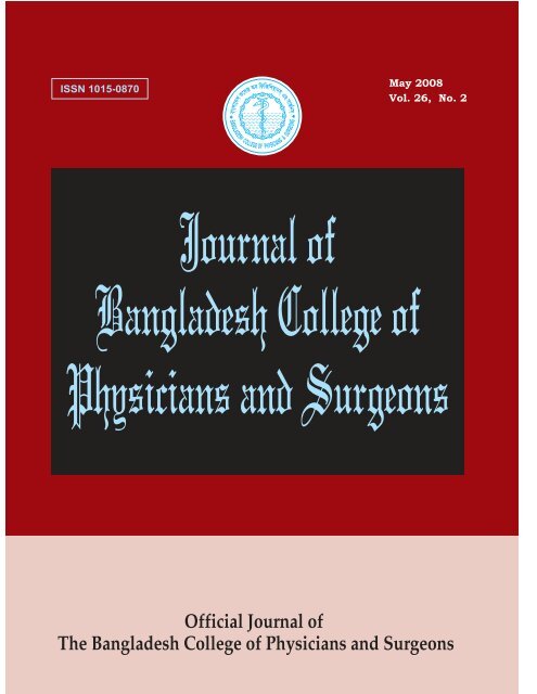
Journal of Bangladesh College ofPhysicians and SurgeonsVol. 26, No. 2, <strong>May</strong> <strong>2008</strong>Official Journal of the Bangladesh College of Physicians and SurgeonsBCPS Bhaban, 67 Shaheed Tajuddin Ahmed SaraniMohakhali, Dhaka-1212, BangladeshEDITORIAL BOARDChairpersonMd. Abul FaizEditor-in-ChiefMd. Rajibul AlamEditorsMd. Harun-ur-RashidK.M.H.S. Sirajul HaqueMd. SalehuddinAbdus SalamMahmuda KhatunShafiqul HaqueKhokan Kanti DasSyed Kamaluddin AhmedProjesh Kumar RoyA.K.M. Khorshed AlamShafquat Hussain KhundkerEmran Bin YunusU.H. Shahera KhatunMd. Abdul MasudMohammed Abu AzharNazneen KabirMd. Mizanur RahmanHarunur RashidA.K.M. Fazlul HaqueSyed Azizul HaqueTahmina BegumNooruddin AhmedMd. Abid Hossain MollaAbdul Wadud ChowdhuryMd. Muzibur Rahman BhuiyanDewan Saifuddin AhmedMd. Azharul IslamNishat BegumMohammad Monir HossainA.K.M. Aminul HoqueHasina AfrozMd. Mujibur Rahman HowladerADVISORY BOARDMobin KhanQuazi Deen MohammadM.A. MajidMd. Abul Kashem KhandakerA.H.M. Towhidul Anowar ChowdhuryT.I.M. Abdullah-Al-FaruqMohammad Saiful IslamMahmud HasanChoudhury Ali KawserMd. Ruhul AminS.A.M. Golam KibriaSayeba AkhterNazmun NaharMd. Sanawar HossainAbdul Kader KhanM.A. MajedTofayel AhmedA.H.M. AhsanullahA.N.M. Atai RabbiEditorial StaffAfsana HuqDilruba PervinPUBLISHED BYMd. Rajibul Alamon behalf of the Bangladesh Collegeof Physicians and SurgeonsPRINTED ATAsian Colour Printing130 DIT Extension Road, FakirerpoolDhaka-1000, Phone : 9357726, 8362258ANNUAL SUBSCRIPTIONTk. 300/- for local and US$ 30for overseas subscribersThe Journal of BangladeshCollege of Physicians andSurgeons is a peer reviewedJournal. It is published threetimes in a year, (January,<strong>May</strong> and September). Itaccepts original articles,review articles, and casereports. Complimentary copiesof the journal are sent tolibraries of all medical andother relevant academic institutionsin the country and selectedinstitutions abroad.While every effort is alwaysmade by the Editorial Boardand the members of the JournalCommittee to avoid inaccurateor misleading informationappearing in the Journal ofBangladesh College of Physiciansand Surgeons, informationwithin the individual article arethe responsibility of its author(s).The Journal of BangladeshCollege of Physicians andSurgeons, its Editorial Boardand Journal Committee acceptno liability whatsoever for theconsequences of any suchinaccurate and misleadinginformation, opinion or statement.ADDRESS OF CORRESPONDENCEEditor-in-Chief, Journal of Bangladesh College of Physicians and Surgeons, BCPS Bhaban, 67, Shaheed Tajuddin Ahmed Sarani, Mohakhali,Dhaka-1212, Tel : 8825005-6, 8856616-7, Fax : 880-2-8828928, E-mail : <strong>bcps</strong>@bdonline.com
INFORMATION FOR AUTHORSThe Journal of Bangladesh College of Physicians andSurgeons agrees to accept manuscript prepared inaccordance with the 'Uniform Requirements Submitted tothe Biomedical Journals' published in the New EnglandJournal of Medicine 1991; 324 : 424-8.Aims and scope:The Journal of Bangladesh College of Physicians andSurgeons is one of the premier clinical and laboratory basedresearch journals in Bangladesh. Its international readershipis increasing rapidly. It features the best clinical andlaboratory based research on various disciplines of medicalscience to provide a place for medical scientists to relateexperiences which will help others to render better patientcare.Conditions for submission of manuscript:● All manuscripts are subject to peer-review.●●●Manuscripts are received with the explicit understandingthat they are not under simultaneous consideration by anyother publication.Submission of a manuscript for publication implies thetransfer of the copyright from the author to the publisherupon acceptance. Accepted manuscripts become thepermanent property of the Journal of Bangladesh Collegeof Physicians and Surgeons and may not be reproducedby any means in whole or in part without the writtenconsent of the publisher.It is the author's responsibility to obtain permission toreproduce illustrations, tables etc. from otherpublications.Ethical aspects:● Ethical aspect of the study will be very carefullyconsidered at the time of assessment of the manuscript.●Any manuscript that includes table, illustration orphotograph that have been published earlier shouldaccompany a letter of permission for re-publication fromthe author(s) of the publication and editor/publisher ofthe Journal where it was published earlier.● Permission of the patients and/or their families toreproduce photographs of the patients where identity isnot disguised should be sent with the manuscript.Otherwise the identity will be blackened out.Preparation of manuscript:Criteria:Information provided in the manuscript are important andlikely to be of interest to an international readership.Preparation:a) Manuscript should be written in English and typed onone side of A4 (290 x 210cm) size white paper.b) Double spacing should be used throughout.c) Margin should be 5 cm for the header and 2.5 cm for theremainder.d) Style should be that of modified Vancouver.e) Each of the following section should begin on separatepage :●●●●●●Title pageSummary/abstractTextAcknowledgementReferencesTables and legends.f) Pages should be numbered consecutively at the upperright hand corner of each page beginning with the titlepage.Title Page :The title page should contain:● Title of the article (should be concise, informative andself-explanatory).●●●●Name of each author with highest academic degreeName of the department and institute where the work wascarried outName and address of the author to whom correspondenceregarding manuscript to be madeName and address of the author to whom request forreprint should be addressedSummary/Abstract :The summary/abstract of the manuscript :● Should be informative● Should be limited to less than 200 words● Should be suitable for use by abstracting journals andinclude data on the problem, materials and method, resultsand conclusion.● Should emphasize mainly on new and important aspectsof the study● Should contain only approved abbreviations
Introduction:The introduction will acquaint the readers with the problemand it should include:●●●●Nature and purpose of the studyRationale of the study/observationStrictly pertinent referencesBrief review of the subject excepting data andconclusionMaterials and method :This section of the study should be very clear and describe:● The selection criteria of the study population includingcontrols (if any).●●●The methods and the apparatus used in the research.The procedure of the study in such a detail so that otherworker can reproduce the results.Previously published methods (if applicable) withappropriate citations.Results:The findings of the research should be described here and itshould be:●●●Presented in logical sequence in the text, tables andillustrations.Described without comment.Supplemented by concise textual description of the datapresented in tables and figures where it is necessaery.Tables:During preparation of tables following principles should befollowed● Tables should be simple, self-explanatory andsupplement, not duplicate the text.● Each table should have a tittle and typed in double spacein separate sheet.● They should be numbered consecutively with romannumerical in order of text. Page number should be in theupper right corner.●If abbreviations are to be used, they should be explainedin footnotes.Illustrations:Only those illustrations that clarify and increase theunderstanding of the text should be used and:●●●All illustrations must be numbered and cited in the text.Print photograph of each illustration should be submitted.Figure number, tittle of manuscript, name ofcorresponding author and arrow indicating the top shouldbe typed on a sticky label and affixed on the back of eachillustration.●Original drawings, graphs, charts and lettering should beprepared on an illustration board or high-grade whitedrawing paper by an experienced medical illustrator.Figures and photographs:The figures and photographs :●●Should be used only where data can not be expressed inany other formShould be unmounted glossy print in sharp focus, 12.7 x17.3 cms in size.● Should bear number, tittle of manuscript, name ofcorresponding author and arrow indicating the top on asticky label and affixed on the back of each illustration.Legend:The legend:● Must be typed in a separate sheet of paper.● Photomicrographs should indicate the magnification,internal scale and the method of staining.Units:● All scientific units should be expressed in SystemInternational (SI) units.● All drugs should be mentioned in their generic form. Thecommercial name may however be used within brackets.Discussion:The discussion section should reflect:●●●The authors' comment on the results and to relate themto those of other authors.The relevance to experimental research or clinicalpractice.Well founded arguments.References:This section of the manuscript :● Should be numbered consecutively in the order in whichthey are mentioned in the text.● Should be identified in the text by superscript in Arabicnumerical.● Should use the form of references adopted by US NationalLibrary of Medicine and used in Index Medicus.Acknowledgements :Individuals, organizations or bodies may be acknowledgedin the article and may include:●Name (or a list) of funding bodies.● Name of the organization(s) and individual(s) with theirconsent.Manuscript submission:Manuscript should be submitted to the Editor-in-Chief andmust be accompanied by a covering letter and followinginclusions:
a) A statement regarding the type of article beingsubmitted.b) A statement that the work has not been published orsubmitted for publication elsewhere.c) A statement of financial or other relationships thatmight lead to a conflict of interests.d) A statement that the manuscript has been read,approved and signed by all authors.e) A letter from the head of the institution where the workhas been carried out stating that the work has beencarried out in that institute and there is no objection toits publication in this journal.f) If the article is a whole or part of the dissertation or thesissubmitted for diploma/degree, it should be mentioned indetail and in this case the name of the investigator andguide must be specifically mentioned.Submissions must be in triplicates with four sets ofillustrations. Text must be additionally submitted in a CD.Editing and peer review:All submitted manuscripts are subject to scrutiny by theEditor in-chief or any member of the Editorial Board.Manuscripts containing materials without sufficientscientific value and of a priority issue, or not fulfilling therequirement for publication may be rejected or it may besent back to the author(s) for resubmission with necessarymodifications to suit one of the submission categories.Manuscripts fulfilling the requirements and found suitablefor consideration are sent for peer review. Submissions,found suitable for publication by the reviewer, may needrevision/ modifications before being finally accepted.Editorial Board finally decides upon the publishability ofthe reviewed and revised/modified submission. Proof ofaccepted manuscript may be sent to the authors, and shouldbe corrected and returned to the editorial office within oneweek. No addition to the manuscript at this stage will beaccepted. All accepted manuscript are edited according tothe Journal's style.Reprints for the author(s):Ten copies of each published article will be provided to thecorresponding author free of cost. Additional reprints maybe obtained by prior request and only on necessary payment.Subscription information:Journal of Bangladesh College of Physicians and SurgeonsISSN 1015-0870Published by the Editor-in-Chief three times a year inJanuary, <strong>May</strong> and SeptemberAnnual SubscriptionLocal BDT = 300.00Overseas $ = 30.00Subscription request should be sent to:Editor-in-ChiefJournal of Bangladesh College of Physicians and Surgeons67, Shaheed Tajuddin Ahmed SaraniMohakhali, Dhaka-1212.Any change in address of the subscriber should be notifiedat least 6-8 weeks before the subsequent issue is publishedmentioning both old and new addresses.Communication for manuscript submission:Communication information for all correspondence isalways printed in the title page of the journal. Anyadditional information or any other inquiry relating tosubmission of the article the Editor-in-Chief or the Journaloffice may be contacted.Copyright :No part of the materials published in this journal may bereproduced, stored in a retrieval system or transmitted inany form or by any means electronic, mechanical,photocopying, recording or otherwise without the priorwritten permission of the publisher.Reprints of any article in the Journal will be available fromthe publisher.
JOURNAL OF BANGLADESH COLLEGE OFPHYSICIANS AND SURGEONSVol. 26, No. 2, Page 58-111 <strong>May</strong> <strong>2008</strong>CONTENTSEDITORIALNew Management Strategies of Hormone Refractory Prostate Cancer (HRPC) 58Prof. M.A. SalamORIGINAL ARTICLESSerum Apoprotein ( ApoA1 and ApoB) in Myocardial infarction 62KA Jhuma, MM HoqueProblems and Immediate Outcome of Infants of Diabetic Mothers 67CB Mahmood, MI KayesRadioiodine (131i) Therapy for Thyrotoxicosis Patients and their Outcome: Experience 73at Center for Nuclear Medicine & Ultrasound, BarisalSK Biswas, N Jahan, KBMA RahmanCo-relation between Sepsis Score and Blood Culture Report in Neonatal Septicaemia 79S Afroza, F BegumREVIEW ARTICLESJuvenile Idiopathic Arthritis Essential Elements of Care 83MR AlamApproach to Subclinical Thyroid Disease 91SR SutradharCASE REPORTSGonadoblastoma: Primary Amenorrhoea with Gonadal Dysgenesis 97H Begum, S Khaton, S JahanHenoch-Schonlein Purpura in an Elderly Women Presenting with Severe GI Bleeding: 100A Case ReportMAJ Chowdhury, SM Arafat, Abed HussainMalignant Melanoma of the Vagina - A Case Report 103N Sultana, CM Ali, RA Khanam, M KhatunCOLLEGE NEWS106
EDITORIALNew Management Strategies of Hormone RefractoryProstate Cancer (HRPC)Carcinoma prostate is the commonest cancer in menand recognized as the commonest killer of men.Prostate cancer incidence is increasing in Bangladeshas the detection technology and people are servingslonger. Prostate cancer progression ends up atHormone Refractory Prostate Cancer (HRPC) orstage D3 status where no endocrine manipulation iseffective. The median survival at this stage of prostatecancer is usually less than 10 months. World wide lifeof the most of the prostate cancer patients areterminated at this stage.Hormone Refractory Prostate Cancer (HRPC) mayoccur due to the fact that prostate cancer cell escapefrom androgen withdrawal-induced apoptosis. In thisdevelopment, enhancement of growth factorstimulation has an essential role in the up regulationof survival signals and constitutive proliferation 1 .The principle of treatment for advanced prostatecancer is endocrine manipulation which includesandrogen deprivation. Unfortunately, at this stage ofprostate cancer most of men become resistant tohormonal manipulation, developing what is definedas hormone-refractory prostate cancer (HRPC). Adecade ago, most clinicians find no answer and felthelples*ecause no Chemotherapy was considered tobe ineffective and associated with unacceptabletoxicity. A review of 26 chemotherapy-based trialsrevealed an overall response rate of 8.7% with amedian survival ranging from. 6 to 10 mo 2 . For thisreason, it was established that a median expectedsurvival for patients with HRPC is 10 months.Therefore, novel therapeutic strategies that target themolecular basis of androgen resistance were required.Role of chemotherapy in HRPC was emphasized In2004. Two pivotal trials of Docitaxel-basedchemotherapy were reported and, for the first time, asurvival benefit was observed for chemotherapy inHRPC. The results from the Southwest OncologyGroup (SWOG )99-16 and TAX327 studies changed theexpectations of treatment outcome these patients 7,8 .Also these trials demonstrated the need forcombination therapies in patients with HRPC.The combination of Docitaxel with estramustineincreases the thrombo embolic risk and necessitates aprimary prophylaxis 7,8 . New combination modelsusing Docitaxel may represent an excitinginvestigational field 9 . In particular, less toxicregimens, provided that the activity can bemaintained, are more attractive.Recently, Di Lorenzo et al 9 presented an interestingproposal using a combination of docetaxel,vinorelbine, and zoledronic acid as first-linetreatment in patients with HRPC. Vinorelbine is avinca alkaloid that inhibits the microtubular apparatusin malignant cells and has shown activity in HRPC 9 .The synergism of docetaxel and vinorelbine has beenconfirmed in preclinical studies and human trials 9 .Moreover, the use of docetaxel in a weekly scheduleappears to minimize myelo suppression and has beenassociated with moderate toxicity 9 .Most HRPC develops bone metastases thatt areresponsible for pain and morbility. Bisphosphonatesshowed an inhibitory effect on prostate cancer bonemetastases by blocking proteolytic activity of thematrix, cell adhesion, and possibly cancer cellgrowth 9 . Multicentric randomised trials of HRPCwith bone metastases showed a significant reductionin skeletal related events using zoledronic acid 9 .Di Lorenzo et al 9 developed a phase 2 study toevaluate the impact of weekly docetaxel andvinorelbine and monthly zoledronic acid on PSAresponse, pain improvement, and toxicity profile in40 men with HRPC. Complete and partial response(PSA reduction) were observed in 18% and 32% ofcases, respectively.The objective of this editorial is to emphasizes twopossible strategies: the first, specifically targeted tothe role of the neuro endocrine (NE) system in
Journal of Bangladesh College of Physicians and Surgeons Vol. 26, No. 2, <strong>May</strong> <strong>2008</strong>hormone-refractory stage development, and thesecond, chemotherapy, not target specific and onlycytotoxic.NE activity is considered one of the factors involvedin the progression from an androgendependent to anandrogen-independent state and may be a possiblenew target therapy. In recent years a marked numberof papers related to NE differentiation in prostateadenocarcinomas has published. The NE componentof prostate Adenocarcinoma is androgen independentand does not produce prostate-specific antigen (PSA).The continuous use of androgen-ablation therapy mayproduce hyperactivation of the NE system in prostatetissue 3 . NE system products can act as immortalisingfactors, blocking the apoptotic process in prostateadenocarcinoma cells and then inducing androgenindependentstatu5 and progression.Several clinical trials have demonstrated impressiveefficacy of somatostatin analogues for varioushypersecretory disorders resistant to standard therapy.They have also proved useful for the management ofsymptoms caused by NE diseases. Chromogranin A(CgA) is considered the best marker of NE activity inthe prostate. In different countries CgA determinationstarted to be used and to be repeated in clinicalpractice for the evaluation of men with prostateadenocarcinoma. The primary effect of somatostatinanalogues is not a Ikect cytotoxic effect on NE cells,but rather inhibition of the release of peptidehormones secreted by NE cells. Clinical trials onsomatostatin analogues as monotherapy for prostatecancer have shownknegative results 4 .The mechanismof action of these drugs may suggest their use not asmonotherapy but rather as combination therapy forprostate cancer. Koutsilieris et al 5 first proposed acombination therapy with dexamethasone andsomatostatin analogues in HRPC. The authorcombined standard luteinising hormonereleasinghormone (LHRH) analogue therapy with somatostatinanalogue and dexamethasone. Median overall survivalreported in this study was 12 mo, with improvement inperformance status and bone pain scores. Di Silverioand Sciarra 6 analysed whether the combination ofethinyloestradiol and lanreotide can offer objectiveresponse or symptomatic improvement in patientswith D3 prostate cancer. Patients with metastaticHRPC discontinued LHRH analogue and started thecombination therapy.The rationale for this combination therapy is: (1) toinhibit the protective antiapoptotic effect of NEsystem on prostate adenocarcinoma cells(somatostatin analogue); (2) to use a new mechanismof castration (oestrogens); and (3) to add a directcytotoxic effect on prostate cells (oestrogens). Nomajor related side-effects were reported(gynaecomastia and breast pain). In this phase 2 trial,95% of cases showed an objective clinical responseas demonstrated by at least a 50% PSA decrease frombaseline; in all cases the PSA response wasaccompanied by a significant improvement in EasternCooperative Oncology Group (ECOG) performancestatus and bone pain score; 70% of cases werewithout disease progression at a median of 16.5 mo offollow-up during therapy. These results suggest theneed for a phase 3 trial to confirm the effectiveness ofthis combination therapy in HRPC.An objective response (liver, lung, and lymph nodes)was observed in 6 of 15 patients with measurabledisease. Stratifying the response in terms of Gleasonscore, primary treatment, and number of osseous sites,no differences were observed among these groups. Notoxic death occurred and the most important grade 3toxicities included neutropenia (25%). Painimprovement was found in 47.5% of cases. Medianprogression-free survival was 7 mo, with a medianoverall survival of 17 mo. The majority of patientsreceived, after progression, a second line ofchemotherapy. The rationale to improve docetaxelefficacy and to reduce the related toxicity using acombination with vinorelbine and zoledronic acid is ofgreat interest.(J Bangladesh Coll Phys Surg <strong>2008</strong>; 26: 58-61)Prof. M.A. SalamProfessor of Uro-Oncology, Department of UrologyBSMMU, Dhaka.References1. Landstrom M, Damber JE, Bergj A. Prostatic tumorregrowth after initially successful castration therapy may berelated after to a decreased apoptotic cell death rate. CancerRes 1994;54:428195.59
New Management Strategies of Hormone Refractory Prostate Cancer (HRPC)M.A. Salam2. Yagoda A, Petrylak D. Cytotoxic chemotherapy foradvanced hormone resistant prostate cancer. Cancer5. Sciarra A, Monti S, Gentile V, Mariotti G, Voria G, DiSilverio F. Variation in chromogranin A serum levels duringintermittent versus continuous androgen deprivation therapyfor prostate adenocarcinoma. Prostate 2003;55:168-79.4. Sciarra A, Bosman C, Monti S, et al. Somatostatinanalogues and estrogens in the treatment of androgenablation refractory prostate adenocarcinoma. J Urol 2004172:1775-83.5. Koutsilieris M, Mitsiades C, Dimopoulos T, lannidis A,Ntounis A, Lambou T. A combination therapy ofdexamethasone and somatostatin analog reintroducesobjective clinical response to LHRH analog in androgenablation-refractory prostate cancer patients. J ClinEndocrinol Metab 2001;86:5729-36.6. Di Silverio F, Sciarra A. Combination therapy ofethinyoestradiol and somatostatin analogue reintroducesobjective clinical responses and decreases chromogranin Ain patients with androgen ablation refractory prostatecancer. J Urol 2003;170:1812-8.7. Tannock IF, de Wit R, Berry W. Docetaxel plus prednisoneor mitoxantrone plus prednisone for advanced prostatecancer. N Engl J Med 2004;351:1502-12.8. Petrylak DP, Tangen CM, Hussain MH. Docetaxel andestramustine compared with mitoxantrone and prednisonefor advanced refractory prostate cancer. New Engl J Med2004;351:1513-20.9. Di Lorenzo G, Autorino R, Perdona' S, et al. Docetaxel,vinorelbine and zoledronic acid as first-line treatment inpatients with hormone refractory prostate cancer: a phase IIstudy. Eur Urol 2007;52:1020-7.10. Saad F, Gleason DM, Murray R. Long term efficacy ofzoledronic acid for the prevention of skeletal complicationsin patients with metastatic hormone regractory prostatecancer. J Nati Cancer Inst 2004; 96: 879-82.60
ORIGINAL ARTICLESSerum Apoprotein ( ApoA1 and ApoB)in Myocardial infarctionKA JHUMA a , MM HOQUE bSummary:30 diagnosed cases (Male26, Female 4) of MI (myocardialinfarction) with the mean age of 55.5±9.8 years (range 40-70 years) were included in a case control study to evaluatetheir apoprotein status. Serum apoA1 and apoB weremeasured and compared with those of age and sexmatched healthy control subjects. Mean serum apoA1concentration found significantly low in MI cases (91.84± 11.2 mg/dl) compared to control ( 123.2±10.5mg/dl ) and that of apoB found significantly high in MIcases( 135.3± 23.0 mg/dl ) compared to control (66.2±10.0mg/dl).Serum apoB/apoA1 ratio of MI cases (1.49±0.3)also found significantly higher than that of control(0.54±0.1) .Since the serum apoA1 and apoBconcentration stand for relatively more comprehensivemeasure of antiatherogenic and atherogenic potentialrespectively rather than the traditional lipid profile ;measurement of this apoprotein and their ratio may bemore robust and specific marker for identification ofindividuals at risk of MI even in individuals with normaltraditional lipid profile.Key word: ApoA1, ApoB, MI(J Bangladesh Coll Phys Surg <strong>2008</strong>; 26: 62-66)IntroductionCoronary artery disease (CAD) is one of the leadingcauses of death from global point of view. Theidentification of subjects at risk of developing CAD isan important public health issue 1 . Atherosclerosis isthe underlying cause in more than half of the patientswith CAD 2 . Dyslipidemia is the corner stone ofatherosclerotic process. Commonly serum totalcholesterol ( TC), triacylglycerol (TAG), High densitylipoprotein cholesterol (HDL-C),Low densitylipoprotein cholesterol (LDL-c) are used foridentification of person at risk of CAD. Howeverserum TC can not discriminate well betweenindividuals developing CAD and those who does not;since the TC as a whole without its functional breakupto atherogenic and antiatherogenic potential is a poorindicator of the atherosclerotic scenario 3 .Traditionally serum LDL-C and HDL-C are regardedas the marker of atherogenic and antiatherogenicmeasure respectively, but it is not infrequent for anindividual to develop CAD with traditional lipida. Dr. Khadija Akther Jhuma, Assistant Professor ofBiochemistry, Medical College for Women & Hospital, Plot-4, Road-9, Sector-1, Uttara Model Town, Dhaka-1230.b. Prof. MM Hoque, Professor of Biochemistry, BangabandhuShiekh Mujib Medical University, Shahbag, Dhaka.Address of Correspondence: Prof. MM Hoque, Professor ofBiochemistry, Bangabandhu Shiekh Mujib Medical University,Shahbag, Dhaka.Received: 19 August, 2007 Accepted: 6 April, <strong>2008</strong>profile well within desired level, because LDL-C andHDL-C are not the complete representation ofatherogenic and antiatherogenic lipoprotein.Important atherogenic lipoprotein are chylomicron(CM), chylomicron ramnant (CMR), very low densitylipoprotein(VLDL), intermediate density lipoprotein(IDL), low density lipoprotein (LDL), andlipoprotein(a) [LP(a)]; all of which contain apoB.Although HDL is treated as antiatherogenic but thedifferent subtypes of HDL have different degree ofantiatherogenic potential; some subtype believed to berather atherogenic and all subtypes do not containingapoA1.It is claimed that HDL contain apoA1 areantiatherogenic since apoA1 stimulate LCAT andthus help in reverse cholesterol transport (RCT) byfacilitating HDL maturation. HDL containing apo-A IIcounter act the RCT since apo-A IIinhibit the LCAT.So the antiatherogenecity of HDL 1which has noapoA1 (contain only apoE) and that of other HDLsubtypes with apoA IIis doubtful. So HDL-C cannotrepresent complete antiatherogenic potential of HDL 4 .With this perspective recently the major apolipoproteinslike apoA1 and apoB have received attention andaddressed as the major determinant of the metabolicfate of different lipoprotein 5 . The CAD has been foundto be positively correlated with apolipoprotein B andinversely correlated with apolipoprotein A 1 6 .ApoA1 stimulate the reverse cholesterol transport(RCT), decrease the extent of lipid deposition andalso inhibit the infiltrating monocyte/macrophages in
Journal of Bangladesh College of Physicians and Surgeons Vol. 26, No. 2, <strong>May</strong> <strong>2008</strong>aortic intima which initiate the initial stage of fattystreak formation 7 . Reduced plasma level of ApoA1was found in AMI patient 8 . ApoA1 level can be usedto predict future CAD among the young and in adultnot yet manifestating the disease 9 . Although HDLconsidered to be rather antiatherogenic but someHDL subtype (e.g. HDL1) have recently beingidentified to be atherogenic and they contain noApoA1 10 . So the individuals with normal or evenraised HDL-C may be found to have CAD dueprobably to the predominance of HDL1 subtype orthe HDL subtypes without apoA1. Therefore ApoA1rather than HDL-C seems to be more reliable assessorof antiatherogenic potential.Increased level of plasma apoB was shown to be a riskfactor for atherosclerosis 2 . Apo B moiety of LDLseems particularly important for its atherogenicity,because lipoproteins without apo-B apparently do notproduce atherosclerosis 11 . LDL are heterogenous insize and density and composed of fifteen differentsubtypes. Smaller and denser LDL subtypes (eg .LDL4 ,5,6 etc ) are lipid depleted particles (less cholesterolcontaining) but more atherogenic than larger LDLsubtypes (eg LDL 1,2,3 etc) which are relatively lipidrich but all LDL subtypes plus other apo-Bcontaining atherogenic lipoproteins contain identical& equal apo-B content irrespective of their variedcholesterol content. CAD patients are likely to havemore smaller and denser LDL particle. ThereforeLDL-C is an inadequate measure of LDLatherogenicity 12,13,14 . Measuring ApoB provides adirect estimate of the total number of LDL particlesirrespective of their sub types 15. So only the LDL-Cmeasurement is a poor reflections of atherogenicity;rather ApoB stands for the complete andcomprehensive picture of atherogenic risk. Becauseapo-B accounts all known atherogenic LP (in additionto LDL) and also the number of various LDL subtypes.The CAD patient had significantly lower ApoA1 buthigher ApoB. Both ApoA1 and B had significantdiscriminative power between CAD patients andnormal individuals. So measurement of serum apoA1& apoB appears to be more judicious clinically.Materials and MethodsThis case control study was carried out from july2003 to June 2004 in the Department ofBiochemistry, Dhaka Medical College, Dhaka incooperation with the immunology Dept , BIRDEM,Dhaka. Ethical clearance was taken from Ethicalcommittee of Dhaka Medical College. A total 50 nonsmoker, non-alcholic subjects free from DM, renaldisease, thyroid disease, liver disease and having nohistory of taking antihypertensive orantihyperlipidemic drugs were studied. Fasting bloodglucose, serum creatinine, TSH, serum bilirubin &ALT were measured in all study subjects to excludeDM, renal diseases, thyroid & liver diseases. Amongthe study subject 30 were the diagnosed MI casescollected from Cardiology ward in Dhaka MedicalCollege and 20 were age and sex matched healthycontrol selected from colleagues & relatives. Allstudy subjects were included after taking theirinformed written consent .MI diagnosis based oncharacteristic chest pain, ECG finding and rise andfall of serum cardiac marker.5 ml fasting venous blood was collected from eachsubject with all aseptic precautions and allowed toclot at room temperature & centrifuged for 10-15minutes at 2500rpm. The separated serum was storedfrozen at -35 0 c until used for the measurement ofapoprotein (apoA1 and apoB).Laboratory Method Serum apoA1 & ApoBconcentration was estimated by immunonephelometricmethed with commercially available kits usingBN system of Dade Behring Marburg GmbH, USA16,17 . Results were express as their mean ± SD(Standard deviation)Statistical Analysis: The result were analysed in SPSSby using unpaired student t-test, P< 0.05 were takenas a level of significance.ResultsStudy subjects were grouped as Group -I (30 MIcases,) and Group-II (20 normal control). Group-Iincluded 26 males (86.6%) and 4female (13.3%) ofage range 40-70 years. In Group-II , 16 male (80%)and 4 female (20%) normal control were selectedwith age range 40-70 years. (Table -I) .Table-II shows the serum apoA1 & apoBconcentration in different Groups. In Group-I (case)mean apoA1 concentration found 91.84± 11.2 mg/dl63
Serum Apoprotein ( ApoA1 and ApoB) in Myocardial Infarctionwith the range 68.0- 105.0 and that of apo Bconcentration found 135.3±23.0 mg/dl with the range106.0-187.0. In Group-II (Normal control) meanapoA1 concentration was 123.2±10.5 mg/dl with therange 105.0- 145.0 and of apoB concentration was66.2± 10.0 mg/dl with the range 55.0-94.9respectively. In MI cases apoB found significantlyKA Jhuma & MM Hoqueincreased and apoA1 found significantly decreasedcompared to control.Table III shows the comparison of ratio ofapoB/apoA1. The ratio in group1 (cases) and group II(normal control) were 1.49±0.30 and 0.54±0.10respectively, which was found significantly elevatedin case compared to normal control.Table1Age & sex distribution of study subjectsStudy Subject Age (year) SexMean ± SD Age range Male FemaleMI cases (Gr-I)(n=30) 55.5 ± 9.8 40-70 26 4Normal Control(Gr-II)(n=20) 52.6 ± 9.6 40-70 16 4Table IISerum A1 and Apo B concentration of the study subjectsParameterMean ± SD Group I (cases) Group II (Control) Level of significancen=30 n=20 (p-Value)Apo A1 (mg/dl) 91.84 ± 11.2 123.2 ± 10.5 0.001(68.0 – 105.0)* (105.0 – 145.0)*Apo B (mg/dl) 135.3 ± 23.0 66.2 ± 10.0 0.001(106.0 – 187.0)* (55.0 – 94.9)*P value reached by unpaired t test* Paranthesis shows rangeTable IIIComparison of the ratio of Apo B / Apo A1 between study subject.Parameter Group I (cases) Group II Level of significance(p-Value)Apo B / Apo A1 1.48 ± .03 0.54 ± 0.1 0.001P value reached by unpaired t test64
Journal of Bangladesh College of Physicians and Surgeons Vol. 26, No. 2, <strong>May</strong> <strong>2008</strong>DiscussionIn this study MI patients found to have serum apoBconcentration significantly increased and apoA1concentration significantly decreased in comparisonto control. A similar phenomenon was reported inmany other studies around the world. 18,19,20,21,1,22,23,24,25 .Atherogenic lipoproteins particles are heterogenouswith respect to their cholesterol content buthomogenous with respect to their apoB content .Soserum apoB more accurately reflects the total numberof circulating atherogenic particles which their totalcholesterol content cannot. For example small denseLDL particles are cholesterol depleted compared tolarge LDL particles but all LDL subtypes contain onemolecule apoB .So the number of circulating LDLparticle is more accurately measured by their apo-Bcontent rather then LDL-C. Although about 70% ofplasma cholesterol is carried by LDL but apart fromLDL, there are number of other highly atherogeniccirculating lipoproteins, all of which contain apo-B.Therefore serum apoB is the more comprehensiveand reliable marker of atherogenicity rather than theLDL-C alone 26,27,28 .HDL is regarded as an anti-atherogenic lipoprotein.Various subtypes of HDL (e.g. HDL1, HDL2, HDL3etc.) has been described which differ from each otherwith respect to their apoprotein andantiatherogenicity. To be a antiatherogenic, HDLneeds to contain apo-A1 which is not true for all HDLsubtypes (e.g. HDL 1contain no apo A1) 10 . Thereforeit mightbe possible for an individual to present withMI having normal HDL-C but decreased serumapoA1 concentration due to predominance of HDL1subtype or the HDL subtypes without apoA1.Conclusion:It can be concluded from this study that; serumApoA1 and ApoB are more reliable tool to assess andevaluate the atherosclerotic disorders specially theCAD. Therefore if accurate precise and affordablestandardized methods be come available for themeasurement of apoA1 and apoB, these apoproteinsmeasurement may be recommended as a routinelaboratory test to evaluate the MI patient & to assessthe risk of MI.Reference1. Genest j, Mcnamara JR, Ordovas JM, Jenner JL, SilbermanSR,Anderson KM and Wilson PWF; Lipoproteinscholesterol, Apolipoprotein A1 and B and lipoprotein (a)abnormalities in men with premature coronary arterydisease; J Am Coll Cardiol; 1992;19:792-802.2. Ginsberg H N & Goldberg I J ; Disorder of intermediarymetabolism; in: Harrisons principles of internal medicine;Braunwald E, Fauci A.S. & Kasper D.L.; (eds)13th edition:McGraw Hill publishers; USA,2001; pp1377-1387.3. Durrington PN, Hunt L, Ishola M & kane J; Serumapolipoproteins A1 and B lipoproteins in middle age menwith and without previous myocardial infarction; Br HeartJ;1986; 56: 206-212.4. Stein EA and Myer GL; Lipid ,lipoprotein andapolipoproteins; in : Teitz fundamentals of clinicalchemistry; ed.Burtis CA and Ashwood ER (eds); 4thedition; Philadelphia WB saundex company; 1996;375.5. Sveger T & Fex G; Apolipoprotein A1 and B levels inadolescents: A trial to define subjects at risk for coronaryheart disease; Acta paediatr Scand; 1983: 72: 499-504.6. Kwiterovich PO & Sniderman AD; Atherosclerosis andapoprotein Band A1; Prev Med, 1983; 12: 815-834.7. She MP,Liang P, huang YD, Cai CB, Ran BF, Wang ZL andXia RY; HDL and apolipoproteinA(apoA1):Their effects onretardation of lipid deposition in aortic intima;Clin medJ.(Engl);1992 <strong>May</strong>;105(5):369-373.8. Franzen J & Fex G; Low serum apolipoprotein A1 in acutemyocardial infarction survivors with normal HDLcholesterol; Atherosclerosis: 1986;59:37-42.9. Stewart GM; The meaning of a new marker for coronaryartery disease; N Engl J Med; 1983;309(7):426-427.10. Stein EA and Myer GL; Lipid, lipoprotein andapolipoproteins: in: Tietz fundamentals of ClinicalChemistry; ed.Burtis CA and Ashwood ER (eds); 4thedition; Philadelphia WB saundex company; 1996;pp375.11. Grundy SC, Vega GL, Kesaniemi YA; Abnormalities inmetabolism of low density lipoproteins associated withcoronary heart disease; Acta Med Scand (suppl) 1985; 701:23-37.12. Hoff HF, Heiderman CL, Gaubatz JW & Titus JL;Quatitation of apoB in human aortic fatty streaks: Acomparison with grossly normal intima and fibrous plaques;Atherosclerosis ; 1978; 30:263-268.13. Sniderman AD & Cianflone K; Measurement ofApoproteins: Time to improve the diagnosis and treatmentof the Atherogenic dyslipoproteinemias; Clin Chem ;1996;42(4): 489-491.14. Gardner CD ,Fortmann SP, krauss RM; ApoB/ApoA1 ratiois more robust an specific marker; JAMA 1996; 276: 875-881.65
Serum Apoprotein ( ApoA1 and ApoB) in Myocardial Infarction15. Dati F,Tate J; ejiFcc vol-13 no3:http//www.Ifcc.org/ejifcc/vol 13 no3/130301003.htm.16. Lopes- virella MF L, Virella G and Evans G;Immunonephelometric Assay of Human apolipoproteinA1;Clin Chem; 1980;26(8):1205-1208.17. Heuck CC & Schlier G; Nephelometer of Apolipoprotein Bin Human Serum ;Clin Chem 1979; 25(1):221-226.18. Avogaro,Bon GB, Cazzolato G and Roral E; Relationshipbetween apolipoproteins and chemical componants oflipoproteins in survivors of myocardial infarction;Atherosclerosis ; 1980;37:69-76.19. BackerG, Hulstaert F, Munck k, Rosseneu M, Vanparijs Land Dramaix M; Serum lipids and apoproteins in a studentwhose parents suffered prematurely of myocardialinfarction; Am Heart J; 1986; 112: 478-484.20. Freedman DS ,Srinivasan SR, Shear CL, Franklin FA,Webber LS and Berenson GS; The relation ofapolipoprotein A1 and B in children to parental myocardialinfarction;N Engl J Med; 1986; 315;721-726.21. Vanstiphout WA HJ, Hofman A, Kruijssen HACM, &Vermeeren R; is the ratio of ApoB/apoA1 an early predictorof coronary Atherosclerosis? Atherosclerosis 1986;62: 179-182.KA Jhuma & MM Hoque22. Kukita H ,Hamada M, Hiwada K& Kokubu T; Clinicalsignificance of measurements of serum apolipoproteinA1,All and B in hyper triglyceridemic male patients withwithout coronary artery disease; Atherosclerosis;1985; 55:143-149.23. Al-Muhtaseb N, Hayet N & Al-Khafaji M; Lipoproteins andapolipoprotein in young male servivors of myocardialinfraction; atherosclerosis; 1989;77 (2-3):131-138.24. Durrington PN, Hunt L, Ishola M & Arrol S;Apolipoproteins(a,)A1and B and parental history in menwith early onset ischemic heart disease; TheLancet;1988;14: 1070-1073.25. Zunic G, Jelic- lvanovic Z, Spasic S, Stojiljkovic A andSingh NM ; Reference values for apolipoproteins A1and Bhealthy subjects by Age; clin chem.; 1992;38(4): 566-569.26. Miremadis S, Sinderman A,& Frohlich J; Clinicalchemistry, 2002;48(3)484-488.27. Berneis KK,Krauss RM, Metabolic origens and clinicalsignificance of LDL hheterogenecity; J lipid Resechers,2002; 43: 1363-79.28. Sinderman AD, Furberg CD, Keech A etal;Lancet,2003,361,777-780.66
Journal of Bangladesh College of Physicians and SurgeonsVol. 26, No. 2, <strong>May</strong> <strong>2008</strong>Problems and Immediate Outcome of Infantsof Diabetic MothersCB MAHMOOD a , MI KAYES bSummary:Objective:The present study was undertaken to evaluatethe problems and immediate outcome of infants ofdiabetic mothers(IDMs) in early neonatal period and tocompare the results between infants of gestational andpregestational diabetic mothers.Design: A hospital based prospective study. Setting: Thestudy was done in Chittagong Medical College Hospital, atertiary hospital in Chittagong city. Method: Within onehour of delivery 52 infants of diabetic [pregastational &gestational] mothers consecutively admitted were enrolledin the study. Study period was January 2002 to August2002.Results: Total number of IDMs were 52.Among them 31were gestational and 21 were of pregestational diabeticmothers.Significant number 82.6% of IDMs were delivered bycaesarean section. The mean birth weight of IDMs wassignificantly high(3212±563g ), 21% of IDMs had birthweight>4000 g. Total 23% of the IDMs developedperinatal asphyxia .The 23% of IDMs developedhypoglycaemia.The incidence of hypoglycaemia washigher in infants of pregestational diabetic mothers ascompared to that of gestational diabetic mothers(38.09%and 12.9% respectively), the difference was statisticallysignificant(P
Problems and Immediate Outcome of Infants of Diabetic MothersInfant of diabetic mothers have a 47% risk ofsignificant hypoglycaemia, 22% risk ofhypocalcaemia,19% risk of hyperbilirubinemia, 34%risk of polycythaemia, 6-9% incidence of majorcongenital anomalies( congenital heart disease,central nervous system & vertebral anomalies) 6 , 4%risk of respiratory distress syndrome, 28% risk ofmacrosomia & cardiomegaly (30%).Among the various metabolic errors these infantssuffer, hypoglycaemia is the commonest and mostdangereous 7 .Infants of diabetic mothers havehyperinsulinism at birth due to increased placentaltransfer of glucose and other nutrients stimulatinghyperplasia of islets of Langerhans in the fetus andincreased insulin secretion, raised amount of C-peptide and free insulin in cord blood. Once thematernal supply of glucose is cut-off by clamping thecord, the excess insulin circulating in the baby’ssystem quickly rids the plasma of the remainingglucose and so blood glucose level may dropprecipitously and alarmingly during the first fewhours of life 8 . Hypoglycaemia is defined as a bloodglucose level less then 2.6mmol/L. Symptoms ofhypoglycaemia, are non specific, such as lethargy,apathy, limpness, apnea, cyanosis, weak or highpitched cry, poor feeding ,vomiting, tremors,jitteriness , irritability,seizures,coma 9 . Neonetalhypocalcaemia may be due to hypoparathyroidism,abnormal vitamin D metabolism andhyperphosphataemia. Neonatal hypocalcaemia isdefined as total serum calcium concentration of lessthan 7 mg/dl and an ionized calcium conc. of less than4 mg/dl 10 . Polycythaemia (haematocrit of more than0.651) occurs in 30 to 60% of IDMs causing theneonatal hyperviscosity syndrome. The main cause ofpolycythaemia is chronic intrauterine hypoxaemia,which occurs as consequence of fetal hyperinsulinismand hyperglycaemia. 11 .Macrosomia (birth weight> 4000 g) may beassociated with increased incidence of primarycaesarean section or obstetric trauma such asfractured clavicle, Erb’s palsy or phrenic nerve palsydue to shoulder dystocia 7, 9 .Hypertropic cardiomyopathy with asymmetric septalhypertrophy has been extensively documented 10 .Thebabies may also develop small left colon syndrome, aCB Mahmood & MI Kayestransient delay in the development of left side ofcolon. 4,5 .Despite improvement in diabetic care, theperinatal mortality still remains four times high thanin nondiabetic women. Predominant causes ofmortality are congenital anomaly, birth trauma,respiratory distress syndrome, prematurity andunexplained still birth 12 .Although in developed countries there has beensignificant improvement in the outcome of diabeticpregnancies largely due to better metabolic controlbefore and during pregnancy and vigorous neonatalcare, the management in our country still poses amajor challenge.Aims of this study were to find out problems of IDMduring early neonatal period that threaten baby’s lifeand with appropriate management to determineimmediate outcome in hospitalized IDM.The study was designed to evaluate the problems andimmediate outcome of infants of diabetic mothers inearly neonatal period and to compare the resultsbetween infants of gestational and pregestationaldiabetic mothers in neonatal unit of ChittagongMedical College Hospital,Chittagong.Materials and Method:This hospital based prospective study was done inneonatal unit of Chittagong Medical College Hospitalin collaboration with Department of Obstetrics andGynecology of this hospital. Within one hour ofdelivery 52 infants of diabetic [pregestational &gestational] mothers consecutively admitted forobservation and further management where enrolledin the study. Exclusion criterion was infants ofdiabetic mothers were admitted as referred case fromother hospitals. Study period was January 2002 toAugust 2002.After taking the verbal consent from the attendant,the relevant information from the history, physicalexamination and investigation findings were recordedin a purposely prepared questionnaire. Investigationsroutinely underwent were capillary blood glucose at0(cord blood), 2, 4, 6,12,24,48 and 72 hours of age byusing glucostix. Peripheral blood glucose wascollected with single puncture non-squeezingprocedure by trained technician and level measuredby Glucose oxidase method in auto analyzer at 6,12,24,48,72 hours of age and whenever any68
Journal of Bangladesh College of Physicians and Surgeons Vol. 26, No. 2, <strong>May</strong> <strong>2008</strong>symptoms suggestive of hypoglycaemia developed.The glucostix(capillary blood glucose) was used forscreening purpose and for prompt diagnosis andmanagement of hypoglycaemia and estimation ofperipheral venous blood glucose level was done forfurther confirmation of diagnosis. Serum calciumlevel were measured routinely at 6,24,48 hours of ageand later if the baby remains hypocalcaemic orsymptomatic. Hematocrit at 1hour & 24 hour of agewas done routinely. Blood samples collected in eachtime in all cases by trained technician, results weremeasured by autoanalyzar and interpreted by expertperson. Among other investigations: platelets count,CXR PA view, plain X ray of lumbosacral spine,Hb%, TC, DC, blood culture, ECG, echocardiographyetc were done as indicated by clinical parameters, notdone routinely in all cases as study population wereadmitted within one hour of delivery for managementand further observation whether any problemdeveloped. Results were analyzed by analyzingsoftware SPSS.Results:Total number of IDMs were 52.Among them31(59.6%) were gestational diabetic mothers and21(40.3%) were of pregestational diabetic mothers(Table-I).92.3% of the IDMs were term as compared to 7.6%preterm delivery and majority of the IDMs(82.6%)were delivered by caesarean section as compared to17.3% normal delivery (Table-II).Macrosomia was found in 21.1 %( Table-III).12(23%) out of 52 IDMs developed perinatalasphyxia. 25.8% of IGDMs developed perinatalasphyxia in comparison to 19% of IPGDMs, althoughthe difference is not significant statistically(P>0.50)as shown in Table-IV. Table-V shows 23% of IDMsdeveloped hypoglycaemia. Among the 12 infants(23%) having hypoglycaemia, only 8 weresymptomatic. Lethergy & jitteriness was mostcommonly observed. Only 2 newborns developedseizure. The occurrence of hypoglycaemia was higherin infants of pregestational diabetic mothers ascompared to that of GDM mothers(38.09% and12.9% respectively) the difference was statisticallysignificant (p0.50).Out of the 10, four had symptoms, mainlyjitteriness (Table-VII).In this study ,25.8% and 9.5% of infants ofgestational and pregestational diabetic mothersdeveloped polycythaemia respectively and thedifference was statistically significant(P< 0.001)shown in Table-VIII.In the present study, 3(5.7%) out of 52 IDMs hadcongenital malformation (each one in number:polydactyly, cleft palate & preauricular skin tag).Inthe study undertaken, 2 (3.8%) of IDMs developedRDS (respiratory distress syndrome), one of whichexpired and another one survived. Out of 52 IDMsone developed meconium aspiration syndrome alsohad birth injury (Erb’s palsy) and hypoglycaemia andexpired within 24 hours of birth. Another IDM hadsevere perinatal asphyxia and expired (Table IX).Total survival of IDM was 49(94.2 %) and dischargedwithin 7 days of admission.Table-IDistribution of neonates according to type ofmaternal diabetes (n=52)Group No. of cases PercentageIGDMs 31 59.61IPGDMs 21 40.38IDMs (n=52)IGDMs = Infants of gestational diabetic mothersIPGDMs = Infants of pregestational diabetic mothersIDMs = Infants of diabetic mothersTable-IIFrequency of gestational age & mode of delivery inIDMs(n=52).Features No. %Gestational age:Term 48 92.3Preterm 04 7.6Mode of delivery:Normal 09 17.3Caesarian 43 82.669
Problems and Immediate Outcome of Infants of Diabetic MothersTable-IIIDistribution of IDMs according tobirth weight (n=52)Birth weight(grams) No. of cases Percentage1500 - 2499 04 7.62500 - 2999 06 11.53000 - 3499 15 28.83500 - 4000 16 30.7>400011 21.1Mean 3212±563Table-IVFrequency of perinatal asphyxia in IGDMs &IPGDMs.Perinatal Perinatalsphyxia asphyxiaPresent absentGroup No % No % P valueIGDMs (n=31) 8 25.80 23 74.19 P>0.50IPGDMs (n=21) 4 19.04 17 80.95IDMs (n=52) 12 23.07 40 76.92Table -VFrequency of hypoglycaemia in IDM:Number 12(23%)IGDMs IPGDMs IDMsn=31 n=21 (Total, n=52)No % No % No %Symptomatic 3 9.6 5 23.8 8 15.3Asymptomatic 1 3.2 8 38.0 12 23.0Total 4 12.9Table -VIFrequency of hypoglycaemia in IGDMs & IPGDMs.Hypoglycaemia HypoglycaemiaPresent absentGroup No % No % P valueIGDMs n=31 4 12.90 27 87.09 P>0.50IPGDMs n=21 13 61.90IDMs (total) n=52 8 38.09 40 76.9212 23.07Table -VIIFrequency of hypocalcaemia in IGDMs & IPGDMs.Hypocalcaemia HypocalcaemiaPresent absentGroup No % No % P valueIGDMs n=31 5 16.12 26 83.87 P>0.50IPGDMs n=21 16 76.19IDMs (total) n=52 5 23.80 42 80.7610 19.23Table -VIIIFrequency of polycythaemia in IDMs.Polycythaemia PolycythaemiaPresent absentGroup No % No % P valueIGDMs n=31 8 25.80 23 74.19 P>0.50IPGDMs n=21 2 9.52 19 90.47IDMs (total) n=52 10 19.23 42 80.76Table – IXCB Mahmood & MI KayesImmediate outcome of IDMs in relation to problem.Problems Total Total Totalsurvival=49 death=3 cases n=52Number % Number % Number %1500-2499 03(6.1) 01(33.3) 04(7.6)Birth weight 2500-2999 05(10.2) 01(33.3) 06(11.5)(gm) 3000-3499 15(30.6) ——- 15(28.8)3500-4000 15(30.6) ——- 16(30.7)>4000 11(22.4) 01(33.3) 11(21.1)Perinatal Yes 11(22.4) 01(33.3) 12(23.0)asphyxiaNo 38(77.5) 02(66.6) 40(76.9)Hypoglycaemia Yes 10(20.4) 02(66.6) 12(23.0)No 39(79.5) 01(33.3) 40(76.9)Hypocalcaemia Yes 10(20.4) ——- 10(19.2)No 39(79.5) 03(100.0) 42(80.7)Polycythaemia Yes 10(20.4) ——— 10(19.2)No 39(79.5) 03(100.0) 42(80.7)Congenital Yes 03(6.1) ——— 03(5.7)malformationsNo 46(93.8) 03(100.0) 49(94.2)Birth injuries Yes 01(2.0) 01(33.3) 02(3.8)No 48(97.9) 02(66.6) 50(96.1)Respiratory distress Yes 01(2.0) 01(33.3) 02(3.8)syndromeNo 48(97.9) 02(66.6) 50(96.1)Meconium Yes 00(0) 01(33.3) 01(1.9)aspirationsyndromeNo 49(100) 02(66.6) 51(98.0)70
Journal of Bangladesh College of Physicians and Surgeons Vol. 26, No. 2, <strong>May</strong> <strong>2008</strong>Discussion:Diabetes mellitus is prevalent among 2.1% people ofBangladesh. 13 Among them a significant number arefemale. GDM (gestational diabetes mellitus)develops among 6.7% of all pregnancies in ourpopulation. 14 . In western world 2 to 3% of allpregnancies are currently being diagnosed as GDM 5 .In this study, the total number of IDMs was 52.Among them 31 were gestational diabetic mothersand 21 were pregestational diabetic mothers. BegumA 15 in a study of 105 newborns reported that 44.4%of diabetic mothers had GDM and remaining werepregestational. Begum N 16 in her study found thatamong 112 diabetic mothers 58.9% had GDM and41% had pregestational diabetes mellitus.93% of the IDMs were term as compared to 7.6%preterm delivery in the present study. IDMs may needto be delivered prematurely due to maternal or fetalproblems. Ranade et al. 17 reported 36% of the IDMsto be preterm. Overall, 26% of the diabetic womendeliver before 37 weeks gestation, compared with10% in general population. 12 . In this study majorityof the IDMs (82.6%) were delivered by caesareansection as compared to 17.3% normal delivery.Mohsin F 18 in her study found that the rate ofcaesarean section (80%) in IDMs. The mean birthweight was significantly high 3212 ± 563 g in thepresent study. Mohsin F 18 and Begum N 16 in theirstudy found the mean birth weight of IDMs to be3038 ± 69 g and 2970 ± 636 g respectively.Macrosomia, that is, a birth weight above the 90 thpercentile for gestational age or weight>4000 g. wasfound in 22.4% in the present study. The incidence ofmacrosomia in IDMs has been reported to be in therange of 20 to 32% by Gabee et al 19 and Elliot et al 20 .Perinatal asphyxia that occurs in IDMs is perhaps aresult of multiple factors: maternal hypertension withresultant reduction of placental blood flow, prematurelabour ,fetal macrosomia and maternalhyperglycaemia within 6 to 8 hours precedingdelivery ,which supposedly reduces placental bloodflow 21 .In the study undertaken, 12(23%) out of 52IDMs developed perinatal asphyxia. Mohsin F 18 andBegum N 16 reported the incidence of perinatalasphyxia 12% and 20.53% respectively. In the presentstudy, 25% of IGDMs developed perinatal asphyxiain comparison to 19% of IPGDMs, that was notsignificant (P>0.05). Out of 12 cases, one wasseverely asphyxiated and expired few hours afterbirth.Among the different metabolic errors these infantssuffer, hypoglycaemia is commonest and mostdangerous. In this study, 12 infants (23%) havinghypoglycaemia, only 8 were symptomatic. Lethergy& jitteriness was most commonly observed. Only 2newborns developed seizure. Ranade et al 17 , Hossainet al 22 and Mountain 8 reported the incidence to be50%, 52.8% and 55.2% respectively. The occurrenceof hypoglycaemia was higher in infants ofpregestational diabetic mothers as compared to that ofGDM mothers(38.09% and 12.9% respectively) thedifference was statistically significant(p0.50).Out of the 10 , four had symptoms, mainlyjitteriness. Marchant et al 24 ,Ranade et al 17 ,Deorari etal 25 , and Mountain 8 reported the incidence ofhypocalcaemia to be 60%,14% ,13% and 25-50%respectively. In this study, total 10(19.2%) of IDMsdeveloped polycythaemia, one case was symptomaticand needed partial exchange transfusion. Mohsin F 18reported incidence of polycythaemia 29% in herstudy. 25.8% and 9.5% of infants of gestational andpregestational diabetic mothers developedpolycythaemia respectively and the difference wasstatistically significant (P< 0.001). In the presentstudy, congenital malformation was noticed in 3(5.7%) IDMs (each one in number: polydactyly,cleftpalate & preauricular skin tag). Congenitalmalformations have been reported to be 2-4 times ascommon in the offspring of diabetic mothers ascompared to non-diabetic mothers 5, 12, 26 . Begum N 16in her study shown the frequency of congenitalmalformation in IDMs was 10.7%.In the studyundertaken, 2 (3.8%) of IDMs developedRDS(respiratory distress syndrome),one of whichexpired and another one survived. Out of 52 IDMsone developed meconium aspiration syndrome alsohad birth injury (Erb’s palsy) and hypoglycaemia andexpired within 24 hours of birth. Another IDM hadsevere perinatal asphyxia and expired. Despiteimprovement in diabetic care, the perinatal mortalitystill remains four times higher than in nondiabetic71
Problems and Immediate Outcome of Infants of Diabetic Motherswomen. Predominant causes of mortality arecongenital anomaly, birth trauma, respiratory distresssyndrome, prematurity and unexplained still birth 12Conclusion:Despite the curbing of our perinatal mortality rate, theIDMs are victims of significant mortality andmorbidity. Among the important problems the presentstudy revealed perinatal asphyxia, hypoglycaemia,hypocalcaemia, polycythaemia top the list. Thesebabies should be delivered at hospitals where specialneonatal care available for management of high risksbabies to reduce the morbidity and mortality.Screening for GDM should be performed in allpregnant women. All diabetic women should haveplanned pregnancy and proper antenatal care in orderto maintain strict glycaemic control and to have asatisfactory outcome in infants of diabetic mothers.References1. World Health Organization. Prevention of diabetes mellitus:Report of a WHO Study group. Technical Report Series844.Geneva: World health organization, 1994.2. Meharban Singh, editor, Care of the newborn, 4th ed, NewDelhi, India, 1991; 70-71.3. Mahtab H, Latif ZA, Pathan MF. Diabetes mellitus: ahandbook for professionals. Dhaka: Diabetic Association ofBangladesh, 1997.4. Tofail A, Rahman H, Karim A, Kabir. Screening ofgestational diabetes mellitus (abstract No. 850).Diabetologia 1997; 40 (Supl 1).5. Kuhl C. Insulin secretion and insulin resistance inpregnancy and GDM. Diabetes 1991; 40: 18-24.6. Cloherty JP, Maternal conditions that affect the fetus:Diabetes mellitus.In: Cloherty JP,Stark. AR,editors.Manualof neonatal care 4th edition, Philadelphia: Lippincott-Raven Publishers; 1998;15-19.7. Merchant RH, Dalvi, R, Vidwans A.Infant of the diabeticmother. Indian Paediatr 1990; 27: 373-9.8. Mountain KR. The infant of the diabetic mother. Bailliere’sClin Obstet Gynaecol 1991; 5: 413-441.9. Cornblath,M.,et al. Disorders of Carbohydrate Metabolism inInfancy(3rd ED.).Cambridge,MA:Blackwell Scientific, 1991.10. Rubin LP. Hypocalcemia, Hypercalcemia andHypermagnesemia. In : Cloherty JP, Sturk A R, editors.Manual of Neonatal Care. 3rd edition.Boston ; Little,Brown and Company; 1991;437-446.11. Lucas A, Morley R, Cole TG. Adverse outcome of moderateneonatal hypoglycaemia. British medical journal 1988;297: 1304-8CB Mahmood & MI Kayes12. Pickup JC, Williams G, editors. Pregnancy and diabetesmellitus. In: Text book of diabetes.2nd ed. London:Blackwell Science Ltd ; 1997 ; 72 :1-28.13. Mahtab H. Latif ZA, Pathan MF. Diabetes mellitus: ahandbook for professionals.Dhaka: Diabetic Association ofBangladesh, 1997.14. Tofail A, Rahman H, Karim A, Kabir. Screening ofgestational diabetes mellitus(abstract No. 850).Diabetologia 1997; 40(Supl 1).15. Begum AJS. Anthropometric measurement of newbornbabies of diabetic and nondiabetic mothers in two selectedhospital of Dhaka (dissertation). Dhaka: National Instituteof Preventive and Social medicine, 1997.16. Begum N. N. Study of congenital malformations in thenewborns of diabetic mothers- A hospital based study (dissertation) Dhaka: Institute of Postgraduate Medicine andResearch, 1998.17. Ranade AY, Marchant RH, Bajaj RT, Joshi NC. Infants ofdiabetic mother: an analysis of 50 cases. Indian Paediatr1989; 26: 366-70.18. Moshin F. Glucose and calcium profile in infants of diabeticmothers- An analysis of 100 Cases- A hospital based study(dissertation). Dhaka: BSMMU, 1999.19. Gabbe SG, Meshman JH, Freeman RK. Management andoutcome of class A diabetes mellitus. Am J Obstet Gynecol1977; 127: 465-70.20. Elliott JP, Garite TJ, Freman RK. Ultrasonic prediction offetal macrosomia in diabetic patients. Obstet Gynecol 1982;60: 159-63.21. Miller. HC, Wilson HM, Siddique T, Khoury J, Tsang RC,Perinatal asphyxia in infants of Insulin-dependent diabeticmothers. J Pediatr 1988; 113: 345-353.22. Hossain MM, Kawser CA, Amin R, Talukder MQR.Neonatal morbidity among infants born to diabetic mothers.Bangladesh J Child Health 1994; 15: 77-83.23. Tsang RC, Chen IW, Fiedman MA. Parathyroid function ininfants of diabetic mother.Journal of Peadiatrics 1975; 86:399-404.24. Aynsley- Green A, Soltesz G. Disorders of blood glucosehomeostasis in the neonate. In: Roberton N R C,editor.Textbook of Neonatology. 2nd ed. London: ChurchillLivingstone; 1992; 777-796.25. Doeorari AK, Kabra SK, Paul VK, Singh M. Perinataloutcome of infants born to diabetic mothers.Indian Paediatr1991; 28:1271-75.26. Aynsley- Green A, Soltesz G. The infant of a diabeticmother. In: Roberton NRC, editor. A textbook ofneonatology. 2nd ed. London: Churchill Livingestone ,1992: 333-7.72
Journal of Bangladesh College of Physicians and SurgeonsVol. 26, No. 2, <strong>May</strong> <strong>2008</strong>Radioiodine ( 131 i) Therapy for Thyrotoxicosis Patientsand their Outcome: Experience at Center for NuclearMedicine & Ultrasound, BarisalSK BISWAS a , N JAHAN b , KBMA RAHMAN cSummary:Radioiodine therapy appears to be an effective means incontrolling thyrotoxicosis and it acts either by destroyingfunctioning thyroid cells or by inhibiting their ability toreplicate. The variable radiosensitivity of the gland meansthat the choice of dose is empirical. Unfortunately allattempts at dosimetry have thus far failed to reliablydeliver a dose that avoids recurrence and does notultimately lead to hypothyroidism. Ninety five patients(female 66 and male 29) with thyrotoxicosis treated withradioiodine at the Center for Nuclear Medicine &Ultrasound, Barisal and their outcome were analyzedfrom January 2000 to December 2004. Before radioiodineadministration clinical features of the patients, palpationIntroduction:Thyrotoxicosis is a clinical condition that results fromhigh level of circulating thyroxine andtriiodothyronine. These patients are usually restless,talk rapidly, and display emotional liability. Otherclassic signs and symptoms include sweating, heatintolerance, palpitations, insomnias, and worm, fineskin. Prominent eyes or a state may be produced byincreased thyroid hormones level, but infiltrative eyesigns signal the presence of Graves’ disease. Graves’disease is the most common (70-85%) cause ofthyrotoxicosis and occurs most frequently in youngwomen. 1 Radioiodine therapy is a promisingtechnique for the patients with thyrotoxicosis. Themajor attraction of radioiodine as a therapeutic agenta. Dr.Shankar Kumar Biswas, Research Fellow, Fujita HealthUniversity, Japan.b. Dr. Nafisa Jahan, Senior Medical Officer, Center for NuclearMedicine& Ultrasound, Barisal.c. Dr. K.B.M. Abdur Rahman, Principal Medical Officer, Centerfor Nuclear Medicine & Ultrasound, Barisal.Address of Correspondence: Dr. Shankar Kumar Biswas,Research Fellow, Fujita Health University, Toyoake, Aichi-470-1192, Japan, Tel: 81+ 080-6915-5577 (Cell); 81+ 562-93-2312(Off), Email: biswas_70@yahoo.comReceived: 8 July, 2007 Accepted: 27 February, <strong>2008</strong>of the thyroid gland and ultrasonogram wereperformed.131 Iwas given as fixed dose method and thedose ranged from 8-12 mCi. Higher doses wereadministered for larger goiter, multinodular goiter and inrelapse cases. Hyperthyroid state was controlled in 85(89%) patients after receiving single dose of radioiodineand 13 (13.6%) patients developed hypothyroidism within3 months of therapy. Radioiodine therapy has proved to becheap and effective method of treatment forthyrotoxicosis.Key words: Thyrotoxicosis, Radioiodine therapy,Hypothyroidism.(J Bangladesh Coll Phys Surg <strong>2008</strong>; 26: 73-78)for thyrotoxicosis lies in its simplicity, relatively lowcost and absence of significant complications. 2 131 Iis administered orally as a single dose and is trappedand organified in the thyroid, the effects of itsradiation is long lasting, with cumulative effects onfollicular cell survival and replication. Carbimazolereduces the efficacy of 131 I therapy because itprevents organification of 131 I in the gland, and soshould be avoided until 48 hrs after radio-iodineadministration. 3 The majority of the patientseventually develop hypothyroidism and the incidenceof hypothyroidism after radioiodine treatment varies,depending on the dose used, individual sensitivity toradioiodine and the length of follow up. The aim ofthe present study was to assess its effectiveness incontrolling the disease as well as the incidence ofhypothyroidism following radioiodine therapy.Material and Methods:Ninety five patients who received radioiodine therapyduring a period of January 2000 through December2004 at the Center for Nuclear Medicine &Ultrasound, Barisal were enrolled for thisretrospective study. All patients were diagnosedhyperthyroidism on the basis of clinical features andthyroid hormone levels. Their total triiodothyronine(T 3) and thyroxine (T 4) level were raised with low
Radioiodine (131i) Therapy for Thyrotoxicosis Patients and their OutcomeSK Biswas et althyroid stimulating hormone (TSH). Serum T 3and T 4were measured by radioimmunoassay (RIA) methodand TSH was measured by immunoradiometric assay(IRMA). The normal range of T 3, T4 and TSH were1.23-3.54 nmol/L, 54- 173 nmol/l and 0.3-5 mIU/Lrespectively in the studied laboratory.All patients were referred by the physicians. Clinicalfeatures and body weight were recorded in apredesigned clinical proforma.Thyroid glands werepalpated and ultrasonogram was performed for eachindividual using 5 MHz curvilinear probe. Theparenchymal echotexcture and the volume of thethyroid gland were measured and noted accordingly.Radionuclide thyroid scan was performed with 99m Tcpertechnate using gmma camera.Before administering radioiodine, the nature of thetreatment and its radiation risk, cost benefit ratio, theimportance of precautionary measures and thenecessity of subsequent follow up were explained tothe patients. In case of female of reproductive age,menstrual history was taken carefully and pregnancywas excluded before radioiodine therapy and the ruleof 10 days were followed.The dose of 131 I was given to the patients as a fixeddose method. The dose given at first time ranged from8-12 mCi with a mean (± SD) of 10.6 ± 1.54 mCi.Higher dose was given for very large andmultinodular goiter. Similarly lower dose wasrequired for relatively smaller and / or diffuse goiter.Clinical features also considered carefully beforeestimating the dose. In case of 2 nd or subsequenttherapy higher doses were required. Antithyroiddrugs (Crbimazole) and β blockers were given topatients who had marked features of thyrotoxicosis,very ill health, and in old age group. Antithyroiddrugs were usually given for 4-6 weeks and stopped 3days prior to radioiodine therapy. That was resumedafter 3 days and advised to continue for another 4-6weeks according to symptoms.Patients were advised to attend the center as requiredor after 3 months whichever is earlier. Next follow upwas performed at three months interval for first yearand six monthly for subsequent years. In each followup clinical features and thyroid hormone levels wereassessed. When the 1 st dose seemed to be ineffective2 nd or subsequent dosage were considered at least 6months after the first dose. Patients who developedhypothyroidism, managed with thyroxine replacementas long as needed.Results and observations:Ninety five thyrotoxicosis patients treated withradioiodine ( 131 I) from January 2000 throughDecember 2004 were available for the analysis oftherapeutic outcome. Among 95 patients, female were66 and male 29 with the age ranged from 18-75 years(Fig-1). The mean (± SD) age of the patients was41.65 ± 11.48 years and the female to male ratio was2.2:1. Most of them had history of weight loss,palpitation, sweating and tremor (Fig-2), and out ofninety five, thirty six patients had exophthalmos. Onultrasonogram 90 patients (94.73 %) had diffuseFig.1: Age and sex distribution of the patients withthyrotoxicosis.Fig.2: Common clinical features among the patientswith thyrotoxicosis.74
Journal of Bangladesh College of Physicians and Surgeons Vol. 26, No. 2, <strong>May</strong> <strong>2008</strong>goiter, 3 (3.15 %) had single nodular goiter and 2 (2%) had multinodular goiter. Their mean (± SD) T 3level was 7.75 ± 2.85 nmol/L, mean ( ± SD) T 4levelwas 272.75 ± 42.39 nmol/L and mean (± SD) TSHlevel was 0.14 ± 0.11 mIU/L.Out of 95, follow up was possible for 60 (63%)patients and 35 (37 %) patients did not attend thecenter after their first follow up, probably theyattended other centers for further follow up or becameeuthyroid. Thirty five (36.8 %) patients developedhypothyroidism following radioiodine therapy. Amongthe total of 95 patients,13 (13.6%) patients developedhypothyroidism within 3 months of radioiodinetherapy; next 60 patients were considered forsubsequent follow up and of them, 15 (25 %) patientsdeveloped hypothyroidism within 6 months, 5 (8.3 %)within 1 year, 2 (3.3 %) after 18 months. Thirty six (38%) patients had markedly raised T 3& T 4and / orcardiovascular problem and they needed pretreatmentwith antithyroid drugs for 4 weeks prior to radioiodinetherapy. Exophthalmos was present in 36 (38 %)patients and for one of them prednisolone was given,which was tapered gradually. Thyrotoxicosis wascontrolled (euthyroid or hypothyroid) in 85 (89 %)patients after receiving single dose of radioiodinetherapy, 7 patients needed 2 nd dose, 2 patients needed3 rd dose, and 4 th dose was needed for 1 patient.Fig.3: Scan image of diffusely enlarged thyroid glandwith intense and uniform radiotracer concentrationallover.Fig.4: Scan image of autonomously functioning toxicnodule in the left lobeDiscussion:Thyrotoxicosis may be managed by antithyroiddrugs, surgery and radioiodine therapy. 4 Radioactiveiodine is established as a simple, cheap and effectivemethod of treating thyrotoxicosis, and in most of thecases represents the treatment of choice. 5 There is noevidence that thyroid carcinoma or leukaemia isinduced by therapeutic dose of 131 I, or that its resultsin an increased frequency of congenitalmalformation among subsequent offspring. 6,7,8Thyrotoxicosis may be divided into two majorcategories for which 131 I treatment may be indicated:(a) the nonautoimmune toxic nodule, including theautonomously functioning nodule and the toxicmultinodular goiter; and (b) the autoimmune causes,notably Graves’ disease (3).Most patients with Graves’ disease are treated withradioactive iodine 131 I, some early after diagnosis andothers 6-12 months later. There is over 50 years ofexperience with use of therapeutic 131 I.Endocrinologists have become comfortable withtreating patients, even children, with 131 I because ofits high efficacy and low incidence of adverse affects.Symptomatic improvement is usually noted by 3weeks after therapy. However, the full therapeuticeffect takes 3-6 months because stored hormone mustfirst be released and used. Some evidence suggests75
Radioiodine (131i) Therapy for Thyrotoxicosis Patients and their OutcomeSK Biswas et althat exacerbation of exophthalmos with 131 I therapy,so steroids are often administered. 9 In case of nodulargoiter isotope is taken up selectively by the toxicnodules, which are then functionally destroyed andrest part of the gland is largely spared from thedamaging effect. So the patient with toxic nodulargoiter is likely to return to euthyroid state after 131 Itherapy. 10 On the other hand in Graves’ diseasepatient, there is relatively uniform global uptake ofthe 131 I, and ultimate progression of hypothyroidismis common. 4 Antithyroid drugs are generallyineffective for long term remission ofhyperthyroidism. Harshman et al found that there isan early relapse or late recurrence of hyperthyroidismin about 50 % patients when treatment is stoppedeven after a prolonged period course of two years ormore, 11 without surgical risk, still it is favored bymany thyroidologists. Surgical management offersrapid resolution of hyperthyroidism, slowerprogression to permanent hypothyroidism, removalof large goiter causing compression and substernalextension. Indeed, isotope treatment of thenonimmune types of the hyperthyroidism wouldappear to be nearly ideal. 12Calculating the dose, based on thyroid volume andiodine uptake, it is possible to reduce the incidence ofearly hypothyroidism. Combining the mostsophisticated ultrasonogram techniques anddosimetry, one may expect better outcome withprompt (within 3-6months) control ofhyperthyroidism and delayed onset ofhypothyroidism. When treatment results inhypothyroidism, the commonly observed symptomsand signs of a slowed metabolism may beaccompanied by headache and generalized muscle andjoint discomfort. The headache is considered to be theresult of pituitary swelling; both of these symptomsclear promptly with thyroid hormone replacement. 4131 I treatment causes significant reduction of thyroidvolume in both toxic and nontoxic goiter. 13,14,15 Themost obvious objective of radioiodine therapy is torender the patient euthyroid and off drug therapy.Routine use of antithyroid drugs prior to radioiodinetherapy is not necessary. However in high riskpatients with severe hyperthyroidism, associated withother complications particularly cardiovasculardisease, and in old age group, it is reasonable andappropriate to bring these patients to euthyroid statewith antithyroid drugs before radioiodine therapy. 16In the present study, for 36 (38%) patients antithyroiddrugs were given.Exacerbation of symptoms may occur because ofrelease of stored hormone from radiation thyroiditisand it may occur any time from 12 hours to 20 daysafter therapy, average 10-14 days. Since carbimazoleblocks the organification of iodine within the thyroid,carbimazole therapy should be discontinued at least48 hours before therapy is undertaken to ensureadequate residence of 131 I within the follicular cells. 17Beta blockers which are also conventionally used formost of the patients to combat the symptoms do notaffect radioiodine therapy. 18 Radioiodine therapy forhyperthyroidism has no significant complications butthe major disadvantage is post treatmenthypothyroidism. Chapman EM found no evidence ofirradiation thyroiditis in patients administering lessthan 15 mCi of131I in a single dose. 19Hypothyroidism is dose related; however, even withlow doses, 76% patients have hypothyroidism by 11years posttreatment. Range in first year variesbetween 5% and 70%. Average approximately 5.55per month for first six months; 13% per month forsecond 6 months. 18 In the present study 13.6%developed hypothyroidism within 3 months ofradioiodine therapy and 25 % within 6 months, whichcorresponds well with the previous study.There are two ways of fixed dose administration: (a)Low dose- 3-5 mCi administered as a fixed dose.Since initial onset of hypothyroidism id dose related,has lower incidence of hypothyroidism. However,cure rate is lowered, as well. (b) High dose- 8-10mCidose given to all patients, Success rate with onetherapy using this dosage range >90%. Incidence ofhypothyroidism within one or two years aftertreatment has a direct relationship with 131 I dosesgiven to the patients 20 but delayed hypothyroidismdevelops at about the same rate regardless of the doseof 131 I used. 21,22All patients who have been treated with 131 I forhyperthyroidism must have long term follow up todetect the hypothyroidism, and motivation is veryimportant in this respect. In the absence of anyspecific symptoms or signs, annual serum T 476
Journal of Bangladesh College of Physicians and Surgeons Vol. 26, No. 2, <strong>May</strong> <strong>2008</strong>estimation is the best and easiest routine follow-upinvestigation for screening. If T 4is below normal,hypothyroidism can be confirmed by measurement ofserum TSH on the same sample. If recurrenthyperthyroidism is suspected clinically, serum T 3should be estimated. The addition of sensitive TSHmeasurement to the follow up protocol improves theeffectiveness of the assessment but increases the cost. 5Persistence of goiter along with hyperthyroidism at 3months after treatment usually means treatmentfailure. However, it is still recommended to wait theadditional 3 months under protection of a betablocker and an antithyroid drug. Transienthypothyroidism may occur after several months. It iswise to delay T 4replacement therapy until anadditional 2-3 months have lapsed, allowing for thepossibility that a rising TSH may raise T 4and T 3tonormal levels. On the other hand, an enlarging goiterin a patient who is euthyroid or hypothyroid at 3months after 131 I therapy may require T 4replacementthat will shrink the goiter and relieve the symptoms. 23In the present study 89 % patients were controlled(euthyroid/hypothyroid) receiving the single dose ofradioiodine therapy which was close to that in otherstudies. 24,25 Recently one long term follow up studywas carried out in the Institute of Nuclear Medicineand Ultrasound, Dhaka considering teenagehyperthyroidism and radioiodine therapy, whichrevealed that the mean administered dose ofradioiodine was 10.69 ± 2.77 mCi and the mean ageof the patients was 16-18 yrs. The effective control ofhyperthyroidism after radioiodine treatment occurredin 60.72 % patients with a single dose, 35.71 %required a second dose and 3.57 % required morethan two doses. Overall incidence rates ofhypothyroidism after 1 year and 5 years ofradioiodine therapy were 32.14 % and 75 %respectively and patients with Graves’ diseaseshowed a greater tendency in the evolution of earlyhypothyroidism. 26 Though the period of our studywas not long enough to assess the outcome ofradioiodine therapy for thyrotoxicosis, still follow upis continuining with the maintenance of their files.Conclusion:Radioiodine therapy for thyrotoxicosis is nowbecoming the treatment of choice due to itssimplicity, relatively low cost and absence ofsignificant complications. By measuring the exactthyroid volume and estimation of the proper dose, theincidence of hypothyroidism following therapy canbe reduced. Motivation and good cooperation withthe patients ensure regular follow up and ultimatelybetter outcome of radioiodine therapy.References:1. Donnell A L, Spaulding SW. Hyperthyroidism: Systemiceffects and differential diagnosis. In: Falk S A ed, Thyroiddiseases: Endocrinology, Surgery, Nuclear Medicine, andRadiotherapy, 2nd ed, Lippincot-Raven 1997: pp 241.2. Solomon B, Glinoer D, Lagasse R, Wartofsky L. Currenttrends in the management of Graves' disease. J ClinEndocrinol Metab 1990; 70:1518-1524.3. Strachan MWJ and Walker BR. Endocrine disease. In :Boon NA, Colledge NR, Walker BR editors. Davidson’sPrinciples and Practice of Medicine, 20th ed, ChurchilLivingstone 2006: pp 7564. Sobel SH, Bramlet R. Iodine 131I treatment ofhyperthyroidism.In: Falk S A ed, Thyroid diseases:Endocrinology, Surgery, Nuclear Medicine, andRadiotherapy, 2nd ed, Lippincot-Raven 1997: pp 300.5. Maisey MN. Endocrine. In: Maisey MN, Britton KE,Collier BD eds. Clinical Nuclear Medicine, 3rd ed Chapmanand Hall Medical 1998:pp 331,334.6. Walker BR, Toft AD. Endocrine disease. In: Haslett C,Chivers ER, Boon NA, Colledge NR.Davidson's principlesand practice of Medicine, 19th ed. Churchill Livingstone2002: pp 695.7. Troncone L, Galli G. Proceeding's international workshopon the role of 131I Metaiodobenzyleguanidine in thetreatment of neural crest tumour.J Nucl Biol Med 1991; 35:177-362.8. Baulieu J L, Guilloteau D, Baulieu F. Therapeuticeffectiveness of 131I MIBG metastases of a nonsecretingparaganglioma. J Nucl Med 1988; 29: <strong>2008</strong>- 2013.9. Ziessman HA, O’Malley JP, Thrall JH. Endocrine system.Nuclear medicine-The Requisites in Radiology. 3rdedition. Elsevier Mosby 2006: pp 10010. Bertelsen J, Herskind A M,Sprogoe JU,Hegedus L. Isstandard 555MBq 131I therapy for hyperthyroidismablative? Thyroidology 1992; 4 (3): 103- 106.11. Hershman JM, Givens JR, Cassidy CE, Astwood EB. Longterm outcome of hyperthyroidism treated with antithyroiddrugs.J Clin Endocrinol Metab 1966;26: 803.12. Huysmana DA, Hermus AR, Corstens FH, Barentsz JO,Kloppenborg PW. Large compressive goiters treated withradioiodine .Ann Intern Med 1994; 121:757-762.77
Radioiodine (131i) Therapy for Thyrotoxicosis Patients and their OutcomeSK Biswas et al13. Hamburger JI, Hamburger SW.Diagnosis and managementof large toxic multinodular goiters.J Nucl Med1985;26:888-892.14. Nygaard B, Faber J, Hegedus L. Acute changes in thyroidvolume and function following 131I therapy ofmultinodular goiter. Clin Endocrinol 1994; 41: 715-718.15. Falk S. In: Thyroid disease: Endocrinology, Surgery,Nuclear Medicine, Radiotherapy. Falk S ed. New York,Raven press 1990:251.16. Beierwaltes WH. The treatment of hyperthyroidism with131I .Semin Nucl Med 1978; 8: 95-103.17. Clarke SEM. Throid disease.In: Maisey MN,Britton KE,Coiller BD eds. Clinical Nuclear Medicine, 3rd ed.Chapmanand Hall Medical 1998: pp 12.18. Datz FL.Endocrine system imaging.Handbook of NuclearMedicine ,2nd ed, Mosby 1993:pp 17-18.19. Chapman EM. Treatment of hyperthyroidism withradioactive iodine.In: Blahd WH ed. Nuclear Medicine ,2nded.New York,Blakistin 1971:730.20. Alevizaki CC, Alevizaki-Harhalaki MC, IkkosDG.Radioiodine-131-treatment of thyrotoxicosis:doserequired for and some factors affecting the early inductionhyperthyroidism.Eur J Nucl Med 1985;10: 450-454.21. Glennon JA,Gordon ES, Sawin CT. Hyperthyroidism afterlow dose 131I treatment of hyperthyroidism .Ann InternMed 1976:721-723.22. Goolden AWG, Steward JSW. Long term results fromgraded low dose radioactive iodine therapy forthyrotoxicosis.Clin Endocrinol 1986; 24: 217-222.23. Beierwalts WH. In: Thyroid disease: Endocrinology,Surgery, Nuclear Medicine, and Radiotherapy. Falk S ed.New York, Raven press 1990: pp 237.24. Hagen GA,Oulette RP,Chapman EM. Comparison of highand low dosage levels of 131I in the treatment ofthyrotoxicosis.N Engl J Med 1967; 177:559-562.25. Ross DS, Daniles GH, De Stefano P, Maloof F,RidgwayEC.Use of adjunctive potassium iodine after radioactiveiodine (131I) treatment of Garaves’ hyperthyroidism. J ClinEndocrinol Metab 1983; 576: 250-253.26. Hussain FA, Nisa L, Hoque M, Jehan AH. Teenagehyperthyroidism and radioiodine therapy. World Journal ofNuclear Medicine 2007; 6 (Vol. supplement1) (abstract)78
Journal of Bangladesh College of Physicians and SurgeonsVol. 26, No. 2, <strong>May</strong> <strong>2008</strong>Co-relation between Sepsis Score and Blood CultureReport in Neonatal SepticaemiaS AFROZA a , F BEGUM bSummary:Objective: To determine the clinical profile and to corelatethe sepsis score with blood culture reports inneonatal septicaemia. Methods: Over a period of 6 months(1st week of June to 1st week of December'2005) 50consecutive newborns with suspected septicaemia wereenrolled for the study. It was a prospective study andsepticaemia was suspected on the basis of clinicalpresentation like reluctance to feed, lethargy, fever,abdominal distension etc. Sepsis scoring was done for allof them immediately after enrollment into the study.Investigations like CBC, CRP and blood culture were sentfor all the enrolled cases. Then the sepsis scores werecompared with their blood culture reports to find out anycorrelation between them. The data analysis was done bySPSS software. Results: Among the 50 studied babies 31were male and rest were female. Most of them weredelivered by vaginal delivery (74%) but no significantdifference was observed among home and institutionaldelivery. During delivery 24 babies experienced someIntroductionNeonatal sepsis is one of the major health problemsthroughout the world. Infections are a frequent andimportant cause of morbidity and mortality innewborn period’. As many as 2% of foetus areinfected in utero and upto 10% of infants are infectedin first month of life’. Data suggests that among fourmain causes of neonatal death, infection topped thelist 2 . Every year an estimated 30 million newbornsacquire infection and 1-2 million of these die 3 .a. Prof. Syeda Afroza, (Appear in the article: S Afroza/Afroza Sas applicable for the journal) MBBS, FCPS, Paed, DMEd(UK), MMEd (UK), Clinical Fellow of Neonatology (UK),FRCP (Edin), Professor (Paediatrics), Curriculum Developmentand Evaluation Centre for Medical Education, Mohakhali,Dhaka-1212.b. Dr. Feroza Begum, MBBS, Asstt. Registrar, Department ofPaediatrics, Institute of Child and Mother Health, MatuailDhaka-1362Address for correspondence: Prof. Syeda Afroza, (Appear in thearticle: S Afroza/Afroza S as applicable for the journal) MBBS,FCPS, Paed, DMEd (UK), MMEd (UK), Clinical Fellow ofNeonatology (UK), FRCP (Edin), Professor (Paediatrics),Curriculum Development and Evaluation Centre for MedicalEducation, Mohakhali, Dhaka-1212.Received: 9 July, 2006 Accepted: 7 March, 2007problems of which 83.3% had perinatal asphyxia. About59% of the studied babies were not exclusively breastfed.Majority of them (62%) presented with reluctance to feedand 54% were preterm low birth weight. Fever andrespiratory distress were present in 19 (38%) and 18(36%) cases respectively. Forty two percent studied babieshad positive sepsis score 5 and above. Regardingcorrelation of blood culture and sepsis score , 70% culturepositive cases had sepsis score 5 and above whereas 35%of culture negative cases had the same score. Sensitivityand specificity of sepsis score was 70 and 65 respectivelywith CI interval 95%. Conclusion: Sepsis score can beconsidered as an useful tool in the diagnosis of neonatalsepticaemia specially where there is lack of investigationalfacilities. Before using this tool further evaluation isneeded involving large sample size.Key words: Neonatal septicaemia, sepsis score, bloodculture.(J Bangladesh Coll Phys Surg <strong>2008</strong>; 26: 79-82)Neonatal sepsis (also called septicaemia) is defined asa clinical syndrome characterized by signs of systemicinfection and documented by a positive blood culturein the first four weeks of life 1,4-7 . Newborns of wholeworld specially those of third world countries are mostvulnerable group for this illness. It is crucial to protectthe newborns from infection as far as it is possible. Ina poor country like ours, it is the responsibility ofpaediatricians (besides neonatologists) to address thisserious neonatal health problems.In developing countries the organisms causingneonatal sepsis and meningitis are different. ABangladeshi study showed that there is considerablechange in bacteriae causing neonatal sepsis andmeningitis in 5 years period.(1998-2003) 8 . In thatstudy Klebsiella pneumoniae remained the leadingpathogen comprising 23% in 1998 and 23.4% in 2003in causing neonatal sepsis. S aureus was 17% and 6%respectively and Acinatobacter 6.7% and 20.4%respectively.A number of prepartum and intrapartum obstetriccomplications are associated with increased79-82
Co-relation between Sepsis Score and Blood Culture Report in Neonatal SepticaemiaS Afroza & F Begumincidence of neonatal sepsis. Examples are:premature onset of labour (24 hours) 7,9,10 ,prolonged labour andexcessive manipulation during labour 9 ° 10 ,intrapartum maternal fever 7,9. Pre-term and lowbirth weight infants are at particular high risk ofinfection 9 . To diagnose a newborn with neonatalsepsis a careful maternal obstetric historyregarding perinatal events should be taken toidentify any risk factors. Sepsis score is an usefulmethod for early and rapid diagnosis of neonatalsepsis. It can be considered as screening test forneonatal sepsis. This is specially useful in ourcontext where there is limited facilities forinvestigations. This score was developed byTollner U in 1982 11 . It has been recommended foreasy application in our situation 12 .MethodologyType of the study: Descriptive study.Place of the study: Neonatal care unit of ICMH.Duration of the study: 6 months (1 st week of June to1 st week of December’2005).Sample size and sampling: Fifty (50) consecutivenewborns by purposive sampling.Inclusion criteria: suspected septicaemia on thebasis of clinical presentation like reluctance to feed,lethargy, fever, abdominal distension.Exclusion criteria: Perinatal asphyxia with featuresof HIE.Sepsis score developed by Tollner U was used. Sepsisscore data acquisition form includes 12 clinical andhaematological parameters. Each component has somescore e.g., skin colouration (Normal= 0, moderatechange= 2, considerable change= 4), microcirculation(Normal=0, impaired=2, considerably impaired =3),thrombocytopenia (No= 0, yes= 2). Each componenthas been clearly explained in the form. Theinterpretation of the score: score 0-4.5 = no sepsis;score ≥ 5 = observation range and suspicion of sepsis.Sepsis score ≥ 5 was considered as positive sepsisscore in the present study. Study procedure: Sepsisscoring was done for all the studied babiesimmediately after enrollment into the study.Investigations like CBC (Complete blood count),CRP(C-reactive protein) and blood culture were sentfor all the enrolled cases. Then the sepsis scores werecompared with their blood culture reports to find outany correlation between them.The data analysis: was done by SPSS software.ResultsAmong the 50 studied babies 31 were male and restwere female. Most of them were delivered byvaginal delivery (74%) but no significant differencewas observed among home and institutionaldelivery (Table I-III). During delivery 24 babiesexperienced some problems, of which 83.3% hadperinatal asphyxia (Table IV). About 59% of thestudied babies were not exclusively breastfed (TableV). Majority of them (62%) presented withreluctance to feed and 54% were preterm low birthweight. Fever and respiratory distress were presentin 19 (38%) and 18 (36%) cases respectively (TableVI). Blood culture was positive in 10 (20%) and aquite good number of patients 21 (42%) had positivesepsis score ie., 5 and above (Table VII). Regardingcorrelation of blood culture and sepsis score , 70%culture positive cases had sepsis score 5 and abovewhich was statistically significant. Among theculture negative cases only 35% had sepsis score 5and above (Table VII). Sensitivity and specificity ofsepsis score was 70 and 65 respectively with CIinterval 95%.Table ISex distributions of studied babies (N=50)Sex Number Percent(%)Male 31 62Female 19 38Table IIMode of delivery (N=50)Mode Number Percent (%)Vaginal 37 74Caesarian 13 26P value=
Journal of Bangladesh College of Physicians and Surgeons Vol. 26, No. 2, <strong>May</strong> <strong>2008</strong>Table IIIPlace of delivery (N=50)Place Number Percent (%)Home 27 54Hospital 23 46P value=
Co-relation between Sepsis Score and Blood Culture Report in Neonatal SepticaemiaS Afroza & F Begum6. Hull D, Johnston DI editors. Essential Paediatrics. 2"dedition, Churchill Livingstone; 1990.7. John P Clohery, Ann R Stark editors. Manual of NeonatalCare. 4`h edition. Lippincott Raven Publishers,Philadelphia; 1998.8. Soman M, Green B, Daling J. Risk factors for early neonatalsepsis. Am J Epiodemiol 1985; 121: 712-19.9. Pyati SP. Foetal and Neonatal Infections. In: Vidyasagar Deditor. Textbook of Neonatology. Interprint, Mehta OffsetWorks, New Delhi 1987; p. 32-50.10. Schlegel RJ, Bellante JA. Increased susceptibility of male toinfection. Lancet 1969;2:826.11. Tollner U. Early diagnosis of septicaemia in the newborn:clinical studies and sepsis score. Euro J Pediatr 1982; 138:331-37.12. Rashid MA. Neonatal Sepsis (An Update Review).Dissertation for FCPS Part II, 1988.13. Purtillo DT, Sullivan JL. Immunological basis for superiorsurvival of females. Am J Dis Child 1979; 128: 407.14. Nyhan WL, Fousek KD. Septicaemia in the newborn.Pediatrics 1958; 22: 268-78. 15. Gandi GM, Roberton NRCeditors. Lecture notes on Neonatology. Blackwell ScientificPublications Ltd. 1987.16. Mokuolu AO, Jiya N, Adesiyun 00. Neonatal septicaemia inIllorin: bacterial pathogens and sensitivity pattern.. Afr JMed Sci 2002; 31: 127-30.17. Kumher GD, Ramachandran VG, Gupta P. Bacterialanalysis of blood culture isolates from neonates in a tertiarycare hospital in India. J Health Popul Nutr. 2002; 20: 343-7.18. Modi N, Carr R. Promising strategies for reducing burden ofneonatal sepsis. Arch Dis Child (Fetal and Neonatal Ed)2000; 83: F 150-53.82
REVIEW ARTICLESJuvenile Idiopathic ArthritisEssential Elements of CareMR AlamSummary:The chronic arthritides in childhood remain a poorlyunderstood group of conditions. Their classification has beena source of much confusion over the years with differencesin terminology used by different research groups. Childhoodarthritis is an important cause of short term morbidity inchildren and can lead to long term joint destruction anddisability. Proper diagnosis and early aggressive interventionIntroduction & Nomenclature:Juvenile idiopathic arthritis (JlA) is a relatively raredisease affecting 1 in 1000 children in UK 1 .Important changes have occurred in the last decaderegarding the course of juvenile idiopathic arthritisand resultant long-term disabilities. Published studiesdemonstrate that at least 50 percent of all childrenwith JIA continue with active disease as they enteradulthood. Persistent synovitis leads to jointdestruction in children much sooner than previouslythought, often within 2 years of the onset of disease.The long-term impacts on the ability to function andthe effects of chronic disability can be profound.Additionally, juvenile idiopathic arthritis can havedetrimental effects on the physical and psychologicalgrowth of a child. There may be disruption of thefamily unit, divorce and other psychological stressesthat affect all members of the family. The aboveconsiderations have prompted pediatricrheumatologists to treat children with juvenileidiopathic arthritis early and aggressively. The currenttreatment goal is resolution of disease with return tonormal growth, development and activities 7 . In orderto do this, patients must be accurately diagnosed asearly as possible and then treated persistently untiltheir disease resolves. It is widely thought that acomprehensive team approach is associated with asuperior outcome. There has been too little awarenessof the major role played by modem treatment regimenin JIA where methotrexate has transformed theoutlook for most children with severe disease 4, 5 .Juvenile idiopathic arthritis is the umbrella term for agroup of chronic childhood arthritis of unknownAddress of correspondence: Prof (Dr) Md Rajibul Alam, MBBS,FCPS, MD, Professor of Medicine, Dhaka Medical CollegeReceived: 17 February, <strong>2008</strong> Accepted: 20 April, <strong>2008</strong>can minimize both the short and long term morbidity of thedisease, thereby improving outcome during childhood as wellas in adulthood. The various sub-types of JIA with theirclinical features, diagnosis and differential diagnosis havebeen described. An outline of current management strategiesand outcome of treatment are given and potential futuredevelopments are highlighted.(J Bangladesh Coll Phys Surg <strong>2008</strong>; 26: 83-90)causes in children below sixteen years of age &persisting for at least six weeks 2, 19, 24 . The earliestformal description of this disease was given by SirGeorge Frederick Still in 1897. this work was donewhen he was a registrar at the hospital for sickchildren, Great Ormond Street, London. In this initialdescription of 19 patients, he identified three patternsof arthritis, one of which came to be known later asStill’s Disease (now known as systemic onset JIA) 67,68 . Subsequently different classifications were givenby researchers.According to American College Of Rheumatology itis called Juvenile rheumatoid arthritis (JRA) lasting atleast six weeks with several subtypes e.g.1. Puciarticular (1 to 4 joints involved)2. Polyarticular (5 or more joints involved)3. Systemic JRA4. SpondyloarthropathiesAccording to European League against RheumaticAssociation it is called JCA (juvenile chronicarthritis) lasting at least 3 months with followingsubtypes●●Puciarticutar (1 to 4 joints involved)Polyarticular (5 or more joints,RF negative)●. Systemic JCA● SpondyloarthropathiesFinally the term JIA (Juvenile Idiopathic Arthritis)was first proposed in 1994 & later revised in 1997 bythe International league against rheumatism ascompromise for the American term JRA & theEuropean term JCA 50,51,52 . Because the American &European classification of the disease were
Juvenile Idiopathic Arthritis Essential Elements of Careconfusing, it was difficult to use the terminterchangeably, in an effort to improve research andtreatment, ILAR has given the name J1A.Howeverregardless of the classification, children who developsymptoms that persist for at least six weeks before theage of sixteen years are considered to have Juvenileidiopathic arthritis. The term idiopalhic meansunknown cause. This classification is gaining favouramong researchers and health professionals but is notyet universally used.JlA (Juvenile Idiopathic Arthritis) is an inflammatorydisorder of connective tissue, characterized by jointswelling & pain or tenderness. It may also involveskin, heart, lungs, liver, spleen, eyes. Depending onthe type the disease can occur as earty as six weeks ofage, but rarely does so before the age of 6 months,peak onsets are usual between the age of one & threeyears and between eight & twelve years. Causeremaining unclear, but genetic factor, viral, bacterialinfection, trauma and emotional stress are said to beresponsible.Difficulty arises in diagnosing cases in some of thesubvarieties e.g. psoriatic arthritis, enthesitis relatedarthritis & systemic onset varieties.Special problems in children:It is important to realize that the symptoms of arthritiscan vary greatly. Many children particularly youngones do not complain when they have pain in joints ormay not admit it when asked. Clues that a child maybe having joints problem include●●●●●●Reluctance to join in physical activitiesUnusual changes in moodUnwillingness to use one limb particularlyUnusually bad behaviorThe morning journey is often difficult because ofearly morning stiffnessHe or she may be able to move less quickly thanothers between classes and sometimes teacherscan play important role in recognition of thecondition and improvement of quality of lifePresentation & Differential Diagnosis :With the exception of systemic variety, children withchronic arthritis usually present with pain or swellingMR Alamof joints. In determining symptoms it must beremembered that age of the child will affect howsymptoms are expressed and age appropriateassessment – must be used.Arthralgia clearly distinguishes from arthritis, wherethere is objective evidence of abnormality onexamination of joints.JIA is diagnosed by presence of chronic persistentarthritis of at least 6 weeks duration on children oradolescences – who are under the age of 16 years.The diagnosis of JIA also requires exclusion of otherdiseases, which may present in a similar manner. AsJIA is an exclusionary diagnosis, it is important to befamiliar with the alternative diagnosis. The requiredsix-week duration of arthritis is an important 1 st stepin excluding common conditions such as viralarthritis, trauma, Henoch-Schonlion purpura andrheumatic fever.Orthopedic conditions such as “Legg-Calve Perthes”disease must be excluded which may have similarpresentations.Septic arthritis needs to be considered when there ismonoarticular arthritis accompanied by fever, severepain and exquisite tenderness.Perhaps one of the most concerning aspects ofdiagnosis of JIA is the recognition that somechildhood malignancies such as leukemia andhaematoblastoma may present with musculo-skeletalpain or arthritis. Elevated ‘lactate dehydrogenase’ isthe only test that can differentiate malignancy fromJIA.Chronic childhood rheumatic diseases like SystemicLupus Erythomatosus; Mixed Connective TissueDiseases; Juvenile Dermatomyosities are importantdifferential diagnoses.Children with growing pains have nocturnal lowerextremity pain that can be relieved by comfort such asmassage.The most common subtype of JIA is oligoarthritis (1-4 joints), which may lead to polyarticular variety incourse of time. One of the recognized associations ofJIA is chronic frequently asymptomatic iritis.Children with involvement of 5 or more joints in the1 st 6 months are classified as polyarticular type.Generally polyarticular type tends to be symmetrical.84
Journal of Bangladesh College of Physicians and Surgeons Vol. 26, No. 2, <strong>May</strong> <strong>2008</strong>Systemic variety is the least common subtype. Thistype of arthritis is considered while fever has beenpresent for at least 2 weeks with rash. Serositis,anemia of chronic disease, lymphadenopathy,hepatosplenomegaly all may be seen. Leucocytosisand thrombocytosis are commonly seen.A careful history should distinguish betweenmechanical, inflammatory and non-organic joint pain.Examination will confirm objective evidence of joininflammation. Once a diagnosis of arthritis has beenreached, the length of history and the exclusion ofother causes of arthritis (e.g. infection, connectivetissue disorder) will lead to a diagnosis of JIA.Radiological and laboratory investigations are notnecessary in making a diagnosis of JIA. Investigationmay be useful in ruling out other pathology,determining the disease subtype and assessing diseaseactivity in some children.Diagnosis:Diagnosis of JIA remains a clinical one & essentiallyone of exclusions in addition to pattern recognition.There are no clinical, laboratory or radiologic teststhat are pathognomonic for this disease.Laboratory investigations –ESR: <strong>May</strong> be normal in oligoarthritis andpolyarticular arthritis, but is usually very high (>60mm/hr) in systemic onset disease. If high in patientswith oligoarthritis, consider infection, underlyingspondyloarthropathy (e.g., IBD, Reiter’s syndrome),or malignancy.WBC: Should be normal in oligoarthritis andpolyarticular juvenile arthritis. Elevated WBC with aleft shift is sometimes seen in systemic onset juvenilearthritis, including leukemoid reaction (>30,000).Remember that a normal peripheral WBC and smearcannot exclude the diagnosis of leukemia.Platelet Count: Usually normal, except in activesystemic onset juvenile arthritis, where it may beelevated (>500,000). If platelet count is low, considermalignancy).Other investigations should be done only to excludeother diagnosis.Ensuring the correct diagnosis is essential for furthermanagement. The misdiagnosis of non-organic jointpain as arthritis will cause immense difficulties to thechild and family and may be very difficult to undo. Adelay in correctly diagnosing a child with JIA willlead to a delay in the child receiving appropriatetherapy that may result in long-term sequele.Management:Management of JIA includes multidisciplinaryapproach like rheumatologist, physician, pediatrician,physical medicine specialists, teachers, socialworkers, psychologists etc. drug treatment includesNSAIDs, DMARD, steroid. The aim of moderntreatment for JIA is rapid induction of disease controlto prevent joint damage, to maximize joint function &to achieve a normal joint function for patients.Methotrexate in JIA:Weekly methotrexate is an established treatment inpediatric rheumatology & its efficacy shown bydifferent randomized control trials 4, 5, 7, 33 . AmongDMARDs, methotrexate has transformed the outlookfor children with JIA. Most of the evidences fromuncontrolled clinical trails suggested thatmethotrexate is an effective agent for treating activeJIA. A more recent randomized controlled doubleblind crossover multi center study by woo, et. allooked at the effectiveness and safety of orallyadministered methotrexate in extended oligoarticular& systemic arthritis. This study used methotrexate atdose of 15 to 20 mg/m 2 /week. A significantimprovement occurred in three of five variables(ESR, physicians and patient’s global assessment).(The study by Giannini et al forms the basis of currentuse of methotrexate in pediatric rheumatologicalpractice). This was a six month randomized, doubleblind controlled multi center study of 12743, 44. 45, 46children with resistant JIA (Mean age 10.1 years,mean disease duration 0.5-1 years). 63% of the grouptreated with 10mg/m 2 /week improved compared with32% of those treated with 5 mg/m 2 /week & 36% ofplacebo group.Mechanisms of action of methotrexate:Methotrexate is a folate analogue with an amino(NH 2) & methyl (CH 3) group. It binds dehydrofolate(DHFR) with high affinity and inhibits synthesis ofthymidylate and purine, which are essentialcompound of DNA.85
Juvenile Idiopathic Arthritis Essential Elements of CareAlthough the primary mechanism of action ofmethotrexate in JIA or adult RA is not clearly known,recent reviews suggest that the anti-inflammatoryeffects of methotrexate seems to be related to theextra cellular adenosine release and its interactionwith specific cell surface receptor 44 .Dose & route of administration:In general, children with JIA, methotrexate therapystarted at a dose of 10 to 15 mg/m 2 /week or 0.3-0.6mg/kg/week. However children seem to toleratemuch higher dose than adult and some series describeusing up to 20-25 mg/m 2 /week in children withrefractory cases, with relative safety in the short term.At doses more than 15 mg/m 2 /week the parental routemay be preferred.A recent multinational, randomized controlled studyby Pediatric Rheumatology International TrialsOrganization (PRINTO) compared 30mg/m 2 /week inchildren with polyarticular JIA who failed to improvewith 8-12.5 mg/m 2 /week. Maximum response wasfound with 15 mg/m 2 /week and there was no addedbenefit of the 30mg/m 2 /week dose over15mg/m 2 /week 47 .Folic acid supplementation:A recent multi center randomized double blindedplacebo controlled trail showed that 2.5-5mg folicacid supplementation 2 days after methotrexatereduced the incidence of increased liver enzyme buthad no effect on the incidence of othergastrointestinal and mucosal side effects 26 .Side effects:Nausea is infrequent and can be lessened by use ofantiemetics like Ondansetron, consideration needs tobe given to be psychological support of children inmethotrexate, in whom habitual nausea maysometimes occur 48, 49 .Sulphasalazine:Three recent studies have confirmed earlier reportsthat Sulphasalazine is effective in oligoarticular &polyarticular varieties of JIA. Usual doses are 40-50mg/kg of body wt/day (maximum 2gm/day). In aplacebo controlled study 10 of 69 patients withdrewdue to side effects, which were reversible 31, 32, 33 .Leflunomide:Leflunomide, an orally administrated inhibitor ofpyrimidine synthesis has been shown to be safe andMR Alameffective long term therapy for adult with rheumatoidarthritis. In a pilot open-label study of children withpolyarticular course JIA, 52% of those receivingleflunomide had a response even though all patientseither had no response to or were intolerant tomethotrexate. To confirm this a total 48 weeksrandomized control multicentre (32 centres in 12countries from march 2002-jan 2003) study wasconducted to compare leflunomide with methotrexatein children (3-17 yrs), with active polyarticular JIA.Of 94 patients, randomized response rate was 89%and 68% in methotrexate and leflunomiderespectively at 16 weeks and improvement wasmaintained at 48 weeks. Methotrexate was used in adose of 0.5 mg/kg/week (25 mg/week) andleflunomide 10-20 mg/day according to body wt.following a bolus dose of 100 mg/day (for l-3 daysaccording to body wt). Methotrexate & leflunomideboth resulted in high rate of improvment in JIApatient (polyarticular type) but at doses used in thatstudy methotrexate was more effective thanleflunomide 62-66 .Monitoring Methotrexate and other DMRDtherapy:Before commencing DMARD therapy baselineinformation regarding CBC, Liver function, renalfunction should be obtained. Full blood count andliver and renal function monitoring is requiredfortnightly until a stable dose is achieved. Thereaftermonthly monitoring for 6 months, increasing to 6weekly is the usual practice 28-30 .TNF α blocker (Etanercept):Tumor necrosis factor was identified in synovial fluidin 45% patient of JIA & found to play aproinflammatory role in pathogenesis.In a randomize double blind multi-centre study, TNFα blocker was found safe, effective in children withpoly articular JIA who did not tolerate or had aninadequate response to methotrexate. At the end ofopen study 74% of patient had a 30% improvement,64% had a 50% improvement & 36% had a 70%improvement 43 .Refractory JIA:Refractory juvenile idiopathic arthritis should beconsidered when the disease does not respond to86
Journal of Bangladesh College of Physicians and Surgeons Vol. 26, No. 2, <strong>May</strong> <strong>2008</strong>high dose of Methotrexate (1 mg/Kg/week,subcutaneously) 23, 56 . Combination of methotrexatewith other DMRDS e.g. sulphasalazine, leflunomideare required in such cases and in some JIA subtypessuch as ehthesitis related and systemic onset JIA 69-73 .Eternercept as monotherapy or in combination withmethotrexate resulted in signifixcant improvement insign and symptom of JIA. More aggressive therapieslike IV methylprednisolone & cyclophosphamide canbe considered in some cases of refractory JIA, sincethe biological agents is not possible for mostpatients 23, 37, 39, 42 .General aspects of management:Nutrition:All children with chronic rheumatic disease aresusceptible to both growth retardation andmalnutrition 7, 8 . Fatigue, non-specific abdominalpain, or worry about poor body image may all causeanorexia, limiting dietary intake. Ensuring anadequate protein, calorie and calcium intake isimportant but supplements including iron, folic acid,and vitamin D may also be indicated 58-60 .Physiotherapy and splints:Physiotherapists ensure that both passive and activeexercise schedules are implemented to maintain jointmovement and improve muscle function.Compliance:Education of children with chronic disease and theirparents about the need to take medication accordingto prescribed regimens is essential. Parents may bewary about giving children about the multiplemedications, which are often necessary. In a usefulreview of factors affecting compliance it was notedthat between 55-95% of medication (including selfadministeredor by parents for younger children) istaken correctly, but adhere with physiotherapyregimens is lower at 46-86%. Where there issuspected lack of compliance with oral therapy,perhaps with adverse social factors, in associationwith poor disease control, the administration ofmethotrexate sub-cutaneously by home care teammay be useful.Written information about arthritis, treatment andsupport groups should be offered to children,adolescents and parents.Remission rate or when to discontinue thetherapy:The question of when, how and by what criteria,attempt should be made to withdraw methotrexatetherapy in JIA is still more a clinical art than ascience. “Remission” is a controversial concept inJIA. The criteria for “remission” or “relapse” havenever been operationally defined and prospectivelytested in JIA. In literature on JIA, the cited criteria forremission are often subjective and have not includedlong-term physical and functional outcomes.However, methotrexate withdrawal may result indisease flare in more than 50% of patients as shownby Ravelli et al, a feature also noted by others 26 . Theease with which remission is achieved whenmethotrexate is re-established is still unclear.Reported rates of “remission” in JIA treated withmethotrexate vary from 6.9% to 45%; the averageduration of methotrexate treatment until “remission”is around one year at a weekly dose 10-15 mg/m 2 .The first phase of remission is the achievement ofinactive disease which is defined as: no joints withactive arthritis; no fever, serositis, splenomegaly, orgeneralized lymphadenopathy attributable to JIA; noactive uveitis; normal ESR or CRP; and a physician’sglobal assessment of disease activity indicating nodisease activity. Clinical remission on medication isdefined as inactive disease on medication for a fullsix months, and clinical remission off medication isachieved when there is inactive disease off ofmedications for a full 12 months. Although manychildren can achieve clinical remission onmedications, most will have a flare of their arthritiswithin three years of discontinuing medications.Once there is complete remission, effectivemedications are continued for 6 to 12 months beforetapering 45 .Complications of JIA:Complication may be local or systemic, diseaserelated or as a consequence of treatment.Localized joint problems can be minimized by good,early control of inflammatory process. Children withinflamed joints will rapidly develop flexiondeformities which may become fixed if inadequatelymanaged. Drug treatment is combined with87
Juvenile Idiopathic Arthritis Essential Elements of Carephysiotherapy and the judicious use of splinting tomaintain correct joint position and function.Persistent inflammation in a joint may lead to bonyovergrowth at that joint. This is seen particularly inchildren with oligoarthritis and involvement of oneknee. If not controlled this may lead to overgrowth ofthat knee and a leg length discrepancy. Undergrowthof the mandible as a consequence oftemporomandibular joint involvement may lead tosignificant functional and cosmetic problems.Disturbance of overall growth is well recognized inchildren with JIA. Many children with JIA developmarked osteopenia. Poor diet, inactivity and steroidsmay contribute but other factors more directly relatedto disease process are clearly involved.Anemia in severe JIA may be a significant problemand detract from the well being of child 60 .Oligoarticular arthritis is associated with chronicuveitis which is asymptomatic and may therefore goundetected for considerable time unless screened for.Amyloidosis is well described in this condition andwas previously reported to occur in around 10% ofEuropean cases 36 .Prognosis:JIA is a chronic disease with perhaps 50% of patientswill have active arthritis in adult years. JIA impactsthe life style of not only the child but also the wholefamily. There is still very little published data topredict which patients will have a prolonged diseasecourse & which medications are likely to be effectivein which type of patients. In general those withinvolvement of few joints do better than those withsystemic disease or RA factor positive JIA. Fifteenyear follow up studies from USA & Italy of 227patients from all subgroups of JIA show thatfrequently the long-term outcome is good, the worstprognostic factors were identified as the severe typeof arthritis score at onset; early hand involvement &symmetrical arthritis with suggestion that ESR mayhave some predictive value related to quality of life 15,16, 61 .Future developments in JIAThe aetiology of JIA remains elusive. It is hoped thatan improved classification system will facilitatefurther research by identifying more homogeneousMR Alampatient groups for study. As our understanding ofthese conditions improves, so the search for a ‘cure’should prove more fruitful.New developments in the field of antirheumatictherapy include biologic agents (such as anti-cytokinedrugs) and new immunosuppressive agents withimproved toxicity profiles. Stem cell transplantationis being increasingly used in the field of autoimmunedisease and several children with severe JIA havebeen successfully transplanted.Conclusion:JIA is the most common group of rheumatic diseasein childhood. Diagnosis is made on the basis ofclinical criteria. The effective treatment needsmultidisciplinary approach. Awareness amongstgeneral pediatricians/ rheumatologist/ physicians,early recognition, prompt introduction of specificDMARD (e.g. methotrexate, Sulphasalazine) therapyeither singly or as a combination at appropriate doses,in addition to other supportive therapies (NSAIDs,Intra articular Steroid etc.) are measures that willimprove outcome and quality of life for thesechildren. Nowadays, parents are more likely torequest for newer therapies & adequate time isneeded to address their concerns about the diseaseand the drugs.Reference:1. Kroll T, Barlow JH. Shaw K. Treatment adherenceinjuvenile rheumatoid arthritis. A review. Scand JRheumatol 1999; 28: 10-282. Petty RE. Southwood TR. Baum J, et al. Revision of theproposed classification criteria for juvenile idiopathicarthriris: Durban 1997. J Rheumatol 1998; 25: 1991-4.3. Cassidy JT. Medical management of children with juvenilerheumatoid arthritis. Drugs 1999; 58: 831-504. Wallace CA. On beyond Methotraxate treatment of severejuvenile arthritis. Clin Exp Rheumatol 1999; 17; 499-504.5. Singsen BH. Goldbach-Mansky R. Methotrexate in thetreatment of juvenile rheumatoid arthritis and otherpediatric rheumatic and nonrheumatic disorders. Rheum DisClin N America 1997; 23: 811-40.6. Rosenberg AM. Treatment of juvenile rheumatoid arthritis:approach to patients who fail standard therapy. J Rheumatol1996; 23: 1652-6.7. Hull R. Guidelines for management of" childhood arthritis.Rheumatol 2001; 40: 1309-12.8. Gregorgy I, Lowe S, Bates CJ, et al; National diet andninrition survey (NDNS) of people aged 4-18 years. VolumeI, HMSO. London. 2000.88
Journal of Bangladesh College of Physicians and Surgeons Vol. 26, No. 2, <strong>May</strong> <strong>2008</strong>9. Ravelli A, Villa S, Migliavacca D, et al; The extendedoligoarticular subtype is the best predictor of methotrexateefficacy in juvenile idiopathic arthritis. J Paediat 1999; 135:316-20.10. Gottleib BS, Keenan GF, Lu T, et at; Discontinuation ormethotrexate treatment in juvenile rheumatoid arthritis. JPediatr 1997; 100: 994-7.11. Feldman BM. Innovative strategies for trial design. JRheumatol 2000; 27, suppl 58:4-7.12. Moroldo MB. Giannini EH. Estimates of the discriminantability of definitions of improvement for juvenilerheumatoid arthritis, J Rheumatol 1998; 25: 986-9.13. Ravelli A. Viola S, Ramenghi B, et al; Radiologicprogression in patients with juvenile chronic arthritiscreated with methotrexate. J Pediatr 1998, 133: 262-5.14. Harel L. Wagner-Weiner L. Pozanski A, et al; Effect ofmethotrexate on radiologic progression in juvenilerheumatoid arthritis. Arthritis Rheum 1993; 36: 1370-4.15. Ruperto N, Levinson JE, Ravelli A, et al; Long-term healthoutcomes and quality of life in American and Italianinception cohorts of patients with juvenile rheumatoidarthritis. 1. Outcome status. J Rheumatol 1997; 24:945-51.16. Ruperto N, Levinson JE, Ravelli A, et al; Long-term healthoutcomes and quality of life in American and Italianinception cohorts of patients with juvenile rheumatoidarthritis. 1I. Early predictors of Outcome. J Rheumatol1997; 24: 952-8.17. Lomater C, Gerloni V, Gattinara M, et al; Systemic onsetjuvenile idiopathic arthritis: A retrospective study of 80consecutive patients followed for 10 years. J Rheumatol2000; 27: 491-6.18. Graham TB, Lovell DJ. Outcome in paediatric rheumaticdisease. Curr Opinion Rheumatol 1997; 9: 434-9.19. Dressler F. Juvenile rheumatoid arthritis andspondyloarthropathies. Curr Opinion Rheumatol 1998; 10:468-74.20. Duffy CM, Tucker L, Burgos-Vargas. Update on functionalassessment tools. J Rheumatol 2000; 27: Suppl 58: 11-14.21. Flatto B, Vinje O, Forre O. Toxicity of anti-rheumatic andanti-inflammatory drugs in children. Clin Rheumatol 1998;17: 505-10.22. Malleson PN. Management of childhood arthritis. Part 2:chronic arthritis. Arch Dis Child 1997; 76: 541-4.23. Adebajo AO, Hall MA. The use of intravenous pulsedmethyl prednisolone in the treatment of systemic onsetjuvenile chronic arthritis. Br J Rheumatol 1998; 37: 1240-2.24. Dent PB, Walker N. Intra-articular corticosteroids in thetreatment of juvenile rheumatoid arthritis. Curr OpinionRheumatol 1998; 10: 475-80.25. Passo MH, Hashkes PJ. Use of methotrexate in children.Bull Rheum Dis 1998; 47(5): 1-5.26. Ravelli A, Migliavacca D, Viola S, et al; Efficacy of folinicacid in reducing methotrexate toxicity in juvenile idiopathicarthritis. Clin Exp Rheumatol 1999; 17: 625-7. (12).27. Wallace CA. The use of methotrexate in childhoodrheumatic diseases. Arthritis Rheum 1998; 41:381-91.28. Hunt PG, Rose CD, McIlvain-Simpson, et al; The effects ofdaily intake of folic acid in the efficacy of methotrexatetherapy in children with juvenile rheumatoid arthritis. Acontrolled study. J Rheumatol 1997; 24:2230-2.29. Hashkes PJ, Balistreri WF, Bove KE, et al; The relationshipof hepatotoxic risk factors and liver histology inmethotrexate therapy for juvenile rheumatoid arthritis. JPediatr 1999; 134: 47-52.30. Cron RQ, Sherry DD, Wallace CA. Methotrexate-inducedhypersensitivity pneumonitis in a child with juvenilerheumatoid arthritis. J Pediatr 1998; 132: 901-2.31. Rossum MAJ van, Fiselier TJW, Franssen MJAM, et al;Sulfasalazine in the treatment of juvenile chronic arthritis.Arthritis Rheum 1998; 41: 808-16.32. Huang J-L, Chen L-C. Sulphasalasine in the treatment ofchildren with chronic arthritis. Clin Rheumatol 1998; 17:359-63.33. Varbanova BB, Dyankov ED. Sulphasalazine. Analternative drug for the second-line treatment of juvenilechronic arthritis. Mallia, Utitto editors: RheumaDerm.Kluwer Academic/Plenum Publ, New York 1999; chapter50, 331-6.34. Brogan PA, Dillon MJ. The use of immunosuppressive andcytotoxic drugs in non-malignant disease. Arch Dis Child2000; 83: 259-64.35. Savolainen HA. Chlorambucil in severe juvenile chronicarthritis: longterm follow with special reference toamyloidosis. J Rheumatol 1999;26: 898-903.36. Schmitzer RG, Ansell BM. Amyloidosis in juvenile chronicpolyarthritis. Arthritis Rheum 1977; 20: 245-52.37. Wallace CA, Sherry DD. Trial of intravenous pulsecyclophosphamide in the treatment of severe systemic-onsetjuvenile rheumatoid arthritis. Arthritis Rheum 1997; 40:1852-5.38. Onel KB. Advances in the medical treatment of juvenilerheumatoid arthritis. Curr Opinion Pediatr 2000; 12: 72-5.39. Giannini EH, Lovell DJ, Silverman ED, et al; Intravenousimmunoglobulin in the treatment of polyarticular juvenilerheumatoid arthritis: A phase I/II study. J Rheumatol 1996;23: 919-24.40. Lovell DJ, Giannini EH, Reiff A, et al; Etanercept inchildren with polyarticular juvenile rheumatoid arthritis.Pediatric Rheumatology Collaborative Study Group. N EnglJ Med 2000; 342: 763-9.41. Wulffraat N, van Royen A, Bierings M, et al; Autologoushaematopoeitic stem-cell transplantation in four patientswith refractory juvenile chronic arthritis. Lancet 1999; 353:550-3.42. Wulffraat NM, Kuis W. Editorial. Treatment of refractoryjuvenile idiopathic arthritis. J Rheumatol 2001; 28: 929-31.43. Lovell DJ, Giannini EH, Reiff A, et al. Etanarcecept inchildren with polyarticular juvenile arthritis. PaediatricRheumatology Collaborative Study Group. N Eng J Med2000; 342:763-9.89
Juvenile Idiopathic Arthritis Essential Elements of Care44. Kremer JM. The mechanism of action of methotrexate inrheumatoid arthritis: the search continues. J Rheumatol1994; 21:1-5. [Medline]45. Woo P, Soulhwood TR, Prieur AM, et al. Randomized,placebo-controlled, crossover trial of low-dose oralmethotrexate in children with extended oligoarlicular orsystemic arthritis. Arthritis Rheum 2000;43:1849-57.46. Ravelli A, Viola S, Ramenghi B, et al. Radiologicprogression in patients with juvenile chronic arthritis treatedwith juvenile chronic arthritis treated with methotrexate. JPediatr 1998;133:262-5. [Medline]47. Ruperto N, Murray KJ, Gerloni V, et al. For the PaedialricRheumatology International Trials Organisation (PRINTO).A randomized trial of methotrexate in medium versus higherdoses in children with juvenile idiopathic arthritis whofailed on standard dose. Ann Rhum Dis 2002;61:60.48. Rose CD, Singsen BH, Eichenfield AH. et al. Safety andefficacy of methotrexate therapy for juvenile rheumatoidarthritis. J Pediatr 1990;117:653-9.[Medline]49. Huang JL. Methotrexate in the treatment of children withchronic arthritis—long-term observations of efficacy andsafety. Br J Clin Pract 1996;50:311-14. [Medline]50. Wood P. Nomenclature and classification of arthritis inchildren In: Munthe E, editor. The Care of RheumaticChildren Basle: EULAR, 1978:47-50.51. Brewer E, Bass J, Baum J, Current proposed revision ofJRA criteria. Arthritis Rheum 1977;20S:195-9.52. Petty RE, Southwood TR, Baum J, et al. Revision of theproposed classification criteria for juvenile idiopathicarthritis: Durban 1997. J Rheumatol 25;1998:1991-4.53. Andersson Gare B. Epidemiology of rheumatic disease inchildren. Curr Opin Rheumatol 1996;8:449-54.54. Laxer RM, Schneider R. Systemic-onset juvenile chronicarthritis, In: Maddison, Isenberg, Woo, Glass, editors.Oxford Textbook of Rheumatology. Oxford UniversityPress, 1998. p. 1114-1131.55. Chylack LT, Dueker DK, Philaja DJ. Ocular manifestationsof juvenile rheumatoid arthritis: pathology, fluorescein irisangiography and patient care patterns. In: Miller, editors.Juvenile Rheumatoid Arthritis. Publishing Science Group,1979. p. 149-6356. Gianni EH, Brewer EJ, Kuzmina N, et al. Methotrexate inresistant juvenile rheumatoid arthritis. Results of the USA-USSR double-blind, placebo-controlled trial. N Engl J Med1992;326:1043-957. Dent PB, Walker N. Intra-articular corticosteroids in thetreatment of juvenile rheumatoid arthritis. Curr OpinRheumatol 1998;10:475-8058. Aitman TJ, Palmer RG, Loftus J, et al. Serum IGF-1 levelsand growth failure in juvenile chronic arthritis. Clin ExpRheumatol 1989;7:557-61.59. Henderson CJ, Lovell DJ. Assessment of protein-energymalnutrition in children and adolescents with juvenilerheumatoid arthritis. Arthritis Care Res 1989;2:108-13.MR Alam60. Koerper MA, Stempel DA, Dallman PR. Anemia in patientswith juvenile rheumatoid arthritis. J Peds 1978;92:930-3.61. Svantesson H, Akesson A, Eberhardt K. et al. Prognosis injuvenile rheumatoid arthritis with systemic onset. A followupstudy. Scand J Rheumatol 1983:12:139-44.62. van Rossum MA, Fiselier TJ, Franssen MJ, et al.Sulfasalazine in the treatment of juvenile chronic arthritis: arandomized, double-blind, placebo-controlled, multicenterstudy. Arthritis Rheum 1998;41:808-816. [CrossRef][ISI][Medline]63. Silverman E, Spiegel L, Hawkins D, et al. Long-term openlabelpreliminary study of the safety and efficacy ofleflunomide in patients with polyarticular course juvenilerheumatoid arthritis (JRA). Arthritis Rheum (in press).64. Emery P. Disease modification in rheumatoid arthritis withleflunomide. Scand J Rheumatol Suppl 1999;112:9-14.[Medline]65. Cohen S, Cannon GW, Schiff M, et al. Two-year, blinded,randomized, controlled trial of treatment of activerheumatoid arthritis with leflunomide compared withmethotrexate. Arthritis Rheum 2001;44:1984-1992.[CrossRef][ISI][Medline]66. Strand V, Cohen S, Schiff M, et al. Treatment of activerheumatoid arthritis with leflunomide compared withplacebo and methotrexate. Arch Intern Med 1999;159:2542-2550.67. Still GF. On a form of chronic joint disease in children. MedChir Trans 1897;80:47–59.68. Bywaters EG. George Frederic Still (1868–1941): his lifeand work. J Med Biogr 1994;2:125–31.[Medline]69. Ruperto N, Murray KJ, Gerloni V, et al. A randomized trialof parenteral methotrexate comparing an intermediate dosewith a higher dose in children with juvenile idiopathicarthritis who failed to respond to standard doses ofmethotrexate. Arthritis Rheum 2004;50:2191-201.70. Niehues T, Horneff G, Michels H, et al. Evidence-based useof methotrexate in children with rheumatic diseases: aconsensus statement of the Working Groups PediatricRheumatology Germany (AGKJR) and PediatricRheumatology Austria. Rheumatol Int 2005;25:169-78.71. Silverman E, Spiegel L, Hawkins D, et al. Long-term openlabelpreliminary study of the safety and efficacy ofleflunomide in patients with polyarticular-course juvenilerheumatoid arthritis. Arthritis Rheum 2005;52:554-62.72. Burgos-Vargas R, Vazquez-Mellado J, Pacheco-Tena C,Hernandez-Garduno A, Goycochea-Robles MV. A 26 weekrandomised, double blind, placebo controlled exploratorystudy of sulfasalazine in juvenile onsetspondyloarthropathies. Ann Rheum Dis 2002;61:941-2.73. Tse SM, Burgos-Vargas R, Laxer RM. Anti-tumor necrosisfactor alpha blockade in the treatment of juvenilespondylarthropathy. Arthritis Rheum 2005;52:2103-8.90
Journal of Bangladesh College of Physicians and SurgeonsVol. 26, No. 2, <strong>May</strong> <strong>2008</strong>Approach to Subclinical Thyroid DiseaseSR SUTRADHARSummary:Subclinical thyroid dysfunction is defined as anabnormal serum thyroid-stimulating hormone level andfree thyroxine and triiodothyronine levels within theirreference ranges. The prevalence of subclinicalhyperthyroidism is about 2 percent. Subclinicalhypothyroidism is found in approximately 4 to 8.5percent of the population. Most national organizationsrecommend against routine screening of asymptomaticpatients, but screening is recommended for high riskpopulations. The management of subclinical thyroiddysfunction is controversial. There is good evidence thatsubclinical hypothyroidism is associated withIntroduction:Subclinical thyroid disease is, by its very nature, alaboratory diagnosis. Patients with subclinical diseasehave few or no definitive clinical signs or symptomIt is a common clinical problem. Some patients willprogress to overt disease, and in some patients,the serum thyroid- stimulating hormone (TSH)concentration will remain stable over time or willspontaneously return to the reference range. 1,2There are many controversial issues regardingscreening, evaluation and management.In 2002, a consensus committee was formed withrepresentatives from the American ThyroidAssociation, the American Association of ClinicalEndocrinologists, and the Endocrine Society. Thecommittee makes recommendations about thecontroversial issues. 3Subclinical hypothyroidism is defined as a serum TSHconcentration above the statistically defined upperlimit of the reference range when serum freethyroxine (FT 4) concentration is within its referencerange. 4 The panel defined the reference range ofnormal serum TSH concentration as 0.45 to 4.5mIU/L 3Address of correspondence: Dr. Satya Ranjan Sutradhar, FCPS(Medicine)MD (Endocrinology) Assistant Professor, Departmentof Medicine, Mymensingh Medical College MymensinghReceived: 24 June, 2007 Accepted: 15 April, <strong>2008</strong>progression to overt disease. Patients with a serumthyroid-stimulating hormone level greater than 10mIU/L have a higher incidence of elevated serum lowdensity lipoprotein cholesterol concentrations; however,evidence is lacking for other associations. There isinsufficient evidence that treatment of subclinicalhypothyroidism is beneficial. A serum thyroidstimulating hormone level of less than 0.1 mIU/L isassociated with progression to overt hyperthyroidism,atrial fibrillation, reduced bone mineral density, andcardiac dysfunction. There is little evidence that earlytreatment alters the clinical course.(J Bangladesh Coll Phys Surg <strong>2008</strong>; 26: 91-96)Subclinical hyperthyroidism is defined as a serumTSH concentration below the statistically definedlower limit of the reference range when serum FT 4and triiodothyronine (T 3) concentrations are withintheir reference ranges. 4Epidemiology of Subclinical Thyroid Disease:The prevalence of subclinical hypothyroidism in theUS adult population is about 4% to 8.5% in thosewithout known thyroid disease. 5,6 The prevalenceincreases with age, and in women older than 60 years,subclinical hypothyroidism is present in up to 20%. 6Subclinical hyperthyroidism is much less commonthan subclinical hypothyroidism. When the lowerlimit of TSH is less than 0.4 mIU/L, 3.2% of thepopulation is defined as having subclinicalhyperthyroidism. 5 If patients with known thyroiddisease are excluded, the prevalence decreases to 2%.Subclinical hyperthyroid disease is more common inwomen than men, in blacks than whites, in the elderly,and in patients with low iodine intake. 7Screening for Thyroid Disease:In January 2004, the U.S. Preventive Services TaskForce concludes that “the evidence is insufficient torecommend for or against routine screening forthyroid disease in adults.” 1The 2002 consensus group’s expert panelrecommended against population-based screening butrecommends “screening asymptomatic person for
Approach to Subclinical Thyroid DiseaseSR SutradharEvaluation of Subclinical Hypothyroidism :thyroid disease should be considered, specially forsymptomatic hypothyroidism. 15 if there is clear symptomatic benefit.those older than 60 years or with risk factors such aswomen with a family history of thyroid disease, priorthyroid dysfunction, symptoms suggestive ofThe TSH measurement should be repeated along withan FT 4measurement at a minimum of 2 weeks, but nolonger than 3 months, after the initial assessment.hyperthyroidism or hypothyroidism, abnormalIf a high serum TSH concentration is confirmed onthyroid gland on examination, type 1 diabetes, or arepeat testing and serum FTpersonal history of autoimmune disorder. 34is within the referencerange, the patient should be evaluated for signs andThe panel found insufficient evidence to recommendfor or against screening pregnant women or womenplanning a pregnancy. 3symptoms of hypothyroidism, previous treatment forhypothyroidism, thyroid gland enlargement, or familyhistory of thyroid disease. Lipid profiles should beThe American College of Physicians (1998),reviewed. Women who are pregnant or hope to becomerecommends screening for women older than 50pregnant in the near future deserve special consideration.years who have symptoms consistent with thyroiddisease. 8Subclinical Hypothyroidism:EtiologyAnti-thyroid peroxidase(Anti-TPO) antibodies are tobe measured because the presence of anti-TPOantibodies predicts a higher risk of developing overthypothyroidism (4.3% per year vs 2.6% per year inantibody-negative individuals). 16Hashimoto’s thyroiditis , protracted recovery fromacute thyroiditis, early hypothalamic disorder,inadequate levothyroxine replacement therapy in apatient with known hypothyroidism.‘ 3Risks of Treating Subclinical Hypothyroidism:The potential risks of therapy are limited to thedevelopment of subclinical hyperthyroidism, whichConsequences of Untreated Subclinicalmay occur in 14% to 21% of individuals treated withlevothyroxine. 17Hypothyroidism:Treatment:Serum lipid levels in subclinical hypothyroidism(SCH) have been reported as either normal 9 or Subclinical Hypothyroidism With Serum TSH of 4.5elevated 10 . In the Tromso study, low densityto 10 mIU/L.lipoprotein – cholesterol (LDL-C) levels were ● Routine levothyroxine treatment is notsignificantly higher. 10 In Suita study, no significantassociation was observed between sub clinicalthyroid dysfunction and lipid metabolism. The suitastudy reported that SCH was associated with lowerrecommended for patients with TSH levelsbetween 4.5 and 10 mIU/L, but thyroid functiontests should be repeated at 6- to 12-monthintervals to monitor for improvement orfasting blood glucose ( FBG). 11worsening in TSH level. 3 Very recently a studyshowed that patient with subclinicalSCH patients have impaired endothelial function,hypothyroidism with TSH > 4 mIU and FTnormal / depressed systolic function, left ventricular4innormal range obtained improvement in theirdiastolic dysfunction at rest, and systolic and diastolicdysfunction on effort. 12cardiovascular rick factor profile and reducedIn two studies, positivetiredness after treatment with Levothyroxine. 18association between arterial stiffness & SCH has beenThyroxin therapy for TSH level between 4.5- 10reported. 12, 13 But no significant association betweenmIU/L should be reserved for patients who haveSCH and intima-media thickness( IMT) was observed goitre, women that are anticipating pregnancy orin Suita study 11 , which suggests that SCH might notbe related to an increased risk of atherosclerosis.Patient may exhibit the feature of systemicare pregnant, patient with depression or dipolardisorder or TPO antibody positive. Thyroxinetherapy may be considered in patients withhypothyroid symptoms 6,14 , neuropsychiatric symptoms of hypothyroidism who have TSHsymptoms 6,14 and may progress to overt, level between 4.5-10 mIU/L and continued only92
Journal of Bangladesh College of Physicians and Surgeons Vol. 26, No. 2, <strong>May</strong> <strong>2008</strong>Subclinical Hypothyroidism With Serum TSHHigher Than 10 mIU/LLevothyroxine therapy is reasonable. The rate ofprogression is 5% in comparison with patients withlower levels of TSH. 3Subclinical Hypothyroidism During Pregnancy. A TSHlevel might be obtained in pregnant women andwomen who wish to become pregnant if they have afamily or personal history of thyroid disease, physicalfindings or symptoms suggestive of goiter orhypothyroidism, type 1 diabetes mellitus, or a personalhistory of autoimmune disorders. Pregnant women orwomen of childbearing potential planning to becomepregnant who are found to have elevated serum TSHshould be treated with levothyroxine to restore theserum TSH concentration to the reference range. 3 Therequirement for Levothyroxine in treated hypothyroidwomen frequently increases during pregnancy.Therefore, the serum TSH concentration should bemonitored every 6 to 8 weeks during pregnancy.Subclinical Hypothyroidism in Treated OvertHypothyroid Individuals.When the serum TSH is in the upper half of thereference range and levothyroxine-treated patientscontinue to note symptoms suggestive ofhypothyroidism, it is reasonable to increase thelevothyroxine dosage to bring the serum TSH into thelower portion of the reference range. Minimal TSHelevations may not require dosage adjustment inpatients who feel well.Subclinical HyperthyroidismEtiologyIt may be transient or persistentPersistent- Exogenous● Iatrogenic- excessive thyroxine replacement● Intentional suppression● Surreptitious- Endogenous● Early graves’ disease● Toxic multi nodular goiter● Autonomous functioning nodulesTransient- De Quervain’s thyroiditis- Postpartum thyroiditisDifferential diagnosis of low TSHHyperthyroidism- Over- SubclinicalSecondary- pituitary insufficiencyEuthyroidism- Physiological (Near end of first trimester)- Elderly patientsNon thyroidal illnessInterpretation of Thyroid laboratory testFT4 level Normal TSH Increased TSH Decreased TSHNormal Normal, euthyroid Subclinical Subclinicalsick syndrome. hypothyroidism hyperthyroidismIncreased Early thyroitites Hyperthyroidism Hyperthyroidism(Pituitary adenoma) (Graves’ disease,toxic nodule)Decreased Late thyroidites Hypothyroidism Hypothyroidism(Primary thyroid (Primary pituitaryfailure)failure)Consequences of Untreated SubclinicalHyperthyroidism:The potential adverse outcomes would be related tothe degree of TSH suppression. Patients with serumTSH levels < 0.1 mIU / L are at higher risk than thosepatients with TSH levels between 0.1 & 0.45 mIU/L. 3Some studies noted, subclinical hyperthyroid patientshave an increase in heart rate 19 , increase in thefrequency of atrial & ventricular premature beats 20 &an increase in left ventricular mass. 19, 21 However, arecent study noted, sub clinical hyperthyroidism wasnot associated with left ventricular hypertrophy. 22Two studies found minimal or no effect on systolicfunction 19, 20 and one showed slightly enhancedsystolic function 23 . Biondi et al. 23 also reported astatistically significant impairment in diastolicfunction with decreased transmitral blood flow due toslowed left ventricular relaxation, but significant19, 20changes were not observed in two other study.Gussekloo et at. 24 found individuals over age 85years with low serum TSH values had the highestrates of mortality. In contrast, two studies found noincreased frequency of coronary artery disease orcardiovascular mortality.25, 26Bone mineral density is lower at all sites in postmenopausal women 27 , in contrast, in premanopausalwomen it appears to be normal. 2893
Approach to Subclinical Thyroid DiseaseIn one report, the risk of vertebral fracture waselevated 4- fold and hip fracture was elevated 3- foldin women of 65 years of age or older with serumTSH values 0.1 mIU / L or less compared withcontrol. 29Recently, two studies described an increase in typicalhyperthyroid symptoms (Palpitation, tremor, heatsensitivity, sweating, and nervousness) in young &middle aged patients with sub clinicalhypothyroidism.19, 23In a community- based study of persons age 65 years& older, there were no significant differences inmood, anxiety or cognition between sub clinicalhyperthyroid persons & those who were euthyroid 30 .One study showed an increased basal oxygenconsumption that decreased to normal after treatmentwith methimazole. 31 In another study, patients withsub clinical hyperthyroidism were found to havedecreased muscle strength compared with control. 32The risk of progression of overt hyperthyroidismvaries. The etiology plays a role in this regard.Woeber 33 observed that serum TSH values normalizedin five of seven patients with Graves’ disease andsubclinical hyperthyroidism followed for 3-19months, whereas it remained subnormal in patientswith multinodular goiters followed for 11-36 months.Evaluation of Subclinical Hyperthyroidism :Individuals With Serum TSH 0.1 to 0.45 mIU/L NotTreated With Levothyroxine. Measurement should berepeated by measuring FT 4and either total T 3or FT 3levels. Repeat testing within 2 weeks is prudent forpatient with atrial fibrillation, cardiac disease, orother serious medical conditions. Repeat testingwithin 3 months is recommended, when these factorsare absent. 3If the repeat serum TSH concentration remains higherthan 0.1 but lower than 0.45 mIU/L, with normal FT 4and T 3concentrations, and the patient has no signs orsymptoms of cardiac disease, atrial fibrillation, orother arrhythmias, retesting should occur at 3- to 12-month intervals, until either serum TSH levelnormalizes or the clinician & patient are confidentthat the condition is stable. 3Individuals With a Serum TSH Lower Than 0.1mIU/L. The measurement is repeated along with anFT 4and a total T 3or FT 3within 4 weeks if the patienthas no signs or symptoms of cardiac disease, atrialfibrillation or other arrhythmia but within a shorterinterval if signs or symptoms of hyperthyroidism arepresent. 3The panel recommendts further evaluation toestablish the etiology of the low serum TSH. 3A radio-active iodine uptake & Thyroid scan candistinguish between destructive thyroiditis &hyperthyroidism due to Graves’ disease or nodularGoiter.Risks of Treatment of Subclinical Hyperthyroidism:The risks of treatment with antithyroid drugs arepotential allergic reactions including agranulocytosis.Radioactive iodine therapy commonly causeshypothyroidism & may cause exacerbation ofhyperthyroidism or Graves’ eye disease. 34Treatment:SR SutradharExogenous Subclinical Hyperthyroidism With TSH0.1 to 0.45 mIU/L.The indication of thyroid hormone therapy should bereviewed. Many patients with thyroid cancer & somepatients with thyroid nodules required TSHsuppression and target TSH level should be reviewedby the treating physician. When prescribed for othercauses the dosage of levothyroxine is decreased toallow serum TSH to increase toward the referencerange. 3Exogenous Subclinical Hyperthyroidism With TSHLower Than 0.1 mIU/L.The indication for thyroid hormone therapy should bereviewed. For patients with thyroid cancer andthyroid nodules, the target serum TSH value shouldbe reviewed by the physician. When levothyroxine isprescribed for hypothyroidism in the absence ofthyroid nodules or thyroid cancer, the panelrecommends decreasing the dosage of levothyroxineto allow serum TSH to increase toward the referencerange. 3Endogenous Subclinical Hyperthyroidism (SerumTSH 0.1-0.45 mIU/L)The panel 3 recommends against routine treatment forall patients whose TSH is mildly decreased (0.1-0.45mIU/L). Because of a possible association with94
Journal of Bangladesh College of Physicians and Surgeons Vol. 26, No. 2, <strong>May</strong> <strong>2008</strong>increased cardiovascular mortality, 35 clinicians mightconsider treatment of elderly individuals and forthose with or at increased risk for heart disease,osteopenia, or osteoporosis (including estrogendeficientwomen), or for those with symptomssuggestive of hyperthyroidism, despite the absence ofsupportive data from intervention trials and notherapy is required for younger patient .Endogenous Subclinical Hyperthyroidism (SerumTSH Lower Than 0.1 mIU/L)The panel 3 recommends that treatment be consideredfor subclinical hyperthyroidism (TSH 10 mIU/L, thyroxine therapy is to be given. If TSH4.5- 10 mIU/L, thyroxine therapy may be given forgoitrous patients, women who are pregnant oranticipating pregnancy, or patient with depression orTPO antibody positive. Postmenopausal women orpatient older then 60 years or with heart disease orosteoporosis or symptoms of hyperthyroidism shouldbe treated if TSH
Approach to Subclinical Thyroid Diseasethe community: a twenty-year follow-up of the WhickhamSurvey. Clin Endocrinol (Oxf). 1995;43:55-68.17. Parle JV, Franklyn JA, Cross KW, Jones SR, Sheppard MC.Thyroxine prescription in the community: serum thyroidstimulating hormone level assays as an indicator ofundertreatment or overtreatment. Br J Gen Pract.1993;43:107-9.18. Razvi S,Ingoe L,Keeka G, Oates C, McMillan C, WeaverJU.The beneficial effect of L-thyroxine on cardiovascularrisk factors, endothelial function and quality of life insubclinical hypothyroidism: randomised, crossover trial.JClin Endocrin Metab.2007;February 13 as dol:10.1210/JC.2006-1869.Epub ahead of print.19. Sgarbi JA, Villaca F, Garbeline B, Villar HE, Romaldini JH.The effects of early antithyroid therapy for endogenoussubclinical hyperthyroidism on clinical and heartabnormalities. J Clin Endocrinol Metab. 2003; 88:1672-720. Petretta M, Bonaduce D, Spinelli L, Vicario MLE, Nuzzo V,Marciano F ,et al.Cardiovascular haemodynamics andcardiac autonomic control in patients with subclinical andovert hyperthyroidism. Eur J Endocrinol. 2001; 145 : 691-96.21. Tamer I, Sargin M, Sargin H, Seker M, Babalik E, Tekce M,et al. The evaluation of left ventricular hypertrophy inhypertensive patients with subclinical hyperthyroidism.Endocr J. 2005; 52: 421-522. Dorr M, Wolff B, Robinson DM, John U, Ludemann J,Meng W, et al. The association of thyroid function withcardiac mass and left ventricular hypertrophy. J ClinEndocrinol Metab. 2005; 90: 673-723. Biondi B, Palmieri EA, Fazio S, Cosco C, Nocera M, SaccaL, el al.Endogenous subclinical hyperthyroidism affectsquality of life and cardiac morphology & function in youngand middle- aged patients. J Clin Endocrinol Metab. 2000;85: 4701-524. Gussekloo J, van Exel E, de Craen AJ, Meinders AE, FrolichM, Westendorp RG. Thyroid status, disability and cognitivefunction and servival in old age. JAMA. 2004; 292: 2591-9.SR Sutradhar25. Walsh JP, Bremner AP, Bulsara MK,O’Leary P, LeedmanPJ, Feddema P, et al.Subclinical thyroid dysfunction as arisk factor for cardiovascular disease.Arch Intern Med.2005;165:2467-72.26. Cappola AR, Fried LP, Arnold AM, Danese MD, Kuller LH,Burke GL, et al. Thyroid status, cardiovascular risk,andmortality in older adults. JAMA. 2006; 295: 1033-4127. Foldes j,Tarjan G, Szathmary M, Varga F,Krasznai I,Horvath C.Bone mineral density in patients withendogenous subclinical hyperthyriodism: is the thyroidstatus a risk factor for osteoporosis?ClinEndocrinol(Oxf).1993;39:512-27.28. Gurlek A, Gedik O.Effect of endogenous subclinicalhyperthyroidism on bone metabolism and bone mineraldensity in premenopausal women.Thyroid. 1999;9:539-43.29. Bauer DC, Ettinger B, Nevitt MC, Stone KL . Risk for thestudy of osteoporotic fractures. Risk for fracture in womenwith low serum levels of thyroid-stimulating hormone. AnnIntern Med .2001;134:561-8.30. Roberts LM, Pattison H, Roalfe A, Franklyn J, Wilson S,Hobbs FD, Parle JV. Is subclinical thyroid dysfunction inthe elderly associated with depression or cognitivedysfunction? Ann Intern Med .2006;145: 573-8131. Kvetny J . Subclinical hyperthyroidism in patients withnodular goitre represents a hypermetabolic state. Exp ClinEndocrinol Diabetes.2005; 113: 122-632. Brennan MD, Powell C, Kaufman KR, Sun PC, Bahn RS,Nair KS . The impact of overt & subclinicalhyperthyroidism on skeletal muscle. Thyroid .2006;16: 375-8033. Woeber KA. Observations concerning the natural history ofsubclinical hyperthyroidism. Thyroid. 2005;15:687-91.34. Weetman AP. Graves' disease. N Engl J Med.2000;343:1236-48.35. Parle JV, Maisonneuve P, Sheppard MC, Boyle P, FranklynJA. Prediction of all-cause and cardiovascular mortality inelderly people from one low serum thyrotropin result: a 10-year cohort study. Lancet. 2001;358:861-5.96
CASE REPORTSGonadoblastoma: Primary Amenorrhoeawith Gonadal DysgenesisH BEGUM a , S KHATON b , S JAHAN cSummary:A seventeen year old unmarried girl presented with nodevelopment of breasts and non establishment ofmenstruation till then. She was with average height &weight, chromosome analysis was 46XY (SwyerSyndrome). Laparoscopy followed by laparotomy showedirregular surfaced gonads with rudimentary uterus.Gonadectomy done & histopathology revealed features ofgonadoblastoma. She had under gone 6 cycles ofcombination chemotherapy and hormone replacementtherapy and showed excellent response.(J Bangladesh Coll Phys Surg <strong>2008</strong>; 26: 97-99)Introduction:Gonadoblastoma is a rare and always a benign formof cancer. It is exclusively found in patients with anunderlying gonadal disorder. It accounts for two-thirdof gonadal tumour in women with an abnormalkaryotype 1,2,3 . Most of the cases, it is highlyassociated with abnormal development of thereproductive system 3,4 . The neoplastic nature ofgonadoblastoma has been questioned because somelesions are small & may undergo complete regressionby hyalinization & calcification 4 . In 1953,gonadoblastoma was first detected in details byScully as a gonadal tumour composed of germ cell &sex cord derivatives 1,2 . Gonadoblastoma occursalmost always in patients with pure gonadaldysgenesis with 46XY karyotype (Swyer Syndrome)7 . Some times it occurs in mixed gonadal dysgenesisor in male pseudohermaphrodites 7,8 . Dysgerninomaoccurs in 50% of patients & it may be associated withmore malignant germ cell tumours 6,7 .Case Report:A 17 year old unmarried girl was admitted ingynaecology & obstetrics department of BSMMU on12 th June, 2006 with non establishment ofa. Dr. Hamida Begum, DGO, FCPS (Gynae & Obs), Asst. Prof.,Dept. of Gynae & Obs, BSMMUb. Prof. Sabera Khaton, FCPS (Gynae & Obs), Head ofOncology Unit, BSMMUc. Prof. Sultana Jahan, Chairman & Head of the Department(Gynae & Obs) BSMMUAddress of correspondence: Dr. Hamida Begum, FCPS, AsstProfessor, Dept. of Gynae & Obs., BSMMU, Shahbag, Dhaka,Contact: 8750996 (landline), 01819243619 (cell), Email:hamidabgm@yahoo.comReceived: 27 August, 2007 Accepted: 3 March, <strong>2008</strong>menstruation and absence of development of breaststill then. She gave no history of periodic lowerabdominal pain, dysuria, frequency or retention ofurine. She had no heat or cold intolerance,constipation and no significant weight loss, visualdisturbance, trauma or tuberculosis. Her mother gaveno history of relevant drug intake during pregnancyand she had no history of difficult labor during herbirth or encephalitis in childhood. There was nofamily history of primary amenorrhea, tuberculosis ordiabetes. She gave no significant drug, medical orsurgical history.She was examined thoroughly and general parameterswere found normal. Her height was 5’ 3’’, weight 52kg, & had masculine type body built. She wasdepressed but co-operative. Her scalp hair was long,axillary & pubic hair was well developed with femaledistribution. Her visual field was normal with normalcolor vision. She gave no history of anosomia & hadno bony abnormality. Her thyroid gland was notenlarged & other lymph nodes were not palpable. Herbreasts were not developed & had widely spacednipples. She had no stigmata of chromosomal or otherendocrine diseases.Her per abdominal examination revealed no palpablemass or abnormality. Pubic hair was well developed& female type in distribution. Vulva including labiamajora, minora & clitoris was well developed. Noswelling was found in the inguinal region. Vaginalintroitus was narrow & per vaginal examination couldnot be done. Per rectal examination was done & anodular firm cord like structure was found in themidline at the apex of the examining finger.All the relevant investigations were done. Herkaryotype was 46XY with no structural or numerical
Gonadoblastoma: Primary Amenorrhoea with Gonadal Dysgenesisabnormality of autosome. Her ultrasonography reportshowed no abnormality of the internal genital organsexcept that the uterus was smaller in size (24mm X12mm X 24mm). Her general biochemicalinvestigation report showed no abnormality.Hormone assays were done which showed low levelof oestrogen & testosterone & high level of FSH &LH. The value of oestrogen & testosterone were 21.1pg/ml & 44 ng/ml respectively.On the other hand thevalue of FSH & LH were 87.40 IU/L & 32.63 IU/Lrespectively. Her clinical diagnosis of primaryamenorrhoea with gonadal dysgenesis was confirmedby these investigation reports. She was then properlycounseled and Examination Under Anesthesia (EUA)& laparoscopy was done on 25 th July, 2006.Laparoscopic examination revealed uterus smaller insize, mobile, anteverted. Cervix & vaginal canalpresent & normal. Both sided gonads were present,size of which were 2cm × 1 cm × .5cm, surfaceirregular. Then laparotomy & bilateral gonadectomywere done. Histopathology of the gonads showed atumor composed of biphasic population of germ cells& stromal cells, arranged in nests, areas ofhyalinization and calcification present.So, finally she was diagnosed as a case of primaryamenorrhoea due to gonadal dysgenesis withgonadoblastoma. She was then referred to medicaloncology department of the university where shereceived 6 cycles of combination chemotherapy asBleomycin, Etioposite & Cisplatin. Simultaneouslyshe received hormone replacement therapy byconjugated equine oestrogen. Oestrogen was given inthe dose of .625mg daily for 21 days. In the last weekof the cycle, progesterone was added in the dose of5mg daily. She had withdrawal bleeding regularly &start development of breasts after 3-6 months ofhormone therapy. Her hormone therapy will becontinued for at least 1 year to 3 years.Discussion:Primary amenorrhoea is defined as non establishmentof menstruation. The case of primary amenorrhoeashould be investigated by the age of 16 in presence ofsecondary sex characteristics & by the age of 14when there is no secondary sex characteristic 5 . Forestablishment of menstruation, 5 criteria must befulfilled: i) she must be chromosomally competentfemale. ie 46XX karyotype. ii) hypothalamo pituitaryH Begumovary axis must be intact & well functioning. iii) musthave responsive endometrium. iv) must have patentoutflow tract. v) active support from thyroid &adrenal gland 13 .Menstruation is the final result of a series of eventswhich results in sexual maturity 5 . Maturation of thehypothalamo pituitary ovary through several years oflate childhood begins a cascade of events whichfinally result in establishment of normal menstrualcycle & menstruation. Amenorrhoea will result whenthere is defect or failure of function in any one of theorgans involved in this cascade 5 .Gonadoblastoma is a gonadal tumour & is composedof combination of germ cells and sex cord stromalcells. This tumour occurs in sexually abnormalindividuals, most commonly affected by gonadaldysgenesis and carrying the Y chromosome (i.e. XYgonadal dysgenesis or XO-XY mosaicism) 4,7 .Sometimes gonadoblastoma occurs in bothphenotypically & chromosomally normal females,even those with normal pregnancies 8,9 .3 cases of gonadoblastoma were reported from astudy, done in department of pathology, TehranUniversity in 1992. All were presented with primaryAmenorrhoea.24 year old patient with complete female phenotypewith 46 XY Karyotyping & small uterus & fibroticovaries (Swyer Syndrome). Bilateral gonadectomyrevealed features of gonadoblastoma.19 years old girl with female phenotype with uterineagenesis with 85% 46 XY & 15% 46 XO pattern.Bilateral small ovaries (8mm) removed and showedgonadoblastoma.19 years old girl with female phenotype with smallinfantile uterus (3cm × 2cm × 5cm), 46 XYkaryotype. Bilateral gonadectomy revealedgonadoblastoma overgrown by dysgerminoma.So all patients with gonadoblastoma showedabnormal karyotype. It is bilateral in one-third cases7 . Hence all patients with primary amenorrhoea ē46XY karyotype must be carefully counseled aboutthe malignant potential of gonads (30%).Gonadectomy must be done at a time whencounseling is completed. Patients & her guardiansmust be informed about the karyotyping & nature ofgonad 5 .98
Journal of Bangladesh College of Physicians and Surgeons Vol. 26, No. 2, <strong>May</strong> <strong>2008</strong>Conclusion:The exact incidence of gonadoblastoma is not knownas poorer section of the community present to doctorsfor proper diagnosis & management. It needs a lot ofsophisticated investigations like karyotype,ultrasonography, hormone profile, sometimeslaparoscopy, directed biopsy and histopathology.They need treatment by chemotherapy. All theseinvestigations & treatment are costly. So, for propermanagement, there should be cost effectivenessfacilities in specialized centre at least in tertiary levelhospitals.Patients with pure gonadoblastoma have excellentprognosis provided both gonads have excised asgonadoblastoma have never been detected withmetastatic lesion & never occur outside the gonads4,5 . The prognosis of patients with gonadoblastomaassociated with dysgerminoma is also good providedcourse of chemotherapy has strictly followed.Reference:1. Scully RE Gonadoblastoma. A review of 74 cases.cancer.1970 jun,25(6):1340-1350.2. eMedicine-Gonadoblastoma: Article by Joseph L Lasky III,MD www.emedicine.com/ped/tepic882.htm-88k-cached3. Gonadoblastoma: Information from answers.comwww.answers.com/topic/gonadoblastoma-in-medicine-40kcached.4. Current Obs & Gynae diagnosis & treatment, 8th edition,edited by Alaw H. Dccherthey, MD. Martin L Peonol MD.Page 320, 5815. Dewheents textbook of obstetrics & Gynaecology for postgraduates, 6th edition, edited by D. Keith Edmonds, page312-3166. Chellam VG, Mathew A, Varghese S. Unilateralgonadoblastoma with dysgerninoma review & report ofcase. Indian J Cancer. 1981 Jun, 18(2):163-1667. Fisher RA, Salam R, Spencer RW. Bilateralgonadoblastoma/ dysgerminoma in a 46XY hormonalstudies. J Clin Pathol. 1982 Apr; 35(4):420-424.8. McDonough PG, Byrd JR, Tho PT, Otken L.Gonadoblastoma in true hermaphrodite obstet gyneclo.1976 Mar; 47(3): 355-3889. De Bacalao EB, Dominguez I. Unilateral gonadoblastomain a pregnant woman. Am J obstet Gynecol.1969; Dec 15.105(8); 1279-128110. Medical Journal, Department of Pathology, TehranUniversity, 1992.11. Landis. SH, Murray T, Bolden S. Wingo PA. Cancerstatistics, 1999. CA-A Cancer J Clin 1999. 49 (1) i:812. Jacobs IJ, Skates SJ, Mc Donald N, et al. Screening forovarian cancer, a pilot randomized controlled trial, lancet.1999; 353: 120799
Journal of Bangladesh College of Physicians and SurgeonsVol. 26, No. 2, <strong>May</strong> <strong>2008</strong>Henoch-Schonlein Purpura in an Elderly WomenPresenting with Severe GI Bleeding: A case reportMAJ CHOWDHURY a , SM ARAFAT b , ABED HUSSAIN cSummary:A 65–year-old lady presented with recent onset ofpurpuric rash over the lower limbs, polyarthritis, severecolicky abdominal pain associated with bloody diarrheafollowing a short episode of upper respiratory tractinfection. Henoch-Schonlein purpura (HSP) wasdiagnosed on the basis of normal platelet count, normalserum complement, leucocytoclastic vasculitis on skinbiopsy and negative search for rheumatoid factor (RF),antinuclear antibody (ANA), hepatitis B and C virusmarkers and other infective causes.(J Bangladesh Coll Phys Surg <strong>2008</strong>; 26: 100-102)IntroductionHenoch-Schonlein purpura (HSP) is a nonthrombocytopenic purpura and systemic vasculitis ofchildhood, 1 that occurs twice as often in males thenin females. Heberden and William first describedHenoch-Schonlein purpura in the early 19 th century. 2This mainly affects children between 4 and 11 years. 3Annual incidence is 14 cases per 100,000 people andoccurs more frequently in the spring and fall. 3’4 Itmay present as a triad of symptoms: palpablepurpuric rash especially on the lower extremities,abdominal pain or renal involvement and arthritis.There are only few reported cases of HSP in adults. Inthis citation an elderly lady who presented withsevere GI bleeding and extensive erythematous skinrash in addition to joint and abdominal pain but withminimal renal involvement is reported.Case HistoryA 65-year-old Bangladeshi woman, well controlledhypertensive for seven years, presented with 5 dayshistory of painful swelling of multiple joints anderythematous rashes coalescing together giving riseto large blotchy patches on her lower extremitieswhich gradually involved the whole body. This wasfollowed by colicky abdominal pain with postprandial exacerbation and passage of profuse bloodya. Dr. M A Jalil Chowdhury FCPS, MD, FACP, AssociateProfessorb. Dr. SM Arafat FCPS, Asst. Professorc. Dr. Abed Hussain MBBS, Medical officerDepartment of Medicine, Bangabandhu Sheikh Mujib MedicalUniversity, Dhaka.Address of Correspondence: Dr. M A Jalil Chowdhury FCPS,MD, FACP, Associate Professor, Department of Medicine,Bangabandhu Sheikh Mujib Medical University, Dhaka.Received: 15 <strong>May</strong>, 2007 Accepted: 6 April, <strong>2008</strong>stool. She had two episodes of haemoptysis withinthree days of her illness.On clinical examination she was afebrile, anxious anduncomfortable with abdominal and joint pain. Skinwas marked by tender palpable erythematousmaculopapular rashes all over the body which werenon-blanching and non-itchy. Both ankle and kneejoints were swollen, tender, warm with mild effusion.Abdomen was diffusely tender without anyorganomegaly.Bowel sound was present. Neitherlymphadenopathy nor bony tenderness was present.Examination of other systems was unrevealing.Funduscopy revealed no abnormality.Laboratory investigation showed raised WBC countof 16700 with 90% neutrophill. Except raised serumIgA other laboratory profile including ESR, plateletcount, liver function test, renal function test,coagulation profile were normal. ANA, antinuclearcytoplasmic antibody (ANCA), RF, HBV and HCVmarkers were negative. Routine urine examinationrevealed mild proteinuria, few red blood cells and puscells but no cast. Twenty four hours urinary totalvolume (UTV), urinary total protein (UTP) andcreatinine clearance rate (CCr) were within normalrange. Stool examination showed plenty RBC per highpower field (HPF). Colonoscopy showed patchyulceration with normal appearing intervening mucosa(Fig. 1). Abdominal ultrasound was unremarkable.Skin biopsy showed granular deposition of IgA andC3 along the dermal capillary wall but no depositionof IgG, IgM or fibrin. With these clinical andlaboratory scenarios this patient was diagnosed as acase HSP and was initially treated with intravenousmethyl prednisolone 1 gm daily for three consecutivedays. There was marked improvement of abdominaland joint pain; skin lesions gradually faded away and
Journal of Bangladesh College of Physicians and Surgeons Vol. 26, No. 2, <strong>May</strong> <strong>2008</strong>GI bleeding was stopped. Oral prednisolone wasstarted 3 days after IV steroid but unfortunately quicktapering of steroid resulted in reappearance ofabdominal pain, skin lesions and passage of fresh perrectal bleeding. At this stage parenteralhydrocortisone was started and continued for 4 daysfollowed by oral prednisolone. With this her conditionwas improved dramatically. Repeat colonoscopyrevealed no more evidence of colitis (Fig. 2). She wasdischarged with gradual tapering of prednisolone andhe is being monitored for development of any possiblecomplications especially of renal origin.Fig.-1: Colonoscopic findings of vasculitic colitis inHSPFig.-2:steroidColonoscopic findings after treatment withDiscussionHSP is a vasculitis syndrome comprising ofcharacteristic skin rash, abdominal colic, joint painand renal involvement. Previously it was known asanaphylactoid purpura 4 , purpura rheumatica andpeliosis rheumatica. 5The syndrome is mainly a disease of early childhoodwith most cases present around 10 years of age. 6 It isinfrequent in adults over the age of 20, though HSP ina lady of 81 years old has been documented. 7 Malesare affected as twice as females 5 . HSP in elderlywomen at the age of her 65 is a rare entity worthreporting. Recent history of respiratory tract infectionwas reported in 90 % of cases as is in our case. 6 Anyof the four major components of the syndrome maypresent in advance of the other but renal diseaseusually presents late. 5Classical vasculitis rash appears over the extensorsurfaces of arms and legs and over the buttocks andelbows. However it had been reported that abdominaland chest wall involvement occurred in 54% ofcases. 8 Individual lesions are mostly less than 1 cm indiameter but they may coalesce to form largediscolored patches and disappearing over two weeks.In more severe cases, hemorrhagic, pupuric ornecrotic lesions may be prominent. It becomes thenmandatory to differentiate these lesions from those ofmeningococcal septicemia, or other septic emboli ortoxic vasculitides, such as seen with drug reactions. 9Joint involvement occurs in 60- 84% of cases 10 andgenerally affects ankles and knees. It is the mostincapacitating part of the illness though may betransient and leave no permanent deformity. 11 Childrenusually had large joint involvement but in adultsinvolvement of the small joints is common. 8Gastrointestinal disease occurs in up to 70% ofpatients 10 varying from colicky abdominal pain,nausea and vomiting to intestinal hemorrhage,intussusceptions, pancreatitis and hydrops of gallbladder. More than 30% of patients experienceddiffuse abdominal pain as described as bowel anginatypically occurring after meal and accompanied bybloody diarrhea. Occasionally, the abdominalsymptoms may mimic an acute surgical abdomen.Though adult HSP is said to be characterized by lowerfrequency of abdominal pain but extensive bowlinfarction has been reported. 12 The extensive lower GIhemorrhage due to colitis associated with vasculitis inthe reported case is an uncommon presentation of HSPin a woman of this age group. There is increased riskof renal disease in those patients with bloody stools. 13The reported incidence of renal disease ranges from20-100%. 14 Renal involvement is often more101
Henoch-Schonlein Purpura in an Elderly Women Presenting with Severe GI BleedingMAJ Chowdhury et al.common and more severe in adult. 15 In 80 % of thosewith renal involvement, it becomes apparent withinthe first four weeks of illness. The remainderpredominantly occurs over the next two monthsalthough a few are further delayed. 16 Haematuriawith or without protienuria is the most commonsymptom. Acute nephritic syndrome may beassociated with renal insufficiency, Nephroticsyndrome or both. The case in vignette had minimalrenal impairment indicating good prognosis. 12Direct immunofluroscence in case of HSP shows IgAdominant immune deposit affecting small vessels anddifferentiates vasculitis of HSP from microscopicpolyangitis or hypersensitivity vasculitis which mayalso present as palpable purpura. The presence of IgAdeposits should be interpreted only in combinationwith clinical criteria since the former are not unique toHSP and can be seen in a variety of clinical situationin different inflammatory and neoplastic process.There is no specific treatment for HSP. Bed rest andsupportive care such as adequate hydration, arehelpful . 7 NSAID can relieve joint and soft tissuediscomfort although there are some controversy as itmay affect the renal function. Corticosteroids havesome use in severe cases especially for patients withsevere abdominal pain. However corticosteroids arenot routinely recommended for treatment of rash, jointpain and renal disease alone. Corticosteroidsadministered during acute phase help to ameliorate thesymptoms of severe abdominal pain, arthralgia, andmay prevent progression of renal disease in somecases. It is important to recognize the parvovirus B-19related cases as the treatment is intravenous IgG andIFN alpha but not the immunosuppressive therapy. Inthe absence of renal and central nervous systeminvolvement the prognosis for patients with HSP areexcellent. One half of the patients experiencerecurrence. A long term follow up is necessary forpatients with renal disease as long term renalcomplication occurs in 5% of patients. 17 Though adultHSP represents a more severe clinical syndrome withworse outcome, 18 the reported case responded well tointravenous methyl prednisolone and hydrocortisone.Conclusion:Henoch-Schonlein Purpura is a vasculitis syndromethat can present with extensive skin lesions andprofuse lower GI bleeding even in the very elderlywomen and responds well to intravenous steroidtherapy.References1. Jennet JC, Falk RJ. Small vessel vasculitis. N Eng J Med1997; 337:1512-232. Sinha A, Sood J, Kumar VP. Henoch-Schonlein purpuraand Anesthesia- A case report. Indian J Anaesth 2005;49(1): 47-483. Trujillo H, Guansekaran TS, Eisenberg GM, Pojman D,Kallen R. Henoch- Schonlein purpura: A diagnosis not to beforgotten. J Family Practice 1996; 495-984. Lie JT. Illustrated histopathologic classification criteria forselected vasculitis syndromes. Arthritis & Rheumatism1990; 33(8): 1074-10875. Haycock GB. The nephritis of Henoch-Schonlein purpura.In: Cameron, Davision, Grunfeld, Kerr, Ritz, eds. OxfordTextbook of Clinical Nephrology. Volume 1: OxfordMedical Publications. 1992: 595-6126. Patrignelli R, Sheikh SH, Shaw- Stiffel TA. Henoch-Schonlein purpura – A multisystem disease also seen inadults. Post Grad Med 1995; 97(5): 123-1347. Mak SK, Au Sy. Henoch-Schonlein purpura in an elderlylady: A case report and literature review. Journal of theHong Kong Geriatrics Society 1999; 9:23-288. Han Y, Naparstek Y. Schonlein- Henoch syndrome in adultsand children. Seminars in Arthritis and Rheumatism 1991;21(2):103-1099. Miller ML, Pachman LM. Vasculitis syndrome. In: BehmanRE, Kligeman RM, Arvin AM, eds. Nelson Textbook ofPaediatrics.15th ed. Philadelphia: Saunders, 1996:677-810. Tizard EJ. Henoch-Schonlein purpura. Arch DisChild.1999; 80:380-38311. Schumacher HR Jr. Primer on the rheumatic diseases. 9thed. Atlanta, Ga: Arthritis Foundation.1988:164-512. Carmichael P, Brun E, Jayawardane S, Abdulkadir A,O’Donnell PJ. A fatal case of bowel and cardiacinvolvement in Henoch Schonlein purpura. Nephrol DialTransplant 2002; 17:497- 49913. Lanzkowsky S, Lanzkowsky L, Lanzkowsky P. HenochSchonlein Purpura. Paediatr Rev 1992; 13:130-714. Meadow SR. The prognosis of Henoch-Schonlein purpurain adult. Clin Nephrol 1978; 9:87-9015. Ly MN, Breza Jr TS. Henoch-Schonlein purpura in an adult.Skin Med 2003; 2(4):262-416. Blotch DA, Michel BA, Hunder GU. The American Collegeof Rheumatology 1990 criteria for classification ofvasculitis patients and methods. Arthritis Rheumatism 1990;33(8): 1068-107317. Kraft DM, Denise Mckee D, Scott C. Henoch-Schonleinpurpura: A Review. American Family Physician. 1998;58(2):1-618. Pillebout E, Thervet E, Hill G, Alberti C, Vanhille P, NochyD. Henoch- Schonlein purpura in adults : outcome andprognostic factors. J Am Nephrol 2002; 13(5):1271-8.102
Journal of Bangladesh College of Physicians and SurgeonsVol. 26, No. 2, <strong>May</strong> <strong>2008</strong>Malignant Melanoma of the Vagina - A Case ReportN SULTANA a , CM ALI b , RA KHANAM c , M KHATUN dSummary:A 52 yrs old post menopausal lady was admitted in-theGynae department of SSMC & Mitford Hospital with asmall mass in the lower vagina, foul smelling dischargeand occasional itching at that site for 1 year. Examinationrevealed a small, irregular, firm, partially necrosed, nontender growth with foul smelling brownish discharge 2cmbelow the external urethral meatus, uterus atrophied,cervix flashed, fornicesfree but few small, black, flatIntroduction :Melanoma of the vagina is rare and carries a poorprognosis five year survival rate is seven percentdepending upon the depth of the epithelial invasion. Itsclinical feature and treatment are similar to those of thesquamous cell carcinoma of the vagina. 1 The anteriorsurface and lower half of the vagina are the most commonsites. Grossly, the tumour are exophytic and described aspolypoid or pedunculated with secondary necrosis. 2Therapeutic irradiation may be a factor in the developmentof this type of lesion in non-sun exposure area like genitaltract. 3 Though it is very rare, but its diagnosis is usuallyeasy if a melanine pigment is present. 4 The natural historyof the vaginal malignant melanoma differs from that of theskin with a more aggressive behaviour as it metastasizesearly through the blood stream. Primary treatment shouldbe wide local excision of the tumour, however treatment isineffective if it is deeply invasive. It does not response tochemotherapy. 1,5a. Dr. Nilufar Sultana, FCPS (GYNAE & OBS.), AssistantProfessor of obstetrics & Gynaecology, Dhaka MedicalCollege, Dhaka.b. DR. CHOWDHURY MD. ALI, DDV (D.U), AssociateProfessor of Dermatology, Begum Khaleda Zia MedicalCollege, Dhaka.c. Dr. Rawshan Ara Khanam, MS (Gynae), Assistant Professorof obstetrics & Gynaecology, Begum Khaleda Zia MedicalCollege, Dhaka.d. Prof. Mahmuda Khatun, FCPS (GYNAE & OBS), Professor& head of the Deptt. of Gynae & Obst., Sir SalimullahMedical College, Mitford Hospital.Address for correspondence: Dr. Nilufar Sultana, FCPS(GYNAE & OBS.), Assistant Professor of obstetrics &Gynaecology, Dhaka Medical College, Dhaka.Received: 15 December, 2003 Accepted: 9 December, 2007nodules scattered in the posterior vaginal wall. She had nohistory of exposure to any radiation or sunlight to thatarea or surgery but only received antitubercular drugs forsix month for pulmonary tuberculosis. After conservativetreatment excision biopsy was taken and histopathologyrevealed Malignant Melanoma. She was referred tocancer Institute for adjuvent radiotherapy .(J Bangladesh Coll Phys Surg <strong>2008</strong>; 26: 103-105)Case reportA 52 years old post menopausal widow was admittedin Gynaecology department of Sir SalimullahMedical College & Mitford Hospital for a growth inthe lower vagina with foul smelling brownish pervaginal discharge for one year. According to herstatement she was menopausal for three years and forone year she felt a small, soft, blackish swelling justbelow the external urethral meatus with occasionalitching but no pain.After that, the growth gradually increased in size withcontinuos brown and occasional blood stain dischargewhich became foul smelling. She also complained asingle episode of vaginal bleeding three months back.Initially the growth was soft but gradually it becamefirm in consistency.On admission, physical examination disclosed anessentially healthy appearance and normal vital signs.Pelvic examination disclosed that there was foulsmelling brownish discharge from the growth whichwas irregular, firm, partially necrosed, non-tender,not bleeds on touch and about 4 cm x 3 cm in sizearising 2 cm below the external urethral meatus in thelower part of the vagina. Uterus was atrophied, cervixflashed with the vagina, fornices were free. There wasalso few small, black, flat surface nodule scattered inthe upper part of the posterior vaginal wall . She hadno history of surgery or radiation else where in thebody. Her skin did not demonstrate any suspiciousmelanotic lesion. Upon further questioning, thepatient denied having had any appreciable sunexposure to the thigh and pelvic areas. Her pastmedical history only revealed that she was treated forpulmonary tuberculosis with anti tubercular drugs for
Malignant Melanoma of the Vagina - A Case Report6 months. All necessary investigation including USGof whole abdomen was done. All reports were foundwithin normal limit except ESR which was 52 mmduring 1 st hour and provisional diagnosis of vaginalcarcinoma was made.Initially a short course of conservative treatment tocontrol secondary infection was given withciprofloxacin, metronidazole and antifungal drugs.After subsidence of local infection a biopsy wastaken from the growth and other black sport, andhistopathological report revealed malignantmelanoma. Then patient was finally treated by widelocal excision of the growth with ⋅5 cm ofsurrounding apparently normal tissue. Thehistopathology of all excised tissue finally concludethe malignant melanoma. The patient made anuneventful immediate post operative recovery andshe was referred to cancer institute for adjuvantradiotherapy.DiscussionMalignant melanoma is a virulent diseasecharacterized by steadily rising incidence andmortality rates. 6 In 1996 an estimated 38.300 newinvasive cases are expected in the United statesresulting 7300 deaths. 7 The incidence of malignantmelanoma of the vagina in the united states has beenestimated to be 0.026 per 100,000 per year, with afive year survival rate of 19%. 8 Epidemiologic andcase control studies suggest that sunlight is the mostimportant environmental factor in the pathogenesis ofthe melanoma, with radiation in the ultraviolet Brange proposed to be the critical component.Furthermore melanoma arising from non sun exposedarea such as the genital tract are uncommon and itsorigin is disputed. Some consider that a vaginaltumour of this type is always secondary to a lesionelse where. Other postulate a primary development asa result of metaplasia or misplacement of mesodermaland epithelial tissue, fewer than 140 primary case wasreported. 1.9 About 5% of the vulval carcinoma aremalignant melanoma. Since 0.1% of all nevi in thewomen are on vulvar skin and most commonly arisein the region of labia minora and clitoris, and there isa tendency to superficial spread towards the urethraand vagina. 2 Neovaginal malignant melanoma of a 71years old caucacian lady following surgery andradiation for vulval squamous cell careinoma alsoreported by a case report. 3 CobellisL, et al reported,N Sultana et al.twenty patient affected by vaginal malignantmelanoma, 15 of which were evaluable for outcome,were observed from 1969 to 1993. All patients died oftheir disease and median overall survival rate was 19months. 5 A review of the literature revealed 22 longterm survivals after treatment of malignant melanomaof the vagina and only four surviving more than 10year. 10 With cytodiagnosis however it is difficult todifferentiate amelanomic melanoma or scantilypigmented melanoma from other conditions.Monoclonal antibody HMB-45, the efficacy of whichhas been established in histological studies was usedin the cytodiagnosis of amelanotic melanoma in thevagina, particularly because it obviated the need fortissue invasion. 4 The sentinel node biopsy has beenestablished as standard procedure in many types ofcancer. Nokogawa-s et al reported successfuldetection of the sentinel node using aradiopharmaceutical directed mapping technique inmalignant melanoma of the vagina. 11 Metastaticovarian malignant melanoma are more common thanprimary ovarian malignant melanoma; to date, about73 cases of malignant melanoma metastatic to ovary,compared to only about 20 cases of primary ovarianmelanoma have been reported in the worldliterature. 12ConclusionNevi rarely occur in the vagina, therefore anypigmented lesion of the vagina should be excised orbiopsied. Melanomas of the vagina metastasize likeepidermoid cancer, although liver and pulmonarymetastasis are more common. In general the prognosisin women with these malignancy is poor regardless oftype of surgery. Depth of the infiltration seems to bethe only important prognostic factors influencing thesurvival. With wide local excision intracavitaryirradiation may be given as adjuvant therapy.Reference:1. Bhatla N. Jeffcoates Principles of Gynaecology,International Edition, Chapter-23, P-442-43.2. Alan H. DeCherney lauren Nather, Current obstetrices andGynaecologic Diagnosis and treatment, InternationalEdition, P-885-889.3. Primo N. Lara, Jr., MD et al Neovaginal malignantmelanoma following surgery and radiation for vulvarsquamous cell carcinoma, Gynaecologic Oncology 1997;65: 520-522.4. Takehara-M et al; HMB-45 staining for cytology of primarymelanoma of the vagina. A case report. Aeta-Cytol, 2000.Nov-Dec; 44: 1077-80.104
Journal of Bangladesh College of Physicians and Surgeons Vol. 26, No. 2, <strong>May</strong> <strong>2008</strong>5. Cobellis-L et al; malignant melanoma of the vagina. Areport of 15 cases. Eur J Gynaecol-Oncol. 2003; 21 : 295-76. Marks R. Prevention and control of melanoma; the publichealth approach CA Cancer J Clin 1996; 46: 199-216.7. Rigel D Malignant melanoma: Perspections on incidenceand its effects on awarness diagnosis and treatment. CACancer J Clin 46;1996: 195-198:8. Weinstock MA. Malignant melanoma of the vulva andvagina in the Unites states: Patterns of incidence andpopulation-based estimates of survival. Am J ObstetGynaecol 1994; 171: 1225-1230.9. Brand E, Yaos, Lagasse L, Berek JS. Vulvovaginalmelanoma: report of seven cases and literature review,Gynaecol Oncol 1989; 33-54-60.10. Panek.-G; Bidzinski M Nasierowska-Guttmeier A. Primaryvaginal and uterine melanoma- A case of long term survivalafter local excision and vaginal brachytherapy. Ginekol Pol,2001; Dec; 72: 1501-611. Nakagawa-S. et al. The evaluation of the sentinel nodesuccessfully conducted in a case of malignant melanoma ofthe vagina. Gynecol-Oncol. 2002:86:387-912. Benjamin Piura, et al. Malignant melanoma of the ovary.Gyanecologic Oncologic 68: 1998; 201-205;105
COLLEGE NEWS(J Bangladesh Coll Phys Surg <strong>2008</strong>; 26: 106-111)Examination News:Result of FCPS Part-I, FCPS Part-II and MCPS Examinations held in January, <strong>2008</strong> are given below:3905 candidates appeared in FCPS Part-I Examination held in January, <strong>2008</strong>, of which 571 candidates came outsuccessful, Subject- wise results are as follows:FCPS Part-I Examination:SL No. Name of the Speciality No. of Candidates No. of Candidates Fail % of PassApperedPassed1 Medicine 1272 291 923 22.882 Surgery 641 60 554 9.363 Paediatrics 397 42 335 10.584 Obst and Gynae 923 93 789 10.085 Otolaryngology 64 1 62 1.566 Opthalmology 109 21 82 19.277 Psychiatry 13 3 10 23.088 Anaesthesiology 44 7 35 15.919 Radiology 60 4 52 6.6710 Radiotherapy 25 2 22 8.0011 Dermatology and Venerelogy 92 11 78 11.9612 Physical Medicine & Rehabiliation 22 2 18 9.0913 Dentistry 183 21 156 11.4814 Family Medicine 4 1 3 25.0015 Haematology 24 10 13 41.6716 Biochemistry 5 1 4 20.0017 Microbiology 11 0 11 0.0018 Histopathology 13 1 11 7.6919 Transfusion Medicine 3 0 2 0.00Grand Total 3905 571 3160 14.62760 candidates appeared in FCPS Part-II Examination in Different subjects, List of candidates who satisfied theboard of examiners is as follows:Roll No. Name of candidate From where Graduated Speciality072-7017 Dr. Md. Rafiqul Hassan Khan Mymensingh Medical College, Mymensingh Anaesthesiology072-7031 Dr. Sabbir Muhammad Shawkat Dhaka Medical College, Dhaka Dermatology &Venereology072-7041 Dr. Mousumi Ahmed MAG Osmani Medical College, Sylhet Histopathology072-7075 Dr. Syeda Adib Sultana Chittagong Medical College, Chittagong Medicine072-7078 Dr. Aparna Das Chittagong Medical College, Chittagong Medicine072-7081 Dr. Dilip Kumar Ghosh Mymensingh Medical College, Mymensingh Medicine072-7094 Dr. Md. Sirajul Islam Dhaka Medical College, Dhaka Medicine072-7107 Dr. A.S.M. Ahsannul Karim Institute of Applied Health Science Under USTC, MedicineChittagong
Journal of Bangladesh College of Physicians and Surgeons Vol. 26, No. 2, <strong>May</strong> <strong>2008</strong>Roll No. Name of candidate From where Graduated Speciality072-7113 Dr. Mohammad shahid Ullah Chittagong Medical College, Chittagong Medicine072-7114 Dr. Mohd Azharul Haque Rajshahi Medical College, Rajshahi Medicine072-7122 Dr. Abu Syed Mohammad Salimullah Mymensingh Medical College, Mymensingh Medicine072-7124 Dr. Md. Zahirul Haque Dhaka Medical College, Dhaka Medicine072-7125 Dr. Saki Md. Jakiul Alam Dhaka Medical College, Dhaka Medicine072-7131 Dr. Md. Wali-ur Rahman Rangpur Medical College, Rangpur Medicine072-7133 Dr. Rowsan Ara Chittagong Medical College, Chittagong Medicine072-7138 Dr. Syed- Zakir-Hossain MAG Osmani Medical College, Sylhet Medicine072-7140 Dr. Ayesha Rafiq Chowdhury MAG Osmani Medical College, Sylhet Medicine072-7147 Dr. Mohammed Nurul Alam Dhaka Medical College, Dhaka Medicine072-7163 Dr. Provat Kumar Podder Sir Salimullah Medical College, Dhaka. Medicine072-7176 Dr. Mohammed Zafarullah Sir Salimullah Medical College, Dhaka. Medicine072-7179 Dr. Md. Royes Uddin Dhaka Medical College, Dhaka Medicine072-7187 Dr. Syeda Aleya Sultana Dhaka Medical College, Dhaka Medicine072-7191 Dr. Maruf Bin Habib Chittagong Medical College, Chittagong Medicine072-7206 Dr. Md. Aminul Islam Dhaka Medical College, Dhaka Medicine072-7208 Dr. Shah Md. Sarwer Jahan Rangpur Medical College, Rangpur Medicine072-7223 Dr. Mushtaque Ahmed Rana Dhaka Medical College, Dhaka Medicine072-7230 Dr. Kaniz Fatema Sher-e-Bangla Medical College, Barisal Medicine072-7236 Dr. Md. Shadiqul Hoque Dhaka Medical College, Dhaka Medicine072-7241 Dr. Quazi Arif Ahmed Dhaka Medical College, Dhaka Medicine072-7243 Dr. Md. Abu Shahin Rajshahi Medical College, Rajshahi Medicine072-7249 Dr. Md. Nure Alom Siddiqui Rajshahi Medical College, Rajshahi Medicine072-7255 Dr. Sofia Andalib Safiullah Sir Salimullah Medical College, Dhaka. Microbiology072-7256 Dr. Chandan Kumar Roy MAG Osmani Medical College, Sylhet Microbiology072-7260 Dr. Jakeya Rashid Dhaka Medical College, Dhaka Obst & Gynae072-7261 Dr. Asma Habib Dhaka Medical College, Dhaka Obst & Gynae072-7264 Dr. Afroza Akther Mazumder Mymensingh Medical College, Mymensingh Obst & Gynae072-7267 Dr. Salma Yasmin MAG Osmani Medical College, Sylhet Obst & Gynae072-7295 Dr. Mohammed Kamal Hossain Dhaka Medical College, Dhaka Obst & Gynae072-7298 Dr. Dalia Rahman Dhaka Medical College, Dhaka Obst & Gynae072-7303 Dr. Ismat Ara Mymensingh Medical College, Mymensingh Obst & Gynae072-7309 Dr. Bilkis Begum Mymensingh Medical College, Mymensingh Obst & Gynae072-7316 Dr. Shahnaz Sigma Rajshahi Medical College, Rajshahi Obst & Gynae072-7324 Dr. Jannath Parvin MAG Osmani Medical College, Sylhet Obst & Gynae072-7328 Dr. Hosna Akter MAG Osmani Medical College, Sylhet Obst & Gynae072-7332 Dr. Khodeza Khatun Sher-E-Bangla Medical College, Barisal Obst & Gynae072-7344 Dr. Parvin Akter Sir Salimullah Medical College, Dhaka. Obst & Gynae072-7363 Dr. Kamrun Nahar MAG Osmani Medical College, Sylhet Obst & Gynae072-7366 Dr. Farzana Rabee Choudhury MAG Osmani Medical College, Sylhet Obst & Gynae072-7371 Dr. Nahid Sultana Rangpur Medical College, Rangpur Obst & Gynae072-7378 Dr. Mosammat Bilkis Parvin Rajshahi Medical College, Rajshahi Obst & Gynae072-7401 Dr. Afroza Begum Rangpur Medical College, Rangpur Obst & Gynae107
College NewsRoll No. Name of candidate From where Graduated Speciality072-7403 Dr. Rehana Begum Rangpur Medical College, Rangpur Obst & Gynae072-7414 Dr. Shahana Begum Rajshahi Medical College, Rajshahi Obst & Gynae072-7425 Dr. Afroza Ferdous Sir Salimullah Medical College, Dhaka. Obst & Gynae072-7429 Dr. Rokshana Rahman Mymensingh Medical College, Mymensingh Obst & Gynae072-7436 Dr. Md. Sanwar Hossain Rajshahi Medical College, Rajshahi Opthalmology072-7443 Dr. Md. Mahmud-Ul-Huda Rangpur Medical College, Rangpur Opthalmology072-7451 Dr. A.K.M Mozammel Hoque Mymensingh Medical College, Mymensingh Opthalmology072-7458 Dr. Salma Parveen Mymensingh Medical College, Mymensingh Opthalmology072-7462 Dr. Mohammad Muklesur Rahman Dhaka Dental College, Dhaka Orthodontics &Dentofacial Orthopaedics072-7470 Dr. Kazi Shameemus Salam Sir Salimullah Medical College, Dhaka. Otolaryngology072-7471 Dr. Mohammad Tawhidul Islam Sir Salimullah Medical College, Dhaka Otolaryngology072-7475 Dr. Md. Abdur Rahman Sir Salimullah Medical College, Dhaka Otolaryngology072-7476 Dr. Md. Mostafizur Rahman Sir Salimullah Medical College, Dhaka Otolaryngology072-7478 Dr. Ashok Kumar Dey MAG Osmani Medical College, Sylhet Otolaryngology072-7479 Dr. Debesh Chandra Talukder Dhaka Medical College, Dhaka Otolaryngology072-7480 Dr. Md. Sailful Islam Dhaka Medical College, Dhaka Otolaryngology072-7485 Dr. Md. Rafiqul Islam Dhaka Medical College, Dhaka Otolaryngology072-7486 Dr. Bithi Bhowmik Rajshahi Medical College, Rajshahi Otolaryngology072-7493 Dr. Kazi Shah Alam Sir Salimullah Medical College, Dhaka Otolaryngology072-7506 Dr. Md. Mostafizur Rahman Dhaka Medical College, Dhaka Paediatrics072-7507 Dr. Sukhendu Shekhar Sen Dhaka Medical College, Dhaka Paediatrics072-7509 Dr. Santosh Kumar Saha Mymensingh Medical College, Mymensingh Paediatrics072-7533 Dr. Md. Ibrahim Khalil MAG Osmani Medical College, Sylhet Paediatrics072-7550 Dr. Nobo Krishna Ghosh Rajshahi Medical College, Rajshahi Paediatrics072-7561 Dr. Ujjal Mitra Dhaka Medical College, Dhaka Paediatrics072-7568 Dr. Ananda Kishore Ghosh Chittagong Medical College, Chittagong Paediatrics072-7593 Dr. Naheed Nabi Mymensingh Medical College, Mymensingh Paediatrics072-7598 Dr. Mohammad Kamrul Hassan Dinajpur Medical College, Dinajpur Paediatrics072-7603 Dr. Ehsanul Haque Khan Mymensingh Medical College, Mymensingh Physical Medicine &Rehabilitation072-7606 Dr. S. Abdullah-Al-Farooq Sir Salimullah Medical College, Dhaka Psychiatry072-7613 Dr. Kamrun Nahar Chittagong Medical College, Chittagong Radiology & Imaging072-7649 Dr. Mohammad Shahidur Rahman Sir Salimullah Medical College, Dhaka Surgery072-7670 Dr. Major Md Neazul Islam Chittagong Medical College, Chittagong Surgery072-7675 Dr. Md. Nabir Hossain Dhaka Medical College, Dhaka Surgery072-7686 Dr. Kishore Kumar Das Dhaka Medical College, Dhaka Surgery072-7706 Dr. A.k.M. Ahsan Ullah Sher-e-Bangla Medical College, Barisal Surgery006-8019 Dr. Mohammed Zahir Uddin Dhaka Medical College, Dhaka Preli-Paediatrics006-8028 Dr. Mst. Masuma Sarker Dhaka Medical College, Dhaka Preli-Surgery006-8034 Dr. Md. Abdul Mannan Sir Salimullah Medical College, Dhaka Preli-Surgery011-8502 Dr. Md. Delwar Hossain Dhaka Medical College, Dhaka Gastroenterology108
Journal of Bangladesh College of Physicians and Surgeons Vol. 26, No. 2, <strong>May</strong> <strong>2008</strong>245 candidates appeared in MCPS Examination in different subjects. List of candidates who satisfied the boardof examiners is as follows:Roll No. Name of candidate From where Graduated Speciality072-9002 Dr. Md. Wakely Mandal Rangpur Medical College, Rangpur Anaesthesiology072-9005 Dr. K.M. Baki Billah Sher-e-Bangla Medical College, Barisal Anaesthesiology072-9008 Dr. Tayeba Haque Rajshahi Medical College, Rajshahi Anaesthesiology072-9019 Dr. Md. Sarul Alam Hafiz Rajshahi Medical College, Rajshahi Clinical Pathology072-9021 Dr. Rezina Jasmine Rajshahi Medical College, Rajshahi Clinical Pathology072-9023 Dr. Md. Emdadul Hoque Dhaka Dental College, Dhaka Dental Surgery072-9123 Dr. Mst Nasima Begum Rangpur Medical College, Rangpur Obst & Gynae072-9124 Dr. Fatheha Ferdous Comilla Medical College, Comilla Obst & Gynae072-9125 Dr. Nurun Nahar Sher-e-Bangla Medical College, Barisal Obst & Gynae072-9127 Dr. Mohammad Abdul Ali Dhaka Medical College, Dhaka Obst & Gynae072-9131 Dr. Rubana Gulshan Mymensingh Medical College,Mymensingh Obst & Gynae072-9142 Dr. Tohura Taj Laizu Shahid Ziaur Rahman Medical College, Bogra Obst & Gynae072-9146 Dr. Nargis Sultana Chittagong Medical College, Chittagong Obst & Gynae072-9149 Dr. Mohammed Shahadat Hossen MAG Osmani Medical College, Sylhet Obst & Gynae072-9150 Dr. Sayeda Fatema Khatun Sir Salimullah Medical College, Dhaka Obst & Gynae072-9161 Dr. Tahminaafreendaise Sir Salimullah Medical College, Dhaka Obst & Gynae072-9176 Dr. Begum Shaira Sharifa Sher-e-Bangla Medical College, Barisal Obst & Gynae072-9182 Dr. Enamul Karim Rajshahi Medical College, Rajshahi Opthalmology072-9184 Dr. Md. Harunur Rashid MAG Osmani Medical College, Sylhet Opthalmology072-9185 Dr. Tanuja Tanzin Chittagong Medical College, Chittagong Opthalmology072-9186 Dr. Taslima Mazid Mymensingh Medical College,Mymensingh Opthalmology072-9194 Dr. Md. Kamal Hossain Jahurul Islam Medical College, Bajitpur Otolaryngology072-9200 Dr. Swapan Kumar Sarkar Sher-e-Bangla Medical College, Barisal Paediatrics072-9208 Dr. Probir Kumar Sarkar Dinajpur Medical College, Dinajpur Paediatrics072-9209 Dr. Md. Jahirul Islam Dhaka Medical College, Dhaka Paediatrics072-9213 Dr. Shah Muhammad Mustaquim Billah Comilla Medical College, Comilla Radiology & Imaging072-9215 Dr. Towhida Khan Mymensingh Medical College,Mymensingh Radiology & Imaging072-9216 Dr. Zebun Nahar MAG Osmani Medical College, Sylhet Radiology & Imaging072-9217 Dr. A.F.M. Nurullah Sher-e-Bangla Medical College, Barisal Radiology & Imaging072-9220 Dr. Syed Monirul Islam Chittagong Medical College, Chittagong Surgery45 candidates appeared in Priliminary FCPS- II Examination in different subjects. List of candidates whosatisfied the board of examiners is as follows:Roll No. Name of candidate From where Graduated Speciality006-8019 Dr. Mohammad Zahir Uddin Dhaka Medical College, Dhaka Preli-Paediatrics006-8028 Dr. Mst. Masuma Sarker Dhaka Medical College, Dhaka Preli-Surgery006-8034 Dr. Md. Abdul Mannan Sir Salimullah Medical College, Dhaka Preli-Surgery109
College NewsContinuing Professionals Development Lectures04-03-08TuesdayDate Time Topic Speaker Chairperson / Moderator11-00am to11-50am11-50 am to12-10pm“ An Update on OralContraceptive Pill.”TEADr. Neaz Tahera ParveenB-21 Arambagh Eastern HousingMirpur, Dhaka-1221.Prof. Ameena Majid(Chairperson)Dr. Fatema Begum(Chairperson)Dr. Anwara Begum(Moderator)Gynae12-10 pm to1-00 pm“Hospital EmergencyIncident CommandSystem (Heics) and itsrole in disaster”Dr. Dewan Ali Hassan ChowdhuryAssistant Professor of SurgerySylhet MAG Osmani MedicalCollegeProf. Abu Ahmed Ashraf Ali(Chairperson)(Surgery)Dr. Md. Kamruzzaman Khan(Chairperson)(Surgery)Dr. A.F.M. Anwar Hossain(Moderator)(Surgery)11-03-08Tuesday11-00 am to11-50 am11-50 am to12-10 pm“ Gene Therapy.”TEAMajor Mah Jabeen AraClassified Specialist in PathologyArmed Forces Institute ofPathologyDhaka Cantt, Dhaka.Prof. Ruhul Amin Miah(Chairperson)(Pathology)Prof. F.M. Siddiqui(Chairperson)(Medicine)Dr. Md. Ayub Al Mamun(Moderator)(Medicine)12-10 pm to1-00 pm“ Endometriosis-Current update”.Dr. Laila Parveen BanuAssistant Prof. Obst. & GynaeFacultyInstitute of Child & MotherHealth (ICMH)Matuail, Dhaka-1362.Prof. Hosne Ara Begum(Chairperson)(Gynae)Dr. Suraiya Begum(Chairperson)(Gynae)Dr. Kishwar Sultana(Moderator)110
Journal of Bangladesh College of Physicians and Surgeons Vol. 26, No. 2, <strong>May</strong> <strong>2008</strong>18-03-08TuesdayDate Time Topic Speaker Chairperson / Moderator11-00am to11-50 am“1. HPV vaccines inprevention ofcervical cancer” and2. “ The first BreastfeedProf. Sameena ChowdhuryProf & head of DepartmentObst. & Gynae FacultyInstitute of Child & MotherHealth (ICMH)Prof. Mahmuda Khatun(Chairperson)(Gynae)Prof. Nasima Begum(Chairperson)(Gynae)11-50 am to12-10 pmTEAProf. Rashida Khatun(Moderator)12-10 pm to1-00 pm“Current trend in themanagement ofGlaucoma- BangladeshPerspective”Dr. Md. Nazrul IslamAssociate Professor ofOphthalmologyBIRDEM Hospital, Shahbag,Dhaka.Prof. Brig. Gen. Nazrul Islam(Chairperson)(Ophthalmology)Dr. Md. Shafiqul Islam(Chairperson)(Ophthalmology)Dr. Shamima Islam(Moderator)25-03-08Tuesday11-00am to11-50 am“Meibomian GlandDysfunction (MGD)- Acommon eyelid disorderand usually missed byophthalmologist in theirclinical practice.”Dr. Md. Abdul QuaderHouse# 12, Road # 11Nikunja # 2, Badda, Dhaka.Professor Ava Hossain(Chairperson)(Ophthalmology)Dr. Md. Hazrat Ali(Chairperson)(Ophthalmology)11-50 am to12-10 pmTEADr. Md. Musharaf Hossain(Moderator)(Ophthalmology)12-10 pm to1-00 pm“ Aged PopulationSceneries in Bangladeshand their rehabilitationConcepts”Dr. Md. Shahidur RahmanDept. of Physical MedicineBSMMU, Shahbag, Dhaka.Prof. Birendra NathBhattacharjee(Chair.P.)(Physical Medicine)Dr. Shamsun Nahar(Chairperson)(Physical Medicine)Dr. Md. Ahsan Ullah(Moderator)(Physical Medicine)111


