- Page 1 and 2: ECG Learning Center Dr. Alan Lindsa
- Page 3 and 4: ECG Introduction sublime to the rid
- Page 5 and 6: ECG Image Index ECG Image Index ECG
- Page 7 and 8: ECG Image Index ecg_446.gif--Wander
- Page 9 and 10: ECG Image Index 6. Ventricular Rhyt
- Page 11 and 12: ECG Image Index ecg_0327.gif--Ventr
- Page 13 and 14: ECG Image Index 13. Odds & Ends ecg
- Page 15 and 16: ECG Introduction ACC/AHA Clinical C
- Page 17 and 18: ECG Introduction ● ● ● ● Le
- Page 19 and 20: ECG Feedback The Alan E. Linday ECG
- Page 21 and 22: Lesson III - Characteristics of the
- Page 23 and 24: Lesson III - Characteristics of the
- Page 25 and 26: Lesson III - Characteristics of the
- Page 27 and 28: Lesson 1: The Standard 12 Lead ECG
- Page 29 and 30: Lesson 1: The Standard 12 Lead ECG
- Page 31 and 32: Lesson II - A "Method of ECG Interp
- Page 33 and 34: Lesson II - A "Method of ECG Interp
- Page 35 and 36: Lesson IV - Abnormalities in the EC
- Page 37 and 38: Lesson IV - Abnormalities in the EC
- Page 39 and 40: Lesson V - ECG Rhythm Abnormalities
- Page 41 and 42: Lesson VI - ECG Conduction Abnormal
- Page 43 and 44: Lesson VI - ECG Conduction Abnormal
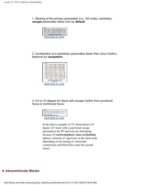
- Page 45: Lesson VI - ECG Conduction Abnormal
- Page 49 and 50: Lesson VI - ECG Conduction Abnormal
- Page 51 and 52: Lesson VI - ECG Conduction Abnormal
- Page 53 and 54: Lesson VII - Atrial Enlargement wav
- Page 55 and 56: Lesson VIII - Ventricular Hypertrop
- Page 57 and 58: Lesson VIII - Ventricular Hypertrop
- Page 59 and 60: Lesson IX - Myocardial Infarction I
- Page 61 and 62: Lesson IX - Myocardial Infarction I
- Page 63 and 64: Lesson IX - Myocardial Infarction c
- Page 65 and 66: Lesson IX - Myocardial Infarction c
- Page 67 and 68: Lesson IX - Myocardial Infarction "
- Page 69 and 70: Lesson X - ST Segment Abnormalities
- Page 71 and 72: Lesson X - ST Segment Abnormalities
- Page 73 and 74: Lesson X - ST Segment Abnormalities
- Page 75 and 76: Lesson XI - T Wave Abnormalities cl
- Page 77 and 78: Lesson XII - Nice Seeing "U" Again
- Page 79 and 80: Lesson V (cont) - ECG Rhythm Abnorm
- Page 81 and 82: Lesson V (cont) - ECG Rhythm Abnorm
- Page 83 and 84: Lesson V - ECG Rhythm Abnormalities
- Page 85 and 86: Lesson V - ECG Rhythm Abnormalities
- Page 87 and 88: Lesson V - ECG Rhythm Abnormalities
- Page 89 and 90: Lesson V - ECG Rhythm Abnormalities
- Page 91 and 92: Lesson V (cont)- Ventricular Arrhyt
- Page 93 and 94: Lesson V (cont)- Ventricular Arrhyt
- Page 95 and 96: Lesson V (cont)- Ventricular Arrhyt
- Page 97 and 98:
Lesson V (cont)- Ventricular Arrhyt
- Page 99 and 100:
Lesson V (cont)- Ventricular Arrhyt
- Page 101 and 102:
Lesson II (cont): Determining the Q
- Page 103 and 104:
ecg_ac.html 60 Cycle Artifact - Mar
- Page 105 and 106:
ecg_tremor.html Muscle Tremor Artif
- Page 107 and 108:
ecg_arrhythmia.html Marked Sinus Ar
- Page 109 and 110:
ecg_0374_mod.html Sino-Atrial Exit
- Page 111 and 112:
ecg_403.html Not All Sore Thumbs Ar
- Page 113 and 114:
ecg_491.html Atrial Tachycardia Wit
- Page 115 and 116:
ecg_vent_pace.html Ventricular Paci
- Page 117 and 118:
ecg_12lead008.html Atrial Flutter W
- Page 119 and 120:
ecg_12lead009z.html Atrial Flutter
- Page 121 and 122:
ecg_12lead011z.html Atrial Flutter
- Page 123 and 124:
ecg_478.html Atrial Flutter With 2:
- Page 125 and 126:
ecg_12lead011.html Atrial Flutter W
- Page 127 and 128:
ecg_12lead008z.html Atrial Flutter
- Page 129 and 130:
ecg_junctional.html Junctional Esca
- Page 131 and 132:
ecg_500.html Junctional Tachycardia
- Page 133 and 134:
ecg_494.html Digitalis Intoxication
- Page 135 and 136:
ecg_0280_mod.html 1st Degree AV Blo
- Page 137 and 138:
ecg_0311_mod.html How long can the
- Page 139 and 140:
ecg_0283_mod.html A Very Subtle 1st
- Page 141 and 142:
http://library.med.utah.edu/kw/ecg/
- Page 143 and 144:
ecg_12lead020.html Left Atrial Enla
- Page 145 and 146:
ecg_12lead030.html Inferior MI: Ful
- Page 147 and 148:
ecg_486.html Giant TU Fusion Waves-
- Page 149 and 150:
ecg_517.html WPW Diagram-KH Frank G
- Page 151 and 152:
ecg_703.html Conceptual Framework:
- Page 153 and 154:
ecg_compens.html Compensatory vs. N
- Page 155 and 156:
ecg_conduct.html RV vs LV PVC's - M
- Page 157 and 158:
ecg_evol.html Diagram: Stages of Ac
- Page 159 and 160:
ecg_lindsay.html Alan E. Lindsay, M
- Page 161 and 162:
ecg_outline39.html The Three Fates
- Page 163 and 164:
ecg_outline42.html Frank G. Yanowit
- Page 165 and 166:
ecg_outlline12.html Diagram: Fronta
- Page 167 and 168:
ecg_torso.html Frontal and Horizont
- Page 169 and 170:
ecg_560.html QRS Axis = -30 degrees
- Page 171 and 172:
ecg_562.html Left Axis Deviation: Q
- Page 173 and 174:
ecg_564.html QRS Axis = +30 degrees
- Page 175 and 176:
ecg_565.html Left Axis Deviation: Q
- Page 177 and 178:
ecg_6lead001.html Frontal Plane QRS
- Page 179 and 180:
ecg_6lead003.html Frontal Plane QRS
- Page 181 and 182:
ecg_6lead005.html Frontal Plane QRS
- Page 183 and 184:
ecg_6lead007.html Frontal Plane QRS
- Page 185 and 186:
ecg_6lead009.html Frontal Plane QRS
- Page 187 and 188:
ecg_6lead011.html Frontal Plane QRS
- Page 189 and 190:
ecg_6lead013.html Indeterminate Fro
- Page 191 and 192:
ecg_6lead017.html Left Axis Deviati
- Page 193 and 194:
ecg_446.html Wandering Atrial Pacem
- Page 195 and 196:
ecg_normal.html Normal Sinus Rhythm
- Page 197 and 198:
ecg_wander.html Wandering Atrial Pa
- Page 199 and 200:
ecg_0226_mod2.html What are those f
- Page 201 and 202:
ecg_0229_mod.html Left Ventricular
- Page 203 and 204:
ecg_0273_mod.html Ventricular Paras
- Page 205 and 206:
ecg_0277_mod.html PVC With Venticul
- Page 207 and 208:
ecg_0315_mod.html Nonconducted And
- Page 209 and 210:
ecg_402.html Identification of PVC'
- Page 211 and 212:
ecg_410.html An Interpolated PAC-KH
- Page 213 and 214:
ecg_418.html A Nonconducted PAC Cau
- Page 215 and 216:
ecg_441.html Atrial Parasystole-KH
- Page 217 and 218:
ecg_457.html Nonconducted and Aberr
- Page 219 and 220:
ecg_485.html Junctional Parasystole
- Page 221 and 222:
ecg_aberrant.html PAC's With and Wi
- Page 223 and 224:
ecg_bigeminy.html Atrial Bigeminy -
- Page 225 and 226:
ecg_inter_pvc.html Interpolated PVC
- Page 227 and 228:
ecg_paired.html PAC Couplet - Marqu
- Page 229 and 230:
ecg_ront.html PVC with R-on-T - Mar
- Page 231 and 232:
ecg_trigem_pvc.html PVCs - Marquett
- Page 233 and 234:
ecg_v_fusion.html Ventricular Fusio
- Page 235 and 236:
ecg_12lead069.html Atrial Fibrillat
- Page 237 and 238:
ecg_487.html Atrial tachycardia Wit
- Page 239 and 240:
ecg_498.html A Very Subtle Atrial T
- Page 241 and 242:
ecg_505.html Atrial Tachycardia Wit
- Page 243 and 244:
ecg_6lead016.html Atrial Flutter wi
- Page 245 and 246:
ecg_atrial_tachy.html Atrial Tachyc
- Page 247 and 248:
ecg_0325_mod.html Accelerated Ventr
- Page 249 and 250:
ecg_12lead057.html Left Ventricular
- Page 251 and 252:
ecg_12lead063.html Right Ventricula
- Page 253 and 254:
ecg_escape.html Ventricular Escape
- Page 255 and 256:
ecg_v_asyst.html Ventricular Asysto
- Page 257 and 258:
ecg_0236_mod.html AV Dissociation b
- Page 259 and 260:
ecg_0246_mod.html Isochronic Ventri
- Page 261 and 262:
ecg_0287_mod.html 2nd Degree AV Blo
- Page 263 and 264:
ecg_0293_mod.html Trifascicular Blo
- Page 265 and 266:
ecg_0295_mod.html Mobitz II 2nd Deg
- Page 267 and 268:
ecg_0298_mod.html 2nd Degree AV Blo
- Page 269 and 270:
ecg_0301_mod.html Complete AV Block
- Page 271 and 272:
ecg_0312_mod.html ECG Of The Centur
- Page 273 and 274:
ecg_411.html Atrial Echos-KH Frank
- Page 275 and 276:
ecg_480.html Second Degree AV Block
- Page 277 and 278:
ecg_507.html 2nd Degree AV Block Wi
- Page 279 and 280:
ecg_second_av1.html 2nd Degree AV B
- Page 281 and 282:
ecg_12lead012.html Left Anterior Fa
- Page 283 and 284:
ecg_12lead013.html Left Bundle Bran
- Page 285 and 286:
ecg_12lead014.html RBBB With Primar
- Page 287 and 288:
ecg_12lead015.html Bifascicular Blo
- Page 289 and 290:
ecg_12lead016z.html RBBB: Precordia
- Page 291 and 292:
ecg_12lead018z.html WPW Type Preexc
- Page 293 and 294:
ecg_12lead034z.html Infero-posterio
- Page 295 and 296:
ecg_12lead036.html Inferior & Anter
- Page 297 and 298:
ecg_12lead043.html Atypical LBBB wi
- Page 299 and 300:
ecg_12lead046.html Infero-posterior
- Page 301 and 302:
ecg_12lead049.html RBBB + LAFB: Bif
- Page 303 and 304:
ecg_12lead068.html WPW and Pseudo-i
- Page 305 and 306:
ecg_476.html Rate-dependent LBBB-KH
- Page 307 and 308:
ecg_706.html Left Anterior Fasicula
- Page 309 and 310:
ecg_first_av1.html Right Bundle Bra
- Page 311 and 312:
ecg_preexcite.html WPW Type Preexci
- Page 313 and 314:
ecg_0327_mod.html Ventricular Paced
- Page 315 and 316:
ecg_12lead045z.html Ventricular Pac
- Page 317 and 318:
ecg_12lead065.html Atrial Pacemaker
- Page 319 and 320:
ecg_12lead067.html AV Sequential Pa
- Page 321 and 322:
ecg_av_pace.html AV Sequential Pace
- Page 323 and 324:
ecg_paced.html Pacemaker Fusion Bea
- Page 325 and 326:
ecg_spikes.html Electronic Ventricu
- Page 327 and 328:
ecg_12lead026z.html Anteroseptal MI
- Page 329 and 330:
ecg_12lead027z.html Extensive Anter
- Page 331 and 332:
ecg_12lead029.html Infero-posterior
- Page 333 and 334:
ecg_12lead031.html Acute Inferopost
- Page 335 and 336:
ecg_12lead032z.html Postero-lateral
- Page 337 and 338:
ecg_12lead037.html Acute Inferopost
- Page 339 and 340:
ecg_12lead038.html Old Infero-poste
- Page 341 and 342:
ecg_12lead040.html Old Inferior MI,
- Page 343 and 344:
ecg_711.html Frontal Plane: Acceler
- Page 345 and 346:
ecg_721.html Inferoposterior MI-KH
- Page 347 and 348:
ecg_12lead020z.html Left Atrial Enl
- Page 349 and 350:
ecg_12lead021z.html Right Axis Devi
- Page 351 and 352:
ecg_12lead022z.html RAE & RVH-KH Fr
- Page 353 and 354:
ecg_12lead025.html LVH and Many PVC
- Page 355 and 356:
ecg_12lead042.html LVH: Limb Lead C
- Page 357 and 358:
ecg_12lead048.html RVH with Right A
- Page 359 and 360:
ecg_12lead054.html LVH - Best seen
- Page 361 and 362:
ecg_705.html http://library.med.uta
- Page 363 and 364:
ecg_12lead002.html Long QT Interval
- Page 365 and 366:
ecg_12lead003z.html Long QT Interva
- Page 367 and 368:
ecg_12lead004z.html Normal Variant:
- Page 369 and 370:
ecg_12lead007.html Inferolateral ST
- Page 371 and 372:
ecg_12lead056.html Long QT: An ECG
- Page 373 and 374:
ecg_12lead061.html Advanced Hyperka
- Page 375 and 376:
ecg_12lead001.html Lead Error: V1 &
- Page 377:
ecg_calibration.html Calibration Si