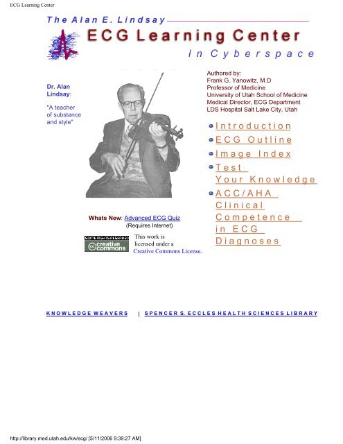
C o m p e t e n c e i n E C G D i a g n o s e s
ECG Learning Center - Student Resources Home Page
ECG Learning Center - Student Resources Home Page
- No tags were found...

You also want an ePaper? Increase the reach of your titles
YUMPU automatically turns print PDFs into web optimized ePapers that Google loves.
ECG Learning Center<br />
Dr. Alan<br />
Lindsay:<br />
"A teacher<br />
of substance<br />
and style"<br />
Whats New: Advanced ECG Quiz<br />
This work is<br />
licensed under a<br />
Creative Commons License.<br />
Authored by:<br />
Frank G. Yanowitz, M.D<br />
Professor of Medicine<br />
University of Utah School of Medicine<br />
Medical Director, ECG Department<br />
LDS Hospital Salt Lake City, Utah<br />
I n t r o d u c t i o n<br />
E C G O u t l i n e<br />
I m a g e I n d e x<br />
T e s t<br />
Y o u r K n o w l e d g e<br />
A C C / A H A<br />
C l i n i c a l<br />
C o m p e t e n c e<br />
i n E C G<br />
D i a g n o s e s<br />
K N O W L E D G E W E A V E R S | S P E N C E R S. E C C L E S H E A L T H S C I E N C E S L I B R A R Y<br />
http://library.med.utah.edu/kw/ecg/ [5/11/2006 9:39:27 AM]
ECG Introduction<br />
THE ALAN E. LINDSAY ECG LEARNING<br />
CENTER<br />
Frank G. Yanowitz, M.D<br />
Professor of Medicine<br />
University of Utah School of Medicine<br />
Medical Director, ECG Department<br />
LDS Hospital<br />
Salt Lake City, Utah<br />
This tutorial is dedicated to the memory of Dr. Alan E. Lindsay, master teacher<br />
of electrocardiography, friend, mentor, and colleague. Many of the excellent<br />
ECG tracings illustrated in this learning program are from Dr. Lindsay's personal<br />
collection of ECG treasures. For many years these ECG's have been used in<br />
the training of medical students, nurses, housestaff physicians, cardiology<br />
fellows, and practicing physicians in Salt Lake City, Utah as well as at many<br />
regional and national medical meetings. It is an honor to be able to provide this<br />
tutorial on the World Wide Web in recognition of Dr. Lindsay's great love for<br />
teaching and for electrocardiography.<br />
This interactive ECG tutorial represents an introduction to clinical<br />
electrocardiography. ECG terminology and diagnostic criteria often vary from<br />
book to book and from one teacher to another. In this tutorial an attempt has<br />
been made to conform to standardized terminology and criteria, although new<br />
diagnostic concepts derived from the recent ECG literature have been included<br />
in some of the sections. Finally, it is important to recognize that the mastery of<br />
ECG interpretation, one of the most useful clinical tools in medicine, can only<br />
occur if one acquires considerable experience in reading ECG's and correlating<br />
the specific ECG findings with the pathophysiology and clinical status of the<br />
patient.<br />
The tutorial is organized in sections based on a recommended "Method" of ECG<br />
interpretation. Each section provides some didactic teaching points, often linked<br />
to illustrations, and an interactive quiz. Beginning students should first go<br />
through the sections in the order in which they are presented. Others may chose<br />
to explore topics of interest in any order they wish. The ECG's range from the<br />
http://library.med.utah.edu/kw/ecg/intro.html (1 of 2) [5/11/2006 9:39:29 AM]
ECG Introduction<br />
sublime to the ridiculous, from simplicity to complexity, and from boring to<br />
fascinating. It is hoped that students will be left with some of the love of<br />
electrocardiography shared by Dr. Lindsay.<br />
Click to return: Home<br />
http://library.med.utah.edu/kw/ecg/intro.html (2 of 2) [5/11/2006 9:39:29 AM]
ECG Outline<br />
TABLE OF CONTENTS<br />
I. The Standard 12 Lead ECG<br />
II. A "Method of ECG Interpretation<br />
III. Characteristics of the Normal ECG<br />
IV. ECG Measurement Abnormalities<br />
V. ECG Rhythm Abnormalities<br />
VI. ECG Conduction Abnormalities<br />
VII. Atrial Enlargement<br />
VIII. Ventricular Hypertrophy<br />
IX. Myocardial Infarction<br />
X. ST Segment Abnormalities<br />
XI. T Wave Abnormalities<br />
XII. U Wave Abnormalities<br />
http://library.med.utah.edu/kw/ecg/ecg_outline/index.html [5/11/2006 9:39:29 AM]
ECG Image Index<br />
ECG Image Index<br />
ECG Categories<br />
The following ECG categories contain hundreds of ECGs that range from the<br />
sublime to the ridiculous, from simplicity to complexity, and from boring to<br />
fascinating. Many of the ECG rhythm strips come from the collection of the late<br />
Dr. Alan Lindsay, master teacher of electrocardiography. Most of the 12- and 6-<br />
lead ECGs were recorded at LDS Hospital in Salt Lake City, Utah. Marquette<br />
Electronics has also given permission to use ECG rhythms and diagrams from<br />
their educational posters. Each of the ECGs has an interpretation and many<br />
have additional explanations that help explain the diagnosis. Feedback is<br />
encouraged using the feedback form provided with this website.<br />
1. Diagrams & Pictures<br />
2. Frontal Plane QRS Axis Examples<br />
3. Sinus, Atrial, and Junctional Rhythms<br />
4. PACs, PJCs, and PVCs<br />
5. Atrial Fibrillation & Flutter, SVT's<br />
6. Ventricular Rhythms and Tachycardias<br />
7. Mischief in the AV Junction (Blocks and Dissociation)<br />
8. Bundle Branch, Fascicular Blocks, and WPW<br />
9. Artificial Pacemakers<br />
10. Myocardial Infarctions<br />
11. Hypertrophies and Enlargements<br />
12. ST-T-U Wave Abnormalities and Long QT<br />
13. Odds & Ends<br />
1. Diagrams & Pictures<br />
ecg_496.gif--Diagram: Digitalis Effect on Rhythm and Conduction<br />
ecg_517.gif--WPW Diagram<br />
ecg_533.gif--ECG Intervals and Waves<br />
ecg_703.gif--Conceptual Framework: Arrhythmias and Conduction Abnormalities<br />
ecg_ccs.gif--Cardiac Conduction System Diagram - Marquette<br />
ecg_compens.gif--Compensatory vs. Non-compensatory Pauses - Marquette<br />
ecg_components.gif--ECG Components Diagram - Marquette<br />
ecg_conduct.gif--RV vs LV PVC's - Marquette<br />
ecg_em_events.gif--Electrical and Mechanical Events Diagram - Marquette<br />
http://library.med.utah.edu/kw/ecg/image_index/index.html (1 of 9) [5/11/2006 9:39:31 AM]
ECG Image Index<br />
ecg_evol.gif--Stages of Acute Q-Wave MI - Marquette<br />
ecg_lead_wire.gif--Pacemaker Lead Wire Placement Diagram - Marquette<br />
ecg_lindsay.gif--Alan E. Lindsay, MD: A Teacher of Substance and Style<br />
ecg_outline38.gif--All About Premature Beats<br />
ecg_outline39.gif--The Three Fates Of PAC's: 1. Normal Conduction; 2. Aberrant<br />
Conduction; 3.Non-conduction<br />
ecg_outline42.gif--Diagram: AV Nodal Reentrant Tachycardia<br />
ecg_outline43.gif--Diagram: Type I vs Type II 2nd Degreee AV Block<br />
ecg_outline12.gif--Diagram: Frontal Plane Leads<br />
ecg_st.gif--ST Segment Diagram - Marquette<br />
ecg_torso.gif--Frontal and Horizontal Plane Lead Diagram<br />
2. Frontal Plane QRS Axis Examples<br />
ecg_559.gif--QRS Axis = +90 degrees<br />
ecg_560.gif--QRS Axis = -30 degrees<br />
ecg_561.gif--QRS Axis = 0 degrees<br />
ecg_562.gif--Left Axis Deviation: QRS Axis = -60 degrees<br />
ecg_563.gif--QRS Axis = +60 degrees<br />
ecg_564.gif--QRS Axis = +30 degrees<br />
ecg_565.gif--Left Axis Deviation: QRS Axis = -45 degrees<br />
ecg_566.gif--Right Axis Deviation: QRS Axis =+130 degrees<br />
ecg_6lead001.gif--Frontal Plane QRS Axis = +90 degrees<br />
ecg_6lead002.gif--Frontal Plane QRS Axis = +75degrees<br />
ecg_6lead003.gif--Frontal Plane QRS Axis = +50 degrees<br />
ecg_6lead004.gif--Frontal Plane QRS Axis = +150 degrees (RAD)<br />
ecg_6lead005.gif--Frontal Plane QRS Axis = 90 degrees<br />
ecg_6lead006.gif--Frontal Plane QRS Axis = +30 degrees<br />
ecg_6lead007.gif--Frontal Plane QRS Axis = +15 degrees<br />
ecg_6lead008.gif--Frontal Plane QRS Axis = 0 degrees<br />
ecg_6lead009.gif--Frontal Plane QRS Axis = -15 degrees<br />
ecg_6lead010.gif--Frontal Plane QRS Axis = -45 degrees<br />
ecg_6lead011.gif--Frontal Plane QRS Axis = -45 degrees<br />
ecg_6lead012.gif--Frontal Plane QRS Axis = -75 degreees<br />
ecg_6lead013.gif--Indeterminate Frontal Plane QRS Axis<br />
ecg_6lead015.gif--Right Axis Deviation<br />
ecg_6lead017.gif--Left Axis Deviation<br />
3. Sinus, Atrial, Junctional Rhythms<br />
ecg_12lead005.gif--Normal ECG<br />
http://library.med.utah.edu/kw/ecg/image_index/index.html (2 of 9) [5/11/2006 9:39:31 AM]
ECG Image Index<br />
ecg_446.gif--Wandering Atrial Pacemaker<br />
ecg_accelerate.gif--Accelerated Junctional Rhythm - Marquette<br />
ecg_arrest.gif--Sinus Pause or Arrest - Marquette<br />
ecg_arrhythmia.gif--Marked Sinus Arrhythmia - Marquette<br />
ecg_brady.gif--Sinus Bradycardia - Marquette<br />
ecg_junctional.gif--Junctional Escape Rhythm<br />
ecg_normal.gif--Normal Sinus Rhythm - Marquette<br />
ecg_sinus_brady.gif--Sinus Bradycardia<br />
ecg_tachy.gif--SinusTachycardia - Marquette<br />
ecg_wander.gif--Wandering Atrial Pacemaker - Marquette<br />
4. PACs, PJCs, and PVCs<br />
ecg_0226_mod.gif--PAC's with RBBB Aberrant Conduction<br />
ecg_0226_mod2.gif--What are those funny looking beats????<br />
ecg_0228_mod.gif--Long QT Mischief<br />
ecg_0229_mod.gif--Long QT Mischief<br />
ecg_0268_mod.gif--Atrial Parasystole<br />
ecg_0273_mod.gif--Ventricular Parasystole<br />
ecg_0274_mod.gif--Ventricular Fusion<br />
ecg_0277_mod.gif--PVC With Venticular Echo<br />
ecg_0286_mod.gif--Nonconducted PACs and Junctional Escapes<br />
ecg_0315_mod.gif-Nonconducted And Conducted PAC's<br />
ecg_401.gif--PAC and PVC: Complete vs. Incomplete Pause<br />
ecg_402.gif--Identification of PVC's and PAC's<br />
ecg_403.gif--Not All Sore Thumbs Are Ventricular In Origin<br />
ecg_404.gif--Nonconducted PAC's: An Unusual Bigeminy<br />
ecg_410.gif--An Interpolated PAC<br />
ecg_414.gif--PAC's With RBBB Aberration<br />
ecg_415.gif--The Three Fates Of PAC's<br />
ecg_418.gif--A Nonconducted PAC Causes An Unexpected Pause<br />
ecg_420.gif--Nonconducted PAC's Slowing The Heart Rate<br />
ecg_441.gif--Atrial Parasystole<br />
ecg_450.gif--Atrial Parasystole<br />
ecg_457.gif--Nonconducted and Aberrantly Conducted PAC's<br />
ecg_463.gif--Sore Thumbs<br />
ecg_485.gif--Junctional Parasystole and Pseudo-AV Block<br />
ecg_508.gif--Premature Junctional Complexes With Retrograde P Waves<br />
ecg_aberrant.gif--PAC's With and Without Aberrant Conduction - Marquette<br />
ecg_bigem_pvs.gif--Ventricular Bigeminy - Marquette<br />
ecg_bigeminy.gif--Atrial Bigeminy - Marquette<br />
ecg_contraction.gif--Nonconducted PAC -Marquette<br />
ecg_cou_pvc.gif--PVC Couplet - Marquette<br />
ecg_inter_pvc.gif--Interpolated PVCs - Marquette<br />
http://library.med.utah.edu/kw/ecg/image_index/index.html (3 of 9) [5/11/2006 9:39:31 AM]
ECG Image Index<br />
ecg_isolated.gif--Isolated PAC - Marquette<br />
ecg_multi.gif--Multifocal PVC's - Marquette<br />
ecg_paired.gif--PAC Couplet - Marquette<br />
ecg_quad_pvc.gif--PVC's - Marquette<br />
ecg_ront.gif--PVC with R-on-T - Marquette<br />
ecg_tri_pvc.gif--PVC Triplet - Marquette<br />
ecg_trigem_pvc.gif--PVCs - Marquette<br />
ecg_unifocal.gif--Unifocal PVCs - Marquette<br />
ecg_v_fusion.gif--Ventricular Fusion Beat - Marquette<br />
5. Atrial Fibrillation, Flutter and SVT's<br />
ecg_12lead008.gif--Atrial Flutter With 2:1 AV Conduction<br />
ecg_12lead008z.gif--Atrial Flutter With 2:1 Conduction: Leads II, III, V1<br />
ecg_12lead009.gif--Atrial Flutter With 3:2 AV Conduction<br />
ecg_12lead009z.gif--Atrial Flutter with 3:2 Conduction Ratio: Frontal Plane Leads<br />
ecg_12lead010.gif--Atrial Flutter With 2:1 AV Conduction<br />
ecg_12lead010z.gif--Atrial Flutter With 2:1 AV Conduction: Lead V1<br />
ecg_12lead011.gif--Atrial Flutter With 2:1 AV Conduction<br />
ecg_12lead011z.gif--Atrial Flutter With 2:1 AV Conduction: Leads II, III, V1<br />
ecg_12lead051.gif--LBBB and Atrial Flutter with 2:1 AV Block<br />
ecg_12lead059.gif--Multifocal Atrial Tachycardia (MAT)<br />
ecg_12lead069.gif--Atrial Fibrillation in Patient with WPW Syndrome<br />
ecg_407.gif--A PAC Initiates Paroxsymal Atrial Fibrillation<br />
ecg_428.gif--Massage Parlor Games<br />
ecg_477.gif--Atrial Flutter With Variable AV Block And Rate-Dependent LBBB<br />
ecg_478.gif--Atrial Flutter With 2:1 and 4:1 Conduction and Rate Dependent LBBB<br />
ecg_487.gif--Atrial tachycardia With 3:2 AV Block<br />
ecg_491.gif--Atrial Tachycardia With 3:2 and 2:1 AV Block<br />
ecg_493.gif--Digitalis Intoxication: Junctional Tachycardia With and Without AV Block493<br />
ecg_494.gif--Digitalis Intoxication: Junctional Tachycardia With and Without Exit Block<br />
ecg_495.gif--Atrial Tachycardia With Exit Block and AV Block<br />
ecg_498.gif--A Very Subtle Atrial Tachycardia With 2:1 Block<br />
ecg_500.gif--Junctional Tachycardia With Exit Block: A Manifestation of Digitalis Intoxication<br />
ecg_505.gif--Atrial Tachycardia With 2:1 AV Block: A Manifestation of Digitalis Intoxication<br />
ecg_6lead014.gif--Atrial Flutter with 2:1 AV block<br />
ecg_6lead016.gif--Atrial Flutter with 2:1 AV Block<br />
ecg_atrial_fib.gif--Atrial Fibrillation With Moderate Ventricular Response - Marquette<br />
ecg_atrial_flutter.gif--Atrial Flutter With Variable AV Block - Marquette ecg_atrial_tachy.gif--<br />
Atrial Tachycardia - Marquette<br />
http://library.med.utah.edu/kw/ecg/image_index/index.html (4 of 9) [5/11/2006 9:39:31 AM]
ECG Image Index<br />
6. Ventricular Rhythms and Tachycardias<br />
ecg_0263_mod.gif--Ventricular Parasystole<br />
ecg_0325_mod.gif--Accelerated Ventricular Rhythm With Retrograde Atrial Capture and Echo<br />
Beats<br />
ecg_0331_mod.gif--Ventricular Tachycardia With Retrograde Wenckebach<br />
ecg_12lead057.gif--Left Ventricular Tachycardia<br />
ecg_12lead058.gif--Ventricular Tachycardia<br />
ecg_12lead063.gif--Right Ventricular Tachycardia<br />
ecg_accel_idio.gif--Accelerated IVR With AV Dissociation - Marquette<br />
ecg_escape.gif--Ventricular Escape Beat - Marquette<br />
ecg_ideo.gif--Idioventricular Escape Rhythm<br />
ecg_v_asyst.gif--Ventricular Asystole - Marquette<br />
ecg_v_fib.gif--Ventricular Fibrillation - Marquette<br />
7. Mischief in the AV Junction (Blocks and Dissociation)<br />
ecg_0233_mod.gif--AV Dissociation by Default<br />
ecg_0236_mod.gif--AV Dissociation by Default<br />
ecg_0238_mod.gif--AV Dissociation by Usurpation<br />
ecg_0246_mod.gif--Isochronic Ventricular Rhythm<br />
ecg_0280_mod.gif--1st Degree AV Block<br />
ecg_0285_mod.gif--2nd Degree AV Block, Type I<br />
ecg_0287_mod.gif--2nd Degree AV Block, Type I, with Junctional Escapes<br />
ecg_0291_mod.gif--LBBB and 2nd degree AV Block, Mobitz Type II<br />
ecg_0293_mod.gif--Trifascicular Block: RBBB, LAFB, and Mobitz II 2nd Degree AV Block<br />
ecg_0294_mod.gif--RBBB plus Mobitz II 2nd Degree AV Block<br />
ecg_0295_mod.gif--Mobitz II 2nd Degree AV Block<br />
ecg_0296_mod.gif--Incomplete AV Dissociation Due To 2nd Degree AV Block<br />
ecg_0298_mod.gif--2nd Degree AV Block, Type I With Escapes and Captures<br />
ecg_0299_mod.gif--3rd Degree AV Block Rx'ed With a Ventricular Pacemaker<br />
ecg_0301_mod.gif--Complete AV Block, Junctional Escape Rhythm, and Ventriculophasic<br />
Sinus Arrhythmia<br />
ecg_0305_mod.gif--2nd Degree AV Block, Type I, With Accelerated Junctional Escapes and<br />
a Ladder Diagram<br />
ecg_0311_mod.gif--ECG Of The Century: A Most Unusual 1st Degree AV Block<br />
ecg_0312_mod.gif--ECG Of The Century - Part II: Dual AV Pathways<br />
ecg_0315_mod.gif--Nonconducted And Conducted PAC's<br />
ecg_0317_mod.gif--Two Wrongs Sometimes Make A Right<br />
ecg_411.gif--Atrial Echos<br />
ecg_425.gif--Second Degree AV Block, Type I, With 3:2 Conduction Ratio<br />
ecg_480.gif--Second Degree AV Block,Type I, With Bradycardia-dependent RBBB<br />
ecg_506.gif--Supernormal Conduction: 2nd Degree AV Block With Rare Captures;<br />
Accelerated Ventricular Rhythm<br />
ecg_507.gif--2nd Degree AV Block With Junctional Escapes And Captures<br />
http://library.med.utah.edu/kw/ecg/image_index/index.html (5 of 9) [5/11/2006 9:39:31 AM]
ECG Image Index<br />
ecg_first_av.gif--Right Bundle Branch Block<br />
ecg_second_av1.gif--2nd Degree AV Block, Type I (Wenckebach)<br />
ecg_third_av1.gif--Complete AV Block (3rd Degree) with Junctional Rhythm<br />
8. Bundle Branch, Fascicular Blocks, and WPW<br />
ecg_12lead012.gif--Left Anterior Fascicular Block(LAFB)<br />
ecg_12lead012z.gif--LAFB: Frontal Plane Leads<br />
ecg_12lead013.gif--Left Bundle Branch Block (LBBB)<br />
ecg_12lead013z.gif--LBBB: Precordial Leads<br />
ecg_12lead014.gif--RBBB With Primary ST-T Wave Abnormalities<br />
ecg_12lead014z.gif--RBBB with Primary ST-T Abnormalities: Precordial Leads<br />
ecg_12lead015.gif--Bifascicular Block: RBBB + LAFB<br />
ecg_12lead016.gif--Bifascicular Block: RBBB + LAFB<br />
ecg_12lead016z.gif--RBBB: Precordial Leads<br />
ecg_12lead018.gif--WPW Type Preexcitation<br />
ecg_12lead018z.gif--WPW Type Preexcitation: Precordial Leads<br />
ecg_12lead018z.gifAbnormalities<br />
ecg_12lead034.gif--Infero-posterior MI & RBBB<br />
ecg_12lead034z.gif--Infero-posterior MI & RBBB: Frontal Plane Leads + V1<br />
ecg_12lead035.gif--Inferior MI and RBBB<br />
ecg_12lead036.gif--Inferior & Anteroseptal MI + RBBB<br />
ecg_12lead036z.gif--Anteroseptal MI With RBBB: Precordial Leads<br />
ecg_12lead043.gif--Atypical LBBB with Q Waves in Leads I and aVL<br />
ecg_12lead044.gif--Atypical LBBB with Primary T Wave Abnormalities<br />
ecg_12lead046.gif--Infero-posterior MI with RBBB<br />
ecg_12lead047.gif--RBBB + LAFB = Bifascicular block<br />
ecg_12lead049.gif--RBBB + LAFB: Bifascicular Block<br />
ecg_12lead050.gif--Right Bundle Branch Block (RBBB)<br />
ecg_12lead068.gif--WPW and Pseudo-inferior MI<br />
ecg_12lead070.gif--WPW with a Pseudo-inferior MI<br />
ecg_476.gif--Rate-dependent LBBB<br />
ecg_482.gif--Bradycardia-dependent LBBB With Carotid Sinus Massage<br />
ecg_706.gif--Left Anterior Fasicular Block: Frontal Plane Leads<br />
ecg_first_av1.gif--Right Bundle Branch Block<br />
ecg_lbbb.gif--Left Bundle Branch Block - Marquette<br />
ecg_preexcite.gif--WPW Type Preexcitation - Marquette<br />
ecg_rbbb.gif--RBBB - Marquette<br />
9. Artificial Pacemakers<br />
http://library.med.utah.edu/kw/ecg/image_index/index.html (6 of 9) [5/11/2006 9:39:31 AM]
ECG Image Index<br />
ecg_0327.gif--Ventricular Paced Rhythm With Retrograde Wenckebach<br />
ecg_12lead045.gif--Ventricular Pacemaker Rhythm<br />
ecg_12lead045z.gif--Ventricular Pacemaker Rhythm: V1-3<br />
ecg_12lead053.gif--Ventricular Pacemaker: Demand mode functioning<br />
ecg_12lead065.gif--Atrial Pacemaker Rhythm<br />
ecg_12lead066.gif--AV Sequential Pacing<br />
ecg_12lead067.gif--AV Sequential Pacing<br />
ecg_atrial_pace.gif--Electronic Atrial Pacing - Marquette<br />
ecg_av_pace.gif--AV Sequential Pacemaker - Marquette<br />
ecg_out_fail.gif--Pacemaker Failure to Pace - Marquette<br />
ecg_paced.gif--Pacemaker Fusion Beat - Marquette<br />
ecg_sense_fail.gif--Pacemaker Failure To Sense - Marquette<br />
ecg_spikes.gif--Electronic Ventricular Pacemaker Rhythm - Marquette<br />
ecg_vent_pace.gif--Ventricular Pacing in Atrial Fibrillation - Marquette<br />
10. Myocardial Infarctions<br />
ecg_12lead026.gif--Anteroseptal MI: Fully Evolved<br />
ecg_12lead026z.gif--Anteroseptal MI, Fully Evolved: Precordial Leads<br />
ecg_12lead027.gif--Extensive Anterior/Anterolateral MI: Recent ecg_12lead027z.gif--<br />
Extensive Anterior/Anterolateral MI: Precordial Leads<br />
ecg_12lead028.gif--Acute Anterior MI<br />
ecg_12lead029.gif--Infero-posterior MI<br />
ecg_12lead030.gif--Inferior MI: Fully Evolved<br />
ecg_12lead030z.gif--Fully Evolved Inferior MI: Frontal Plane<br />
ecg_12lead031.gif--Acute Inferoposterior MI<br />
ecg_12lead032.gif--Postero-lateral MI: Fully Evolved<br />
ecg_12lead032z.gif--Postero-lateral MI: Precordial Leads<br />
ecg_12lead033.gif--Diffuse Anterolateral T Wave Abnormalities<br />
ecg_12lead034.gif--Infero-posterior MI & RBBB<br />
ecg_12lead034z.gif--Infero-posterior MI & RBBB: Frontal Plane Leads + V1<br />
ecg_12lead035.gif--Inferior MI and RBBB<br />
ecg_12lead036.gif--Inferior & Anteroseptal MI + RBBB<br />
ecg_12lead036z.gif--Anteroseptal MI With RBBB: Precordial Leads<br />
ecg_12lead037.gif--Acute Inferoposterior MI with Right Ventricular MI<br />
ecg_12lead037z.gif--True Posterior MI and Right Ventricular MI<br />
ecg_12lead038.gif--Old Infero-posterior MI<br />
ecg_12lead039.gif--Old Inferior MI<br />
ecg_12lead040.gif--Old Inferior MI, PVCs, and Atrial Fibrillation<br />
ecg_12lead041.gif--Old Inferior MI<br />
ecg_12lead043.gif--Atypical LBBB with Q Waves in Leads I and aVL<br />
ecg_12lead044.gif--Atypical LBBB with Primary T Wave Abnormalities<br />
ecg_12lead046.gif--Infero-posterior MI with RBBB<br />
ecg_12lead055.gif--High Lateral Wall MI (seen in aVL)<br />
http://library.med.utah.edu/kw/ecg/image_index/index.html (7 of 9) [5/11/2006 9:39:31 AM]
ECG Image Index<br />
ecg_711.gif--Frontal Plane: Accelerated Junctional Rhythm and Inferior MI<br />
ecg_720.gif--Inferoposterior MI<br />
ecg_721.gif--Inferoposterior MI<br />
11. Hypertrophies and Enlargements<br />
ecg_12lead019.gif--Left Atrial Abnormality & 1st degree AV Block<br />
ecg_12lead019z.gif--Left Atrial Abnormality & 1st Degree AV Block: Leads II and V1<br />
ecg_12lead020.gif--Left Atrial Enlargement & Nonspecific ST-T Wave Abnormalities<br />
ecg_12lead020z.gif--Left Atrial Enlargement: Leads II and V1<br />
ecg_12lead021.gif--Right Ventricular Hypertrophy (RVH) & Right Atrial Enlargement (RAE)<br />
ecg_12lead021z.gif--Right Axis Deviation & RAE (P Pulmonale): Leads I, II, III<br />
ecg_12lead022.gif--Right Atrial Enlargement (RAE) & Right Ventricular Hypertrophy (RVH)<br />
ecg_12lead022z.gif--RAE & RVH ecg_12lead023.gif--Voltage Criteria for LVH<br />
ecg_12lead024.gif--LVH with "Strain"<br />
ecg_12lead025.gif--LVH and Many PVCs<br />
ecg_12lead025z.gif--LVH & PVCs: Precordial Leads<br />
ecg_12lead042.gif--LVH: Limb Lead Criteria<br />
ecg_12lead042z.gif--LVH: Limb Lead Criteria<br />
ecg_12lead048.gif--RVH with Right Axis Deviation<br />
ecg_12lead052.gif--LVH: Strain pattern + Left Atrial Enlargement<br />
ecg_12lead054.gif--LVH - Best seen in the frontal plane leads!<br />
ecg_12lead064.gif--Severe RVH<br />
ecg_705.gif--Left Atrial Enlargement<br />
12. ST-T and U Wave Abnormalities and Long QT<br />
ecg_12lead002.gif--Long QT Interval and Giant Negative T Waves<br />
ecg_12lead003.gif--Long QT Interval<br />
ecg_12lead003z.gif--Long QT Interval<br />
ecg_12lead004.gif--Normal Variant: Early Repolarization<br />
ecg_12lead004z.gif--Normal Variant: Early Repolarization<br />
ecg_12lead006.gif--ST Segment Depression<br />
ecg_12lead006z.gif--ST Segment Depression: Precordial Leads<br />
ecg_12lead007.gif--Inferolateral ST Segment Elevation<br />
ecg_12lead007z.gif--ST Segment Elevation: Frontal Plane Leads<br />
ecg_12lead056.gif--Long QT: An ECG Marker For Sudden Cardiac Death<br />
ecg_12lead060.gif--Hyperkalemia and Old Inferior MI<br />
ecg_12lead061.gif--Advanced Hyperkalemia<br />
ecg_486.gif--Giant TU Fusion Waves<br />
ecg_6lead018.gif--Hypothermia: J-waves or Osborne Waves<br />
http://library.med.utah.edu/kw/ecg/image_index/index.html (8 of 9) [5/11/2006 9:39:31 AM]
ECG Image Index<br />
13. Odds & Ends<br />
ecg_12lead001.gif--Lead Error: V1 & V3 are Transposed<br />
ecg_12lead001z.gif--Lead Error: V1 and V3 are Transposed!<br />
ecg_ac.gif--60 Cycle Artifact - Marquette<br />
ecg_baseline.gif--Wandering Baseline Artifact - Marquette<br />
ecg_calibration.gif--Calibration Signal - Marquette<br />
ecg_tremor.gif--Muscle Tremor Artifact - Marquette<br />
http://library.med.utah.edu/kw/ecg/image_index/index.html (9 of 9) [5/11/2006 9:39:31 AM]
Test your knowledge - ECG Quizzes<br />
ECG Quizzes Lessons 1-12<br />
Frank G. Yanowitz, MD<br />
Professor of Medicine<br />
University of Utah School of Medicine<br />
Co-author: Steve Parsons, MS II University of Utah School of Medicine<br />
Basic Quizzes<br />
1. The 12 Lead ECG and Method of Interpretation (Lessons I and II)<br />
2. Normal ECG Characteristics and Measurement Abnormalities (Lessons III and IV)<br />
3. Arrhythmias (Lesson V)<br />
4. Conduction Abnormalities (Lesson VI)<br />
5. Atrial Enlargement and Ventricular Hypertrophy (Lessons VII and VIII)<br />
6. Myocardial Infarctions (Lesson IX)<br />
7. ST, T, and U Waves (Lessons X, XI, and XII)<br />
Advanced Quizzes<br />
1. Quiz 1<br />
http://library.med.utah.edu/kw/ecg/tests/index.html [5/11/2006 9:39:31 AM]
ECG Introduction<br />
ACC/AHA Clinical Competence in ECG Diagnoses<br />
The following list of ECG diagnoses was derived from a recently published<br />
statement of the American College of Cardiology/American Heart Association<br />
(ACC/AHA) Committee to Develop a Clinical Competence Statement on<br />
Electrocardiography and Ambulatrory Electrocardiography (J Am Coll Cardiol<br />
2001;38:2091-2100). These diagnoses are considered to be the minimum<br />
knowledge necessary for competence in interpreting 12-lead ECGs. Items in the<br />
list are linked to topics in the ECG Outline of the Alan E. Lindsay ECG Learning<br />
Center or to ECG examples in the Image Index.<br />
Normal Tracing<br />
●<br />
Normal ECG<br />
Technical Problem<br />
●<br />
●<br />
Lead Misplaced<br />
Artifact<br />
❍ Normal Variants or Artifacts<br />
❍ 60 Cycle Artifact<br />
❍ Wandering Baseline Artifact<br />
❍ Muscle Tremor Artifact<br />
Sinus Rhythms/Arrythmias<br />
●<br />
●<br />
●<br />
●<br />
●<br />
●<br />
Sinus rhythm (50-90 bpm)<br />
Sinus tachycardia (>90 bpm)<br />
Sinus bradycardia (
ECG Introduction<br />
❍<br />
❍<br />
❍<br />
❍<br />
❍<br />
❍<br />
❍<br />
❍<br />
❍<br />
❍<br />
❍<br />
Atrial Flutter With 3:2 AV Conduction<br />
Atrial Flutter with 3:2 Conduction Ratio<br />
Atrial Flutter With Variable AV Block And Rate-Dependent LBBB<br />
Atrial Flutter With 2:1 AV Conduction<br />
LBBB and Atrial Flutter with 2:1 AV Block<br />
Atrial Flutter With 2:1 and 4:1 Conduction and Rate Dependent LBBB<br />
Atrial Flutter With 2:1 AV Conduction<br />
Atrial Flutter With 2:1 AV Conduction<br />
Atrial Flutter With Variable AV Block<br />
Atrial Flutter With 2:1 Conduction<br />
Atrial Flutter with 2:1 Block<br />
●<br />
●<br />
●<br />
●<br />
●<br />
Junctional prematures<br />
Junctional escapes or rhythms<br />
Accelerated Junctional rhythms<br />
Junctional tachycardia<br />
❍ Junctional Tachycardia With Exit Block<br />
❍ Junctional Tachycardia With and Without AV Block<br />
❍ Junctional Tachycardia With and Without Exit Block<br />
Paroxysmal supraventricular tachycardia<br />
Ventricular Arrythmias<br />
●<br />
●<br />
●<br />
●<br />
●<br />
●<br />
PVC's<br />
Ventricular escapes or rhythm<br />
Accelerated ventricular rhythm<br />
Ventricular tachycardia (uniform)<br />
Ventricular tachycardia (polymorphous or torsade)<br />
Ventricular fibrillation<br />
AV Conduction<br />
●<br />
●<br />
●<br />
●<br />
●<br />
●<br />
●<br />
●<br />
1st degree AV block<br />
❍ 1st Degree AV Block<br />
❍ A Most Unusual 1st Degree AV Block<br />
❍ Left Atrial Abnormality & 1st degree AV Block<br />
❍ Left Atrial Abnormality & 1st Degree AV Block<br />
❍ A Very Subtle 1st Degree AV Block<br />
Type I 2nd degree AV block (Wenckebach)<br />
Type II 2nd degree AV block (Mobitz)<br />
AV block, advanced (high grade)<br />
3rd degree AV block (junctional escape rhythm)<br />
3rd degree AV block (ventricular escape rhythm)<br />
AV dissociation (default)<br />
❍ Subsidiary escape pacemaker takes over by default<br />
AV dissociation (usurpation)<br />
❍ Incomplete AV Dissociation due to accelerated ventricular rhythm<br />
Intraventricular Conduction<br />
●<br />
●<br />
Complete LBBB, fixed or intermittent<br />
Incomplete LBBB<br />
http://library.med.utah.edu/kw/ecg/ACC_AHA.html (2 of 4) [5/11/2006 9:39:33 AM]
ECG Introduction<br />
●<br />
●<br />
●<br />
●<br />
Left anterior fascicular block (LAFB)<br />
Left posterior fascicular block (LPFB)<br />
Nonspecific IVCD<br />
WPW preexcitation pattern<br />
QRS Axis and Voltage<br />
● Right axis deviation (+90 to +180)<br />
● Left axis deviation (-30 to -90)<br />
● Bizarre axis (-90 to -180)<br />
● Indeterminate axis<br />
● Low voltage frontal plane (
ECG Introduction<br />
●<br />
●<br />
Chronic pulmonary disease pattern<br />
Suggests hypokalemia<br />
❍ Giant TU Fusion Waves<br />
●<br />
●<br />
●<br />
●<br />
Suggests hyperkalemia<br />
Suggests hypocalcemia<br />
Suggests hypercalcemia<br />
Suggests digoxin effect<br />
http://library.med.utah.edu/kw/ecg/ACC_AHA.html (4 of 4) [5/11/2006 9:39:33 AM]
ECG Feedback<br />
The Alan E. Linday ECG Learning Center Feedback Form<br />
Please let us know how we're doing!<br />
How useful is the online Electrocardiography information?<br />
Vital Useful Not Useful<br />
Check all that apply:<br />
Patient Student Resident Physician<br />
Other: (fill in)<br />
Email address:<br />
(please provide your name and e-mail address if you would like a reply)<br />
Comments:<br />
http://library.med.utah.edu/kw/ecg/feedback/feedback.html (1 of 2) [5/11/2006 9:39:33 AM]
ECG Feedback<br />
Thank you for your input!<br />
http://library.med.utah.edu/kw/ecg/feedback/feedback.html (2 of 2) [5/11/2006 9:39:33 AM]
Lesson III - Characteristics of the Normal ECG<br />
III. Characteristics of the Normal ECG<br />
Frank G. Yanowitz, MD<br />
Professor of Medicine<br />
University of Utah School of Medicine<br />
It is important to remember that there is a wide range of normal variability in the<br />
12 lead ECG. The following "normal" ECG characteristics, therefore, are not<br />
absolute. It takes considerable ECG reading experience to discover all the<br />
normal variants. Only by following a structured "Method of ECG Interpretation"<br />
(Lesson II) and correlating the various ECG findings with the particular patient's<br />
clinical status will the ECG become a valuable clinical tool.<br />
Topics for Study:<br />
1. Measurements<br />
2. Rhythm<br />
3. Conduction<br />
4. Waveform description<br />
1. Measurements<br />
Heart Rate: 60 - 90 bpm<br />
How to calculate the heart rate on ECG paper<br />
PR Interval: 0.12 - 0.20 sec<br />
QRS Duration: 0.06 - 0.10 sec<br />
QT Interval (QT c
Lesson III - Characteristics of the Normal ECG<br />
Bazett's Formula: QT c = (QT)/SqRoot RR (in seconds)<br />
Poor Man's Guide to upper limits of QT: For HR = 70 bpm, QT
Lesson III - Characteristics of the Normal ECG<br />
Click to view<br />
P Wave<br />
It is important to remember that the P wave represents the sequentialactivation of the right<br />
and left atria, and it is common to see notched or biphasic P waves of right and left atrial<br />
activation.<br />
P duration < 0.12 sec<br />
P amplitude < 2.5 mm<br />
Frontal plane P wave axis: 0 o to +75 o<br />
May see notched P waves in frontal plane<br />
QRS Complex<br />
The QRS represents the simultaneousactivation of the right and left ventricles, although most<br />
of the QRS waveform is derived from the larger left ventricular musculature.<br />
QRS duration < 0.10 sec<br />
QRS amplitude is quite variable from lead to lead and from person to person.<br />
Two determinates of QRS voltages are:<br />
Size of the ventricular chambers (i.e., the larger the chamber, the<br />
larger the voltage)<br />
Proximity of chest electrodes to ventricular chamber (the closer,<br />
the larger the voltage)<br />
Frontal plane leads:<br />
The normal QRS axis range (+90 o to -30 o ); this implies that the<br />
QRS be mostly positive (upright) in leads II and I.<br />
Normal q-waves reflect normal septal activation (beginning on<br />
the LV septum); they are narrow (
Lesson III - Characteristics of the Normal ECG<br />
Precordial leads: (see Normal ECG)<br />
Small r-waves begin in V1 or V2 and progress in size to V5. The<br />
R-V6 is usually smaller than R-V5.<br />
In reverse, the s-waves begin in V6 or V5 and progress in size to<br />
V2. S-V1 is usually smaller than S-V2.<br />
The usual transition from S>R in the right precordial leads to<br />
R>S in the left precordial leads is V3 or V4.<br />
Small "septal" q-waves may be seen in leads V5 and V6.<br />
ST Segment and T wave<br />
In a sense, the term "ST segment" is a misnomer, because a discrete ST segment distinct from the T wave is usually<br />
absent. More often the ST-T wave is a smooth, continuous waveform beginning with the J-point (end of QRS), slowly<br />
rising to the peak of the T and followed by a rapid descent to the isoelectric baseline or the onset of the U wave. This<br />
gives rise to an asymmetrical T wave. In some normal individuals, particularly women, the T wave is symmetrical and a<br />
distinct, horizontal ST segment is present.<br />
The normal T wave is usually in the same direction as the QRS except in the right precordial leads. In the normal ECG<br />
the T wave is always upright in leads I, II, V3-6, and always inverted in lead aVR.<br />
Normal ST segment elevation: this occurs in leads with large S waves (e.g., V1-3), and the normal<br />
configuration is concave upward. ST segment elevation with concave upward appearance may also be seen<br />
in other leads; this is often called early repolarization, although it's a term with little physiologic meaning<br />
(see example of "early repolarization" in leads V4-6):<br />
Click to view<br />
Convex or straight upward ST segment elevation (e.g., leads II, III, aVF) is<br />
abnormal and suggests transmural injury or infarction:<br />
http://library.med.utah.edu/kw/ecg/ecg_outline/Lesson3/index.html (4 of 6) [5/11/2006 9:39:35 AM]
Lesson III - Characteristics of the Normal ECG<br />
Click to view<br />
ST segment depression is always an abnormal finding, although often<br />
nonspecific (see ECG below):<br />
Click to view<br />
ST segment depression is often characterized as "upsloping", "horizontal", or<br />
"downsloping".<br />
Click to view<br />
The normal U Wave: (the most neglected of the ECG waveforms)<br />
U wave amplitude is usually < 1/3 T wave amplitude in same lead<br />
U wave direction is the same as T wave direction in that lead<br />
U waves are more prominent at slow heart rates and usually best seen in the<br />
right precordial leads.<br />
Origin of the U wave is thought to be related to afterdepolarizations which<br />
interrupt or follow repolarization.<br />
http://library.med.utah.edu/kw/ecg/ecg_outline/Lesson3/index.html (5 of 6) [5/11/2006 9:39:35 AM]
Lesson III - Characteristics of the Normal ECG<br />
http://library.med.utah.edu/kw/ecg/ecg_outline/Lesson3/index.html (6 of 6) [5/11/2006 9:39:35 AM]
Lesson 1: The Standard 12 Lead ECG<br />
I. The Standard 12 Lead ECG<br />
Frank G. Yanowitz, MD<br />
Professor of Medicine<br />
University of Utah School of Medicine<br />
The standard 12-lead electrocardiogram is a representation of the heart's<br />
electrical activity recorded from electrodes on the body surface. This section<br />
describes the basic components of the ECG and the lead system used to record<br />
the ECG tracings.<br />
Topics for study:<br />
1. ECG Waves and Intervals<br />
2. Spatial Orientation of the 12 Lead ECG<br />
click to view<br />
This diagram illustrates ECG waves and<br />
intervals as well as standard time and voltage<br />
measures on the ECG paper.<br />
1. ECG Waves and Intervals:<br />
What do they mean?<br />
P wave: the sequentialactivation (depolarization) of the right and left atria<br />
QRS complex: right and left ventricular depolarization (normally the ventricles are<br />
activated simultaneously)<br />
ST-T wave: ventricular repolarization<br />
http://library.med.utah.edu/kw/ecg/ecg_outline/Lesson1/index.html (1 of 3) [5/11/2006 9:39:35 AM]
Lesson 1: The Standard 12 Lead ECG<br />
U wave: origin for this wave is not clear - but probably represents "afterdepolarizations" in<br />
the ventricles<br />
PR interval: time interval from onset of atrial depolarization (P wave) to onset of ventricular<br />
depolarization (QRS complex)<br />
QRS duration: duration of ventricular muscle depolarization<br />
QT interval: duration of ventricular depolarization and repolarization<br />
RR interval: duration of ventricular cardiac cycle (an indicator of ventricular rate)<br />
PP interval: duration of atrial cycle (an indicator of atrial rate)<br />
2. Orientation of the 12 Lead ECG<br />
It is important to remember that the 12-lead ECG provides spatial information<br />
about the heart's electrical activity in 3 approximately orthogonal directions:<br />
Right<br />
Superior<br />
Anterior<br />
Left<br />
Inferior<br />
Posterior<br />
Each of the 12 leads represents a particular orientation in space, as indicated<br />
below (RA = right arm; LA = left arm, LF = left foot):<br />
Bipolar limb leads (frontal plane):<br />
Lead I: RA (-) to LA (+) (Right Left, or lateral)<br />
Lead II: RA (-) to LF (+) (Superior Inferior)<br />
Lead III: LA (-) to LF (+) (Superior Inferior)<br />
Augmented unipolar limb leads (frontal plane):<br />
Lead aVR: RA (+) to [LA & LF] (-) (Rightward)<br />
Lead aVL: LA (+) to [RA & LF] (-) (Leftward)<br />
Lead aVF: LF (+) to [RA & LA] (-) (Inferior)<br />
http://library.med.utah.edu/kw/ecg/ecg_outline/Lesson1/index.html (2 of 3) [5/11/2006 9:39:35 AM]
Lesson 1: The Standard 12 Lead ECG<br />
Unipolar (+) chest leads (horizontal plane):<br />
Leads V1, V2, V3: (Posterior Anterior)<br />
Leads V4, V5, V6:(Right Left, or lateral)<br />
Click here to see: Lead Placement Diagrams (Requires an Internet connection)<br />
http://library.med.utah.edu/kw/ecg/ecg_outline/Lesson1/index.html (3 of 3) [5/11/2006 9:39:35 AM]
Lesson II - A "Method of ECG Interpretation<br />
II. A "Method" of ECG Interpretation<br />
Frank G. Yanowitz, MD<br />
Professor of Medicine<br />
University of Utah School of Medicine<br />
This "method" is recommended when reading all 12-lead ECG's. Like the<br />
physical examination, it is desirable to follow a standardized sequence of steps<br />
in order to avoid missing subtle abnormalities in the ECG tracing, some of which<br />
may have clinical importance. The 6 major sections in the "method" should be<br />
considered in the following order:<br />
1. Measurements<br />
2. Rhythm Analysis<br />
3. Conduction Analysis<br />
4. Waveform Description<br />
5. Ecg Interpretation<br />
6. Comparison with Previous ECG (if any)<br />
1. Measurements (usually made in frontal plane leads):<br />
Click to view<br />
Heart rate (state atrial and ventricular, if different)<br />
PR interval (from beginning of P to beginning of QRS)<br />
QRS duration (width of most representative QRS)<br />
http://library.med.utah.edu/kw/ecg/ecg_outline/Lesson2/index.html (1 of 4) [5/11/2006 9:39:36 AM]
Lesson II - A "Method of ECG Interpretation<br />
QT interval (from beginning of QRS to end of T)<br />
QRS axis in frontal plane (go to: "How To Determine Axis")<br />
Go to: ECG Measurement Abnormalities (Lesson IV)for description of normal and abnormal<br />
measurements<br />
2. Rhythm Analysis<br />
State basic rhythm (e.g., "normal sinus rhythm", "atrial fibrillation", etc.)<br />
Identify additional rhythm events if present (e.g., "PVC's", "PAC's", etc)<br />
Consider all rhythm events from atria, AV junction, and ventricles<br />
Go to: ECG Rhythm Abnormalities (Lesson V)for description of arrhythmias<br />
3. Conduction Analysis<br />
"Normal"conduction implies normal sino-atrial (SA), atrio-ventricular (AV), and<br />
intraventricular (IV) conduction.<br />
Click to view<br />
The diagram<br />
illustrates the<br />
normal cardiac<br />
conduction<br />
system.<br />
The following conduction abnormalities are to be identified if present:<br />
SA block (lesson VI): 2nd degree (type I vs. type II)<br />
AV block (lesson VI): 1st, 2nd (type I vs. type II), and 3rd degree<br />
IV blocks (lesson VI): bundle branch, fascicular, and nonspecific blocks<br />
Exit blocks: blocks just distal to ectopic pacemaker site<br />
(Go to ECG Conduction Abnormalities (Lesson VI) for a description of<br />
conduction abnormalities)<br />
http://library.med.utah.edu/kw/ecg/ecg_outline/Lesson2/index.html (2 of 4) [5/11/2006 9:39:36 AM]
Lesson II - A "Method of ECG Interpretation<br />
4. Waveform Description<br />
Carefully analyze the 12-lead ECG for abnormalities in each of the waveforms in the order<br />
in which they appear: P-waves, QRS complexes, ST segments, T waves, and... Don't forget<br />
the U waves.<br />
P waves (lesson VII): are they too wide, too tall, look funny (i.e., are they<br />
ectopic), etc.?<br />
QRS complexes: look for pathologic Q waves (lesson IX), abnormal voltage<br />
(lesson VIII), etc.<br />
ST segments (lesson X): look for abnormal ST elevation and/or depression.<br />
T waves (lesson XI): look for abnormally inverted T waves.<br />
U waves (lesson XII): look for prominent or inverted U waves.<br />
5. ECG Interpretation<br />
This is the conclusion of the above analyses. Interpret the ECG as "Normal", or<br />
"Abnormal". Occasionally the term "borderline"is used if unsure about the significance of<br />
certain findings. List all abnormalities. Examples of "abnormal"statements are:<br />
Inferior MI, probably acute<br />
Old anteroseptal MI<br />
Left anterior fascicular block (LAFB)<br />
Left ventricular hypertrophy (LVH)<br />
Nonspecific ST-T wave abnormalities<br />
Any rhythm abnormalities<br />
Example:<br />
http://library.med.utah.edu/kw/ecg/ecg_outline/Lesson2/index.html (3 of 4) [5/11/2006 9:39:36 AM]
Lesson II - A "Method of ECG Interpretation<br />
Click to view<br />
6. Comparison with previous ecg<br />
If there is a previous ECG in the patient's file, the current ECG should be compared with it<br />
to see if any significant changes have occurred. These changes may have important<br />
implications for clinical management decisions.<br />
Test your knowledge on lessons I and II by clicking here (Internet connection required)<br />
http://library.med.utah.edu/kw/ecg/ecg_outline/Lesson2/index.html (4 of 4) [5/11/2006 9:39:36 AM]
Lesson IV - Abnormalities in the ECG Measurements<br />
IV. Abnormalities in the ECG Measurements<br />
Frank G. Yanowitz, MD<br />
Professor of Medicine<br />
University of Utah School of Medicine<br />
Click on the measurement abnormality you would like to study<br />
1. Heart Rate<br />
2. PR Interval<br />
3. QRS Duration<br />
4. QT Interval<br />
5. QRS Axis<br />
1. Heart Rate<br />
In normal sinus rhythm, a resting heart rate of below 60 bpm is called<br />
bradycardia and a rate of above 90 bpm is called tachycardia.<br />
2. PR Interval<br />
(measured from beginning of P to beginning of QRS in the frontal plane)<br />
Normal: 0.12 - 0.20s<br />
Short PR:
Lesson IV - Abnormalities in the ECG Measurements<br />
(called the "Kent" bundle) connects the right atrium to the right<br />
ventricle (see diagram below) or the left atrium to the left ventricle,<br />
and this permits early activation of the ventricles (delta wave) and a<br />
short PR interval.<br />
Click to view<br />
LGL (Lown-Ganong-Levine): An AV nodal bypass track into the<br />
His bundle exists, and this permits early activation of the ventricles<br />
without a delta-wave because the ventricular activation sequence is<br />
normal.<br />
AV Junctional Rhythms with retrograde atrial activation (inverted P waves in<br />
II, III, aVF): Retrograde P waves may occur before the QRS complex (usually<br />
with a short PR interval), in the QRS complex (i.e., hidden from view), or after<br />
the QRS complex (i.e., in the ST segment).<br />
Ectopic atrial rhythms originating near the AV node (the PR interval is short<br />
because atrial activation originates close to the AV node; the P wave<br />
morphology is different from the sinus P)<br />
Normal variant<br />
Prolonged PR: >0.20s<br />
First degree AV block (PR interval usually constant)<br />
Intra-atrial conduction delay (uncommon)<br />
Slowed conduction in AV node (most common site)<br />
Slowed conduction in His bundle (rare)<br />
Slowed conduction in bundle branch (when contralateral bundle<br />
is blocked)<br />
http://library.med.utah.edu/kw/ecg/ecg_outline/Lesson4/index.html (2 of 5) [5/11/2006 9:39:37 AM]
Lesson IV - Abnormalities in the ECG Measurements<br />
Second degree AV block (PR interval may be normal or prolonged; some P<br />
waves do not conduct)<br />
Type I (Wenckebach): Increasing PR until nonconducted P wave<br />
occurs<br />
Type II (Mobitz): Fixed PR intervals plus nonconducted P waves<br />
AV dissociation: Some PR's may appear prolonged, but the P waves and<br />
QRS complexes are dissociated (i.e., not married, but strangers passing in the<br />
night).<br />
3. QRS Duration<br />
(duration of QRS complex in frontal plane):<br />
Normal: 0.06 - 0.10s<br />
Prolonged QRS Duration (>0.10s):<br />
QRS duration 0.10 - 0.12s<br />
Incomplete right or left bundle branch block<br />
Nonspecific intraventricular conduction delay (IVCD)<br />
Some cases of left anterior or posterior fascicular block<br />
QRS duration > 0.12s<br />
Complete RBBB or LBBB<br />
Nonspecific IVCD<br />
Ectopic rhythms originating in the ventricles (e.g., ventricular<br />
tachycardia, pacemaker rhythm)<br />
http://library.med.utah.edu/kw/ecg/ecg_outline/Lesson4/index.html (3 of 5) [5/11/2006 9:39:37 AM]
Lesson IV - Abnormalities in the ECG Measurements<br />
4. QT Interval<br />
(measured from beginning of QRS to end of T wave in the frontal plane)<br />
Normal: heart rate dependent (corrected QT = QT c = measured QT ÷ sq-root RR in seconds; upper<br />
limit for QT c = 0.44 sec)<br />
Long QT Syndrome - "LQTS" (based on upper limits for heart rate; QT c > 0.47 sec for males and > 0.48<br />
sec in females is diagnostic for hereditary LQTS in absence of other causes of increased QT)<br />
This abnormality may have important clinical implications since it usually indicates a state of<br />
increased vulnerability to malignant ventricular arrhythmias, syncope, and sudden death. The<br />
prototype arrhythmia of the Long QT Interval Syndromes (LQTS) is Torsade-de-pointes, a<br />
polymorphic ventricular tachycardia characterized by varying QRS morphology and amplitude<br />
around the isoelectric baseline. Causes of LQTS include the following:<br />
Drugs (many antiarrhythmics, tricyclics, phenothiazines, and others)<br />
Electrolyte abnormalities ( K + , Ca ++ , Mg ++ )<br />
CNS disease (especially subarrachnoid hemorrhage, stroke, trauma)<br />
Hereditary LQTS (e.g., Romano-Ward Syndrome)<br />
Coronary Heart Disease (some post-MI patients)<br />
5. Frontal Plane QRS Axis<br />
Click here for brief tutorial in Measuring QRS Axis<br />
Normal: -30 degrees to +90 degrees<br />
Abnormalities in the QRS Axis:<br />
Left Axis Deviation (LAD): > -30 o (i.e., lead II is mostly 'negative')<br />
Left Anterior Fascicular Block (LAFB): rS complex in leads II, III, aVF, small<br />
q in leads I and/or aVL, and axis -45 o to -90 o<br />
Some cases of inferior MI with Qr complex in lead II (making lead<br />
II 'negative')<br />
Inferior MI + LAFB in same patient (QS or qrS complex in lead II)<br />
Some cases of LVH<br />
http://library.med.utah.edu/kw/ecg/ecg_outline/Lesson4/index.html (4 of 5) [5/11/2006 9:39:37 AM]
Lesson IV - Abnormalities in the ECG Measurements<br />
Some cases of LBBB<br />
Ostium primum ASD and other endocardial cushion defects<br />
Some cases of WPW syndrome (large negative delta wave in<br />
lead II)<br />
Right Axis Deviation (RAD): > +90 o (i.e., lead I is mostly 'negative')<br />
Left Posterior Fascicular Block (LPFB): rS complex in lead I, qR<br />
in leads II, III, aVF (however, must first exclude, on clinical basis,<br />
causes of right heart overload; these will also give same ECG<br />
picture of LPFB)<br />
Many causes of right heart overload and pulmonary hypertension<br />
High lateral wall MI with Qr or QS complex in leads I and aVL<br />
Some cases of RBBB<br />
Some cases of WPW syndrome<br />
Children, teenagers, and some young adults<br />
Bizarre QRS axis: +150 o to -90 o (i.e., lead I and lead II are both negative)<br />
Consider limb lead error (usually right and left arm reversal)<br />
Dextrocardia<br />
Some cases of complex congenital heart disease (e.g.,<br />
transposition)<br />
Some cases of ventricular tachycardia<br />
Test your knowledge on lessons III and IV by<br />
clicking here (Internet connection required)<br />
http://library.med.utah.edu/kw/ecg/ecg_outline/Lesson4/index.html (5 of 5) [5/11/2006 9:39:37 AM]
Lesson V - ECG Rhythm Abnormalities<br />
V. ECG Rhythm Abnormalities<br />
Frank G. Yanowitz, MD<br />
Professor of Medicine<br />
University of Utah School of Medicine<br />
Topics for Study:<br />
1. Introduction to rhythm analysis<br />
2. Supraventricular arrhythmias<br />
Premature atrial complexes<br />
Premature junctional complexes<br />
Atrial fibrillation<br />
Atrial flutter<br />
Ectopic atrial tachycardia and rythm<br />
Multifocal atrial tachycardia<br />
Paroxysmal supraventricular tachycardia<br />
Junctional rhythms and tachycardias<br />
3. Ventricular arrhythmias<br />
Premature ventricular complexes (PVCs)<br />
Aberrancy vs. ventricular ectopy<br />
Ventricular tachycardia<br />
Differential diagnosis of wide QRS tachycardias<br />
Accelerated ventricular rhythms<br />
Idioventricular rhythm<br />
Ventricular parasystole<br />
http://library.med.utah.edu/kw/ecg/ecg_outline/Lesson5/index.html (1 of 2) [5/11/2006 9:39:37 AM]
Lesson V - ECG Rhythm Abnormalities<br />
Test your knowledge on lesson V by clicking here (Internet connection required)<br />
http://library.med.utah.edu/kw/ecg/ecg_outline/Lesson5/index.html (2 of 2) [5/11/2006 9:39:37 AM]
Lesson VI - ECG Conduction Abnormalities<br />
VI. ECG Conduction Abnormalities<br />
Frank G. Yanowitz, MD<br />
Professor of Medicine<br />
University of Utah School of Medicine<br />
Topics for Study<br />
1. Introduction<br />
2. Sino-Atrial Exit Block<br />
3. Atrio-Ventricular (AV) Block<br />
1st Degree AV Block<br />
Type I (Wenckebach) 2nd Degree AV Block<br />
Type II (Mobitz) 2nd Degree AV Block<br />
Complete (3rd Degree) AV Block<br />
AV Dissociation<br />
4. Intraventricular Blocks<br />
Right Bundle Branch Block<br />
Left Bundle Branch Block<br />
Left Anterior Fascicular Block<br />
Left Posterior Fascicular Block<br />
Bifascicular Blocks<br />
Nonspecific Intraventricular Block<br />
Wolff-Parkinson-White Preexcitation<br />
http://library.med.utah.edu/kw/ecg/ecg_outline/Lesson6/index.html (1 of 11) [5/11/2006 9:39:39 AM]
Lesson VI - ECG Conduction Abnormalities<br />
1. Introduction:<br />
click here to view<br />
This section considers all the important disorders of impulse conduction that<br />
may occur within the cardiac conduction system illustrated in the above<br />
diagram. Heart block can occur anywhere in the specialized conduction system<br />
beginning with the sino-atrial connections, the AV junction, the bundle branches<br />
and their fascicles, and ending in the distal ventricular Purkinje fibers. Disorders<br />
of conduction may manifest as slowed conduction (1 st degree), intermittent<br />
conduction failure (2 nd degree), or complete conduction failure (3 rd degree). In<br />
addition, 2 nd degree heart block occurs in two varieties: Type I (Wenckebach)<br />
and Type II (Mobitz). In Type I block there is decremental conduction which<br />
means that conduction velocity progressively slows down until failure of<br />
conduction occurs. Type II block is all or none. The term exit block is used to<br />
identify conduction delay or failure immediately distal to a pacemaker site. Sinoatrial<br />
(SA) block is an exit block. This section considers conduction disorders in<br />
the anatomical sequence that defines the cardiac conduction system; so lets<br />
begin . . .<br />
2. Sino-Atrial Exit Block (SA Block):<br />
2 nd Degree SA Block: this is the only degree of SA block that can be recognized on the<br />
surface ECG (i.e., intermittent conduction failure between the sinus node and the right<br />
atrium). There are two types, although because of sinus arrhythmia they may be hard to<br />
differentiate. Furthermore, the differentiation is electrocardiographically interesting but not<br />
clinically important.<br />
Type I (SA Wenckebach): the following 3 rules represent the classic rules of<br />
Wenckebach, which were originally described for Type I AV block. The rules are<br />
the result of decremental conduction where the increment in conduction delay<br />
for each subsequent impulse gets smaller until conduction failure finally occurs.<br />
This declining increment results in the following findings:<br />
PP intervals gradually shorten until a pause occurs (i.e., the<br />
blocked sinus impulse fails to reach the atria)<br />
The pause duration is less than the two preceding PP intervals<br />
http://library.med.utah.edu/kw/ecg/ecg_outline/Lesson6/index.html (2 of 11) [5/11/2006 9:39:39 AM]
Lesson VI - ECG Conduction Abnormalities<br />
The PP interval following the pause is greater than the PP<br />
interval just before the pause<br />
Differential Diagnosis: sinus arrhythmia without SA block. The<br />
following rhythm strip illustrates SA Wenckebach with a ladder<br />
diagram to show the progressive conduction delay between SA<br />
node and the atria. Note the similarity of this rhythm to marked<br />
sinus arrhythmia. (Remember, we cannot see SA events on the<br />
ECG, only the atrial response or P waves.)<br />
click here to view<br />
Type II SA Block:<br />
PP intervals fairly constant (unless sinus arrhythmia present)<br />
until conduction failure occurs.<br />
The pause is approximately twice the basic PP interval<br />
Image not available<br />
3. Atrio-Ventricular (AV) Block<br />
Possible sites of AV block:<br />
AV node (most common)<br />
His bundle (uncommon)<br />
Bundle branch and fascicular divisions (in presence of already existing<br />
complete bundle branch block)<br />
1 st Degree AV Block: PR interval >0.20 sec; allP waves conduct to the ventricles.<br />
http://library.med.utah.edu/kw/ecg/ecg_outline/Lesson6/index.html (3 of 11) [5/11/2006 9:39:39 AM]
Lesson VI - ECG Conduction Abnormalities<br />
click here to view<br />
Wenckebach) and Type II AV block.<br />
click here to view<br />
In "classic" Type I (Wenckebach) AV block the PR interval gets longer (by<br />
shorter increments) until a nonconducted P wave occurs. The RR interval of<br />
the pause is less than the two preceding RR intervals, and the RR interval after<br />
the pause is greater than the RR interval before the pause. These are the<br />
classic rules of Wenckebach (atypical forms can occur). In Type II (Mobitz)<br />
AV block the PR intervals are constant until a nonconducted P wave occurs.<br />
There must be two consecutive constant PR intervals to diagnose Type II AV<br />
block (i.e., if there is 2:1 AV block we can't be sure if its type I or II). The RR<br />
interval of the pause is equal to the two preceding RR intervals.<br />
Type I (Wenckebach) AV block (note the RR intervals in ms duration):<br />
click here to view<br />
Type I AV block is almost always located in the AV node,<br />
which means that the QRS duration is usually narrow, unless<br />
there is preexisting bundle branch disease.<br />
Type II (Mobitz) AV block(note there are two consecutive constant PR<br />
intervals before the blocked P wave):<br />
click here to view<br />
http://library.med.utah.edu/kw/ecg/ecg_outline/Lesson6/index.html (4 of 11) [5/11/2006 9:39:39 AM]
Lesson VI - ECG Conduction Abnormalities<br />
Type II AV block is almost always located in the bundle<br />
branches, which means that the QRS duration is wide indicating<br />
complete block of one bundle; the nonconducted P wave is<br />
blocked in the other bundle. In Type II block several<br />
consecutive P waves may be blocked as illustrated below:<br />
click here to view<br />
Complete (3 rd Degree) AV Block<br />
Usually see complete AV dissociation because the atria and ventricles are<br />
each controlled by separate pacemakers.<br />
Narrow QRS rhythm suggests a junctional escape focus for the ventricles with<br />
block above the pacemaker focus, usually in the AV node.<br />
Wide QRS rhythm suggests a ventricular escape focus (i.e., idioventricular<br />
rhythm). This is seen in ECG 'A' below; ECG 'B' shows the treatment for 3 rd<br />
degree AV block; i.e., a ventricular pacemaker. The location of the block may<br />
be in the AV junction or bilaterally in the bundle branches.<br />
click here to view<br />
AV Dissociation(independent rhythms in atria and ventricles):<br />
Not synonymous with 3 rd degree AV block, although AV block is one of the<br />
causes.<br />
May be complete or incomplete. In complete AV dissociation the atria and<br />
ventricles are always independent of each other. In incomplete AV dissociation<br />
there is either intermittent atrial capture from the ventricular focus or ventricular<br />
capture from the atrial focus.<br />
There are three categories of AV dissociation (categories 1 & 2 are always<br />
incomplete AV dissociation):<br />
http://library.med.utah.edu/kw/ecg/ecg_outline/Lesson6/index.html (5 of 11) [5/11/2006 9:39:39 AM]
Lesson VI - ECG Conduction Abnormalities<br />
1. Slowing of the primary pacemaker (i.e., SA node); subsidiary<br />
escape pacemaker takes over by default:<br />
click here to view<br />
2. Acceleration of a subsidiary pacemaker faster than sinus rhythm;<br />
takeover by usurpation:<br />
click here to view<br />
3. 2 nd or 3 rd degree AV block with escape rhythm from junctional<br />
focus or ventricular focus:<br />
click here to view<br />
In the above example of AV dissociation (3 rd<br />
degree AV bock with a junctional escape<br />
pacemaker) the PP intervals are alternating<br />
because of ventriculophasic sinus arrhythmia<br />
(phasic variation of vagal tone in the sinus node<br />
depending on the timing of ventricular<br />
contractions and blood flow near the carotid<br />
sinus).<br />
4. Intraventricular Blocks<br />
http://library.med.utah.edu/kw/ecg/ecg_outline/Lesson6/index.html (6 of 11) [5/11/2006 9:39:39 AM]
Lesson VI - ECG Conduction Abnormalities<br />
Right Bundle Branch Block (RBBB):<br />
"Complete" RBBB has a QRS duration >0.12s<br />
Close examination of QRS complex in various leads reveals that the terminal<br />
forces (i.e., 2 nd half of QRS) are oriented rightward and anteriorly because the<br />
right ventricle is depolarized after the left ventricle. This means the following:<br />
Terminal R' wave in lead V1 (usually see rSR' complex) indicating<br />
late anterior forces<br />
Terminal S waves in leads I, aVL, V6 indicating late rightward<br />
forces<br />
Terminal R wave in lead aVR indicating late rightward forces<br />
The frontal plane QRS axis in RBBB should be in the normal range (i.e., -30<br />
to +90 degrees). If left axis deviation is present, think about left anterior<br />
fascicular block, and if right axis deviation is present, think about left posterior<br />
fascicular block in addition to the RBBB.<br />
"Incomplete" RBBB has a QRS duration of 0.10 - 0.12s with the same terminal<br />
QRS features. This is often a normal variant.<br />
The "normal" ST-T waves in RBBB should be oriented opposite to the direction<br />
of the terminal QRS forces; i.e., in leads with terminal R or R' forces the ST-T<br />
should be negative or downwards; in leads with terminal S forces the ST-T<br />
should be positive or upwards. If the ST-T waves are in the same direction as<br />
the terminal QRS forces, they should be labeled primary ST-T wave<br />
abnormalities.<br />
The ECG below illustrates primary ST-T wave abnormalities (leads I, II, aVR, V5,<br />
V6) in a patient with RBBB. ST-T wave abnormalities such as these may be<br />
related to ischemia, infarction, electrolyte abnormalities, medications, CNS<br />
disease, etc. (i.e., they are nonspecific and must be correlated with the patient's<br />
clinical status).<br />
click here to view<br />
Left Bundle Branch Block(LBBB)<br />
http://library.med.utah.edu/kw/ecg/ecg_outline/Lesson6/index.html (7 of 11) [5/11/2006 9:39:39 AM]
Lesson VI - ECG Conduction Abnormalities<br />
"Complete" LBBB" has a QRS duration >0.12s<br />
Close examination of QRS complex in various leads reveals that the terminal<br />
forces (i.e., 2 nd half of QRS) are oriented leftward and posteriorly because the<br />
left ventricle is depolarized after the right ventricle.<br />
Terminal S waves in lead V1 indicating late posterior forces<br />
Terminal R waves in lead I, aVL, V6 indicating late leftward<br />
forces; usually broad, monophasic R waves are seen in these leads<br />
as illustrated in the ECG below; in addition, poor R progression<br />
from V1 to V3 is common.<br />
click here to view<br />
The "normal" ST-T waves in LBBB should be oriented opposite to the<br />
direction of the terminal QRS forces; i.e., in leads with terminal R or R' forces the<br />
ST-T should be downwards; in leads with terminal S forces the ST-T should be<br />
upwards. If the ST-T waves are in the same direction as the terminal QRS<br />
forces, they should be labeled primary ST-T wave abnormalities. In the above<br />
ECG the ST-T waves are "normal" for LBBB; i.e., they are secondary to the<br />
change in the ventricular depolarization sequence.<br />
"Incomplete" LBBB looks like LBBB but QRS duration = 0.10 to 0.12s, with<br />
less ST-T change. This is often a progression of LVH.<br />
Left Anterior Fascicular Block(LAFB)... the most common intraventricular conduction<br />
defect<br />
Left axis deviation in frontal plane, usually -45 to -90 degrees<br />
rS complexes in leads II, III, aVF<br />
Small q-wave in leads I and/or aVL<br />
R-peak time in lead aVL >0.04s, often with slurred R wave downstroke<br />
QRS duration usually
Lesson VI - ECG Conduction Abnormalities<br />
Usually see poor R progression in leads V1-V3 and deeper S waves in leads<br />
V5 and V6<br />
May mimic LVH voltage in lead aVL, and mask LVH voltage in leads V5 and<br />
V6.<br />
click here to view<br />
In this ECG, note -75 degree QRS axis, rS complexes in II, III,<br />
aVF, tiny q-wave in aVL, poor R progression V1-3, and late S<br />
waves in leads V5-6. QRS duration is normal, and there is a<br />
slight slur to the R wave downstroke in lead aVL.<br />
Left Posterior Fascicular Block(LPFB).... Very rare intraventricular defect!<br />
Right axis deviation in the frontal plane (usually > +100 degrees)<br />
rS complex in lead I<br />
qR complexes in leads II, III, aVF, with R in lead III > R in lead II<br />
QRS duration usually
Lesson VI - ECG Conduction Abnormalities<br />
click here to view<br />
The above ECG shows classic RBBB (note rSR' in V1) plus<br />
LAFB (note QRS axis = -45 degrees, rS in II, III, aVF; and<br />
small q in aVL).<br />
Nonspecific Intraventricular Conduction Defects (IVCD)<br />
QRS duration >0.10s indicating slowed conduction in the ventricles<br />
Criteria for specific bundle branch or fascicular blocks not met<br />
Causes of nonspecific IVCD's include:<br />
Ventricular hypertrophy (especially LVH)<br />
Myocardial infarction (so called periinfarction blocks)<br />
Drugs, especially class IA and IC antiarrhythmics (e.g., quinidine,<br />
flecainide)<br />
Hyperkalemia<br />
Wolff-Parkinson-White Preexcitation<br />
Although not a true IVCD, this condition causes widening of QRS complex<br />
and, therefore, deserves to be considered here<br />
QRS complex represents a fusion between two ventricular activation fronts:<br />
Early ventricular activation in region of the accessory AV<br />
pathway (Bundle of Kent)<br />
Ventricular activation through the normal AV junction, bundle<br />
branch system<br />
ECG criteria include all of the following:<br />
●<br />
Short PR interval (
Lesson VI - ECG Conduction Abnormalities<br />
●<br />
●<br />
●<br />
Initial slurring of QRS complex (delta wave) representing<br />
early ventricular activation through normal ventricular muscle<br />
in region of the accessory pathway<br />
Prolonged QRS duration (usually >0.10s)<br />
Secondary ST-T changes due to the altered ventricular<br />
activation sequence<br />
click here to view<br />
QRS morphology, including polarity of delta wave depends on the particular<br />
location of the accessory pathway as well as on the relative proportion of the<br />
QRS complex that is due to early ventricular activation (i.e., degree of fusion).<br />
Delta waves, if negative in polarity, may mimic infarct Q waves and result in<br />
false positive diagnosis of myocardial infarction.<br />
Test your knowledge on lesson VI by clicking here (Requires and Internet connection)<br />
http://library.med.utah.edu/kw/ecg/ecg_outline/Lesson6/index.html (11 of 11) [5/11/2006 9:39:39 AM]
Lesson VII - Atrial Enlargement<br />
VII. Atrial Enlargement<br />
Frank G. Yanowitz, MD<br />
Professor of Medicine<br />
University of Utah School of Medicine<br />
Topics for study:<br />
1. Right Atrial Enlargement (RAE)<br />
2. Left Atrial Enlargement (LAE)<br />
3. Bi-Atrial Enlargement (BAE)<br />
1. Right Atrial Enlargement (RAE)<br />
P wave amplitude >2.5 mm in II and/or >1.5 mm in V1 (these criteria are not very specific<br />
or sensitive)<br />
Better criteria can be derived from the QRS complex; these QRS changes are due to both<br />
the high incidence of RVH when RAE is present, and the RV displacement by an enlarged<br />
right atrium.<br />
QR, Qr, qR, or qRs morphology in lead V1 (in absence of coronary heart<br />
disease)<br />
QRS voltage in V1 is 6 (Sensitivity = 50%;<br />
Specificity = 90%)<br />
click here to view<br />
In the above ECG, note the tall P waves in Lead II, and the Qr<br />
http://library.med.utah.edu/kw/ecg/ecg_outline/Lesson7/index.html (1 of 2) [5/11/2006 9:39:39 AM]
Lesson VII - Atrial Enlargement<br />
wave in Lead V1.<br />
2. Left Atrial Enlargement (LAE)<br />
P wave duration >0.12s in frontal plane (usually lead II)<br />
Notched P wave in limb leads with the inter-peak duration > 0.04s<br />
Terminal P negativity in lead V1 (i.e., "P-terminal force") duration >0.04s,<br />
depth >1 mm.<br />
Sensitivity = 50%; Specificity = 90%<br />
click here to view<br />
3. Bi-Atrial Enlargement (BAE)<br />
Features of both RAE and LAE in same ECG<br />
P wave in lead II >2.5 mm tall and>0.12s in duration<br />
Initial positive component of P wave in V1 >1.5 mm tall andprominent P-terminal force<br />
http://library.med.utah.edu/kw/ecg/ecg_outline/Lesson7/index.html (2 of 2) [5/11/2006 9:39:39 AM]
Lesson VIII - Ventricular Hypertrophy<br />
VIII. Ventricular Hypertrophy<br />
Frank G. Yanowitz, MD<br />
Professor of Medicine<br />
University of Utah School of Medicine<br />
Topics for study:<br />
1. Introduction<br />
2. Left Ventricular Hypertrophy (LVH)<br />
3. Right Ventricular Hypertrophy (RVH)<br />
4. Biventricular Hypertrophy<br />
1. Introductory Information:<br />
The ECG criteria for diagnosing right or left ventricular hypertrophy are very<br />
insensitive(i.e., sensitivity ~50%, which means that ~50% of patients with ventricular<br />
hypertrophy cannot be recognized by ECG criteria). However, the criteria are very<br />
specific(i.e., specificity >90%, which means if the criteria are met, it is very likely that<br />
ventricular hypertrophy is present).<br />
2. Left Ventricular Hypertrophy (LVH<br />
)<br />
GeneralECG features include:<br />
> QRS amplitude (voltage criteria; i.e., tall R-waves in LV leads, deep S-<br />
waves in RV leads)<br />
http://library.med.utah.edu/kw/ecg/ecg_outline/Lesson8/index.html (1 of 5) [5/11/2006 9:39:40 AM]
Lesson VIII - Ventricular Hypertrophy<br />
Delayed intrinsicoid deflection in V6 (i.e., time from QRS onset to peak R is<br />
>0.05 sec)<br />
Widened QRS/T angle (i.e., left ventricular strain pattern, or ST-T oriented<br />
opposite to QRS direction)<br />
Leftward shift in frontal plane QRS axis<br />
Evidence for left atrial enlargement (LAE) (lessonVII)<br />
ESTESCriteria for LVH ("diagnostic", >5 points; "probable", 4 points)<br />
+ECG Criteria<br />
Voltage Criteria (any of):<br />
a. R or S in limb leads >20 mm<br />
b. S in V1 or V2 > 30 mm<br />
c. R in V5 or V6 >30 mm<br />
ST-T Abnormalities:<br />
Without digitalis<br />
With digitalis<br />
Left Atrial Enlargement in V1<br />
Left axis deviation<br />
QRS duration 0.09 sec<br />
Delayed intrinsicoid deflection in V5 or V6 (>0.05 sec)<br />
Points<br />
3 points<br />
3 points<br />
1 point<br />
3 points<br />
2 points<br />
1 point<br />
1 point<br />
CORNELLVoltage Criteria for LVH (sensitivity = 22%, specificity = 95%)<br />
S in V3 + R in aVL > 24 mm (men)<br />
S in V3 + R in aVL > 20 mm (women)<br />
Other Voltage Criteria for LVH<br />
Limb-lead voltage criteria:<br />
R in aVL >11 mm or, if left axis deviation, R in aVL >13 mm plus<br />
S in III >15 mm<br />
R in I + S in III >25 mm<br />
Chest-lead voltage criteria:<br />
http://library.med.utah.edu/kw/ecg/ecg_outline/Lesson8/index.html (2 of 5) [5/11/2006 9:39:40 AM]
Lesson VIII - Ventricular Hypertrophy<br />
S in V1 + R in V5 or V6 > 35 mm<br />
Example 1: (Limb-lead Voltage Criteria; e.g., R in aVL >11 mm; note wide<br />
QRS/T angle)<br />
click here to view<br />
Example 2: (ESTES Criteria: 3 points for voltage in V5, 3 points for ST-T<br />
changes)<br />
click here to view<br />
(Note also the left axis deviation of -40 degrees, and left atrial<br />
enlargement)<br />
3. Right Ventricular Hypertrophy<br />
GeneralECG features include:<br />
Right axis deviation (>90 degrees)<br />
Tall R-waves in RV leads; deep S-waves in LV leads<br />
Slight increase in QRS duration<br />
ST-T changes directed opposite to QRS direction (i.e., wide QRS/T angle)<br />
May see incomplete RBBB pattern or qR pattern in V1<br />
Evidence of right atrial enlargement (RAE) (lessonVII)<br />
http://library.med.utah.edu/kw/ecg/ecg_outline/Lesson8/index.html (3 of 5) [5/11/2006 9:39:40 AM]
Lesson VIII - Ventricular Hypertrophy<br />
SpecificECG features (assumes normal calibration of 1 mV = 10 mm):<br />
Any one or more of the following (if QRS duration 90 degrees) in presence of disease<br />
capable of causing RVH<br />
R in aVR > 5 mm, or<br />
R in aVR > Q in aVR<br />
Any one of the following in lead V1:<br />
R/S ratio > 1 and negative T wave<br />
qR pattern<br />
R > 6 mm, or S < 2mm, or rSR' with R' >10 mm<br />
Other chest lead criteria:<br />
R in V1 + S in V5 (or V6) 10 mm<br />
R/S ratio in V5 or V6 < 1<br />
R in V5 or V6 < 5 mm<br />
S in V5 or V6 > 7 mm<br />
ST segment depression and T wave inversion in right precordial leads is<br />
usually seen in severe RVH such as in pulmonary stenosis and pulmonary<br />
hypertension.<br />
Example #1:(note RAD +105 degrees; RAE; R in V1 >6 mm; R in aVR >5 mm)<br />
click here to view<br />
http://library.med.utah.edu/kw/ecg/ecg_outline/Lesson8/index.html (4 of 5) [5/11/2006 9:39:40 AM]
Lesson VIII - Ventricular Hypertrophy<br />
Example #2:(more subtle RVH: note RAD +100 degrees; RAE; Qr complex in V1 rather<br />
than qR is atypical)<br />
click here to view<br />
Example #3:(note: RAD +120 degrees, qR in V1; R/S ratio in V6 6 mm<br />
RAD (>90 degrees)<br />
Other suggestive ECG findings:<br />
Criteria for LVH and RVH both met<br />
LVH criteria met and RAD or RAE present<br />
Test your knowledge on lessons VII and VIII by clicking here (Requires Internet)<br />
http://library.med.utah.edu/kw/ecg/ecg_outline/Lesson8/index.html (5 of 5) [5/11/2006 9:39:40 AM]
Lesson IX - Myocardial Infarction<br />
IX. Myocardial Infarction<br />
Frank G. Yanowitz, MD<br />
Professor of Medicine<br />
University of Utah School of Medicine<br />
Topics for study:<br />
1. Introduction (Read this first)<br />
2. Inferior Q-Wave MI Family<br />
3. Anterior Q-Wave MI Family<br />
4. MI + Bundle Branch Block<br />
5. Non Q-Wave MI<br />
6. The Pseudoinfarctions<br />
7. Miscellaneous QRS Abnormalities<br />
1. Introduction to ECG Recognition of Myocardial Infarction<br />
When myocardial blood supply is abruptly reduced or cut off to a region of the heart, a<br />
sequence of injurious events occur beginning with subendocardial or transmural ischemia,<br />
followed by necrosis, and eventual fibrosis (scarring) if the blood supply isn't restored in an<br />
appropriate period of time. Rupture of an atherosclerotic plaque followed by acute coronary<br />
thrombosis is the usual mechanism of acute MI. The ECG changes reflecting this sequence<br />
usually follow a well-known pattern depending on the location and size of the MI. MI's<br />
resulting from total coronary occlusion result in more homogeneous tissue damage and are<br />
usually reflected by a Q-wave MI patternon the ECG. MI's resulting from subtotal occlusion<br />
result in more heterogeneous damage, which may be evidenced by a non Q-wave MI pattern<br />
on the ECG. Two-thirds of MI's presenting to emergency rooms evolve to non-Q wave<br />
MI's, most having ST segment depression or T wave inversion.<br />
Most MI's are located in the left ventricle. In the setting of a proximal right coronary artery<br />
occlusion, however, up to 50% may also have a component of right ventricular infarctionas<br />
well. Right-sided chest leads are necessary to recognize RV MI.<br />
In general, the more leads of the 12-lead ECG with MI changes (Q waves and ST<br />
http://library.med.utah.edu/kw/ecg/ecg_outline/Lesson9/index.html (1 of 10) [5/11/2006 9:39:42 AM]
Lesson IX - Myocardial Infarction<br />
elevation), the larger the infarct size and the worse the prognosis. Additional leads on the<br />
back, V7-9 (horizontal to V6), may be used to improve the recognition of true posterior MI.<br />
The left anterior descending coronary artery (LAD) and it's branches usually supply the<br />
anterior and anterolateral walls of the left ventricle and the anterior two-thirds of the septum.<br />
The left circumflex coronary artery (LCX) and its branches usually supply the posterolateral<br />
wall of the left ventricle. The right coronary artery (RCA) supplies the right ventricle, the<br />
inferior (diaphragmatic) and true posterior walls of the left ventricle, and the posterior third of<br />
the septum. The RCA also gives off the AV nodal coronary artery in 85-90% of individuals; in<br />
the remaining 10-15%, this artery is a branch of the LCX.<br />
Usual ECG evolution of a Q-wave MI; not all of the following patterns may be seen; the<br />
time from onset of MI to the final pattern is quite variable and related to the size of MI, the<br />
rapidity of reperfusion (if any), and the location of the MI.<br />
A. Normal ECG prior to MI<br />
B. Hyperacute T wave changes - increased T wave amplitude and width; may<br />
also see ST elevation<br />
C. Marked ST elevation with hyperacute T wave changes (transmural injury)<br />
D. Pathologic Q waves, less ST elevation, terminal T wave inversion<br />
(necrosis)<br />
(Pathologic Q waves are usually defined as duration >0.04 s or<br />
>25% of R-wave amplitude)<br />
E. Pathologic Q waves, T wave inversion (necrosis and fibrosis)<br />
F. Pathologic Q waves, upright T waves (fibrosis)<br />
click here to view<br />
2. Inferior MI Family of Q-wave MI's<br />
(includes inferior, true posterior, and right ventricular MI's)<br />
http://library.med.utah.edu/kw/ecg/ecg_outline/Lesson9/index.html (2 of 10) [5/11/2006 9:39:42 AM]
Lesson IX - Myocardial Infarction<br />
Inferior MI<br />
Pathologic Q waves and evolving ST-T changes in leads II, III, aVF<br />
Q waves usually largest in lead III, next largest in lead aVF, and smallest in<br />
lead II<br />
Example #1: frontal plane leads with fully evolved inferior MI (note Q-waves,<br />
residual ST elevation, and T inversion in II, III, aVF)<br />
click here to view<br />
Example #2: Old inferior MI (note largest Q in lead III, next largest in aVF, and<br />
smallest in lead II)<br />
click here to view<br />
True posterior MI<br />
ECG changes are seen in anterior precordial leads V1-3, but are the mirror<br />
image of an anteroseptal MI:<br />
Increased R wave amplitude and duration (i.e., a "pathologic R<br />
wave" is a mirror image of a pathologic Q)<br />
R/S ratio in V1 or V2 >1 (i.e., prominent anterior forces)<br />
Hyperacute ST-T wave changes: i.e., ST depression and large,<br />
inverted T waves in V1-3<br />
Late normalization of ST-T with symmetrical upright T waves in<br />
V1-3<br />
http://library.med.utah.edu/kw/ecg/ecg_outline/Lesson9/index.html (3 of 10) [5/11/2006 9:39:42 AM]
Lesson IX - Myocardial Infarction<br />
Often seen with inferior MI (i.e., "inferoposterior MI")<br />
Example #1: Acute inferoposterior MI (note tall R waves V1-3, marked ST<br />
depression V1-3, ST elevation in II, III, aVF)<br />
click here to view<br />
Example #2: Old inferoposterior MI (note tall R in V1-3, upright T waves and<br />
inferior Q waves)<br />
click here to view<br />
Example #3: Old posterolateral MI (precordial leads): note tall R waves and<br />
upright T's in V1-3, and loss of R in V6<br />
click here to view<br />
Right Ventricular MI (only seen with proximal right coronary occlusion; i.e., with inferior<br />
family MI's)<br />
ECG findings usually require additional leads on right chest (V1R to V6R,<br />
analogous to the left chest leads)<br />
ST elevation, >1mm, in right chest leads, especially V4R (see below)<br />
http://library.med.utah.edu/kw/ecg/ecg_outline/Lesson9/index.html (4 of 10) [5/11/2006 9:39:42 AM]
Lesson IX - Myocardial Infarction<br />
click here to view<br />
3. Anterior Family of Q-wave MI's<br />
Anteroseptal MI<br />
Q, QS, or qrS complexes in leads V1-V3 (V4)<br />
Evolving ST-T changes<br />
Example: Fully evolved anteroseptal MI (note QS waves in V1-2, qrS complex<br />
in V3, plus ST-T wave changes)<br />
click here to view<br />
Anterior MI (similar changes, but usually V1 is spared; if V4-6 involved call it<br />
"anterolateral")<br />
Example: Acute anterior or anterolateral MI (note Q's V2-6 plus hyperacute<br />
ST-T changes)<br />
http://library.med.utah.edu/kw/ecg/ecg_outline/Lesson9/index.html (5 of 10) [5/11/2006 9:39:42 AM]
Lesson IX - Myocardial Infarction<br />
click here to view<br />
High Lateral MI (typical MI features seen in leads I and/or aVL)<br />
Example: note Q-wave, slight ST elevation, and T inversion in lead aVL<br />
click here to view<br />
(Note also the slight U-wave inversion in leads II, III, aVF, V4-<br />
6, a strong marker for coronary disease)<br />
4. MI with Bundle Branch Block<br />
MI + Right Bundle Branch Block<br />
Usually easy to recognize because Q waves and ST-T changes are not<br />
altered by the RBBB<br />
Example #1: Inferior MI + RBBB (note Q's in II, III, aVF and rSR' in lead V1)<br />
click here to view<br />
Example #2: Anteroseptal MI with RBBB (note Q's in leads V1-V3, terminal R<br />
wave in V1, fat S wave in V6)<br />
http://library.med.utah.edu/kw/ecg/ecg_outline/Lesson9/index.html (6 of 10) [5/11/2006 9:39:42 AM]
Lesson IX - Myocardial Infarction<br />
click here to view<br />
MI + Left Bundle Branch Block<br />
Often a difficult ECG diagnosis because in LBBB the right ventricle is<br />
activated first and left ventricular infarct Q waves may not appear at the<br />
beginning of the QRS complex (unless the septum is involved).<br />
Suggested ECG features, not all of which are specific for MI include:<br />
Q waves of any size in two or more of leads I, aVL, V5, or V6<br />
(See below: one of the most reliable signs and probably indicates<br />
septal infarction, because the septum is activated early from the<br />
right ventricular side in LBBB)<br />
click here to view<br />
Reversal of the usual R wave progression in precordial leads<br />
(see above )<br />
Notching of the downstroke of the S wave in precordial leads to<br />
the right of the transition zone (i.e., before QRS changes from a<br />
predominate S wave complex to a predominate R wave complex);<br />
this may be a Q-wave equivalent.<br />
Notching of the upstroke of the S wave in precordial leads to the<br />
right of the transition zone (another Q-wave equivalent).<br />
rSR' complex in leads I, V5 or V6 (the S is a Q-wave equivalent<br />
occurring in the middle of the QRS complex)<br />
RS complex in V5-6 rather than the usual monophasic R waves<br />
seen in uncomplicated LBBB; (the S is a Q-wave equivalent).<br />
http://library.med.utah.edu/kw/ecg/ecg_outline/Lesson9/index.html (7 of 10) [5/11/2006 9:39:42 AM]
Lesson IX - Myocardial Infarction<br />
"Primary" ST-T wave changes (i.e., ST-T changes in the same<br />
direction as the QRS complex rather than the usual "secondary" ST-<br />
T changes seen in uncomplicated LBBB); these changes may<br />
reflect an acute, evolving MI.<br />
5. Non-Q Wave MI<br />
Recognized by evolving ST-T changes over time without the formation of<br />
pathologic Q waves (in a patient with typical chest pain symptoms and/or<br />
elevation in myocardial-specific enzymes)<br />
Although it is tempting to localize the non-Q MI by the particular leads<br />
showing ST-T changes, this is probably only valid for the ST segment elevation<br />
pattern<br />
Evolving ST-T changes may include any of the following patterns:<br />
Convex downward ST segment depression only (common)<br />
Convex upwards or straight ST segment elevation only<br />
(uncommon)<br />
Symmetrical T wave inversion only (common)<br />
Combinations of above changes<br />
Example: Anterolateral ST-T wave changes<br />
click here to view<br />
6. The Pseudoinfarcts<br />
These are ECG conditions that mimic myocardial infarction either by<br />
simulating pathologic Q or QS waves or mimicking the typical ST-T changes of<br />
acute MI.<br />
WPW preexcitation (negative delta wave may mimic pathologic<br />
Q waves)<br />
IHSS (septal hypertrophy may make normal septal Q waves<br />
http://library.med.utah.edu/kw/ecg/ecg_outline/Lesson9/index.html (8 of 10) [5/11/2006 9:39:42 AM]
Lesson IX - Myocardial Infarction<br />
"fatter" thereby mimicking pathologic Q waves)<br />
LVH (may have QS pattern or poor R wave progression in leads<br />
V1-3)<br />
RVH (tall R waves in V1 or V2 may mimic true posterior MI)<br />
Complete or incomplete LBBB (QS waves or poor R wave<br />
progression in leads V1-3)<br />
Pneumothorax (loss of right precordial R waves)<br />
Pulmonary emphysema and cor pulmonale (loss of R waves V1-<br />
3 and/or inferior Q waves with right axis deviation)<br />
Left anterior fascicular block (may see small q-waves in anterior<br />
chest leads)<br />
Acute pericarditis (the ST segment elevation may mimic acute<br />
transmural injury)<br />
Central nervous system disease (may mimic non-Q wave MI by<br />
causing diffuse ST-T wave changes)<br />
7. Miscellaneous Abnormalities of the QRS Complex:<br />
The differential diagnosis of these QRS abnormalities depend on other ECG<br />
findings as well as clinical patient information<br />
Poor R Wave Progression - defined as loss of, or no R waves in leads V1-3<br />
(R ≤2mm):<br />
Normal variant (if the rest of the ECG is normal)<br />
LVH (look for voltage criteria and ST-T changes of LV "strain")<br />
Complete or incomplete LBBB (increased QRS duration)<br />
Left anterior fascicular block (should see LAD in frontal plane)<br />
Anterior or anteroseptal MI<br />
Emphysema and COPD (look for R/S ratio in V5-6
Lesson IX - Myocardial Infarction<br />
Prominent Anterior Forces - defined as R/S ration >1 in V1 or V2<br />
Normal variant (if rest of the ECG is normal)<br />
True posterior MI (look for evidence of inferior MI)<br />
RVH (should see RAD in frontal plane and/or P-pulmonale)<br />
Complete or incomplete RBBB (look for rSR' in V1)<br />
WPW preexcitation (look for delta waves, short PR)<br />
Test your knowledge on lesson IX by clicking here (Requires Internet)<br />
http://library.med.utah.edu/kw/ecg/ecg_outline/Lesson9/index.html (10 of 10) [5/11/2006 9:39:42 AM]
Lesson X - ST Segment Abnormalities<br />
X. ST Segment Abnormalities<br />
Frank G. Yanowitz, MD<br />
Professor of Medicine<br />
University of Utah School of Medicine<br />
Topics for study:<br />
1. General Introduction to ST-T and U Wave Abnormalities<br />
2. ST Segment Elevation<br />
3. ST Segment Depression<br />
1. General Introduction to ST, T, and U wave abnormalities<br />
Basic Concept:the specificityof ST-T and U wave abnormalities is provided more by the<br />
clinical circumstancesin which the ECG changes are found than by the particular changes<br />
themselves. Thus the term, nonspecific ST-T wave abnormalities, is frequently used when the<br />
clinical data are not available to correlate with the ECG findings. This does not mean that the<br />
ECG changes are unimportant! It is the responsibility of the clinician providing care for the<br />
patient to ascertain the importance of the ECG findings.<br />
Factors affecting the ST-T and U wave configuration include:<br />
Intrinsic myocardial disease (e.g., myocarditis, ischemia, infarction, infiltrative<br />
or myopathic processes)<br />
Drugs (e.g., digoxin, quinidine, tricyclics, and many others)<br />
Electrolyte abnormalities of potassium, magnesium, calcium<br />
Neurogenic factors (e.g., stroke, hemorrhage, trauma, tumor, etc.)<br />
Metabolic factors (e.g., hypoglycemia, hyperventilation)<br />
http://library.med.utah.edu/kw/ecg/ecg_outline/Lesson10/index.html (1 of 5) [5/11/2006 9:39:42 AM]
Lesson X - ST Segment Abnormalities<br />
Atrial repolarization (e.g., at fast heart rates the atrial T wave may pull down<br />
the beginning of the ST segment)<br />
Ventricular conduction abnormalities and rhythms originating in the ventricles<br />
"Secondary" ST-T Wave changes (these are normalST-T wave changes solely due to<br />
alterations in the sequence of ventricular activation)<br />
ST-T changes seen in bundle branch blocks (generally the ST-T polarity is<br />
opposite to the major or terminal deflection of the QRS)<br />
ST-T changes seen in fascicular block<br />
ST-T changes seen in nonspecific IVCD<br />
ST-T changes seen in WPW preexcitation<br />
ST-T changes in PVCs, ventricular arrhythmias, and ventricular paced beats<br />
"Primary" ST-T Wave Abnormalities (ST-T wave changes that are independent of changes<br />
in ventricular activation and that may be the result of global or segmental pathologic<br />
processes that affect ventricular repolarization)<br />
Drug effects (e.g., digoxin, quinidine, etc)<br />
Electrolyte abnormalities (e.g., hypokalemia)<br />
Ischemia, infarction, inflammation, etc<br />
Neurogenic effects (e.g., subarrachnoid hemorrhage causing long QT)<br />
2. Differential Diagnosis of ST Segment Elevation<br />
Normal Variant "Early Repolarization" (usually concave upwards, ending with symmetrical,<br />
large, upright T waves)<br />
Example #1: "Early Repolarization": note high take off of the ST segment in<br />
leads V4-6; the ST elevation in V2-3 is generally seen in most normal ECG's; the<br />
ST elevation in V2-6 is concave upwards, another characteristic of this normal<br />
variant.<br />
http://library.med.utah.edu/kw/ecg/ecg_outline/Lesson10/index.html (2 of 5) [5/11/2006 9:39:42 AM]
Lesson X - ST Segment Abnormalities<br />
click here to view<br />
Ischemic Heart Disease (usually convex upwards, or straightened)<br />
Acute transmural injury - as in this acute anterior MI<br />
click here to view<br />
Persistent ST elevation after acute MI suggests ventricular aneurysm<br />
ST elevation may also be seen as a manifestation of Prinzmetal's (variant)<br />
angina (coronary artery spasm)<br />
ST elevation during exercise testing suggests extremely tight coronary artery<br />
stenosis or spasm (transmural ischemia)<br />
Acute Pericarditis<br />
Concave upwards ST elevation in most leads except aVR<br />
No reciprocal ST segment depression (except in aVR)<br />
Unlike "early repolarization", T waves are usually low amplitude, and heart<br />
rate is usually increased.<br />
May see PR segment depression, a manifestation of atrial injury<br />
http://library.med.utah.edu/kw/ecg/ecg_outline/Lesson10/index.html (3 of 5) [5/11/2006 9:39:42 AM]
Lesson X - ST Segment Abnormalities<br />
Other Causes:<br />
Left ventricular hypertrophy (in right precordial leads with large S-waves)<br />
Left bundle branch block (in right precordial leads with large S-waves)<br />
Advanced hyperkalemia<br />
Hypothermia (prominent J-waves or Osborne waves)<br />
3. Differential Diagnosis of ST Segment Depression<br />
Normal variants or artifacts:<br />
Pseudo-ST-depression (wandering baseline due to poor skin-electrode<br />
contact)<br />
Physiologic J-junctional depression with sinus tachycardia (most likely due to<br />
atrial repolarization)<br />
Hyperventilation-induced ST segment depression<br />
Ischemic heart disease<br />
Subendocardial ischemia (exercise induced or during angina attack - as<br />
illustrated below)<br />
click here to view<br />
Note: "horizontal" ST depression in lead V6<br />
ST segment depression is often characterized as "horizontal",<br />
"upsloping", or "downsloping"<br />
http://library.med.utah.edu/kw/ecg/ecg_outline/Lesson10/index.html (4 of 5) [5/11/2006 9:39:42 AM]
Lesson X - ST Segment Abnormalities<br />
click here to view<br />
Note: "Upsloping" ST depression is not an<br />
ischemic abnormality<br />
Non Q-wave MI<br />
Reciprocal changes in acute Q-wave MI (e.g., ST depression in leads I & aVL<br />
with acute inferior MI)<br />
Nonischemic causes of ST depression<br />
RVH (right precordial leads) or LVH (left precordial leads, I, aVL)<br />
Digoxin effect on ECG<br />
Hypokalemia<br />
Mitral valve prolapse (some cases)<br />
CNS disease<br />
Secondary ST segment changes with IV conduction abnormalities (e.g.,<br />
RBBB, LBBB, WPW, etc)<br />
http://library.med.utah.edu/kw/ecg/ecg_outline/Lesson10/index.html (5 of 5) [5/11/2006 9:39:42 AM]
Lesson XI - T Wave Abnormalities<br />
XI. T Wave Abnormalities<br />
Frank G. Yanowitz, MD<br />
Professor of Medicine<br />
University of Utah School of Medicine<br />
INTRODUCTION:<br />
The T wave is the most labile wave in the ECG. T wave changes including lowamplitude<br />
T waves and abnormally inverted T waves may be the result of many<br />
cardiac and non-cardiac conditions. The normal T wave is usually in the same<br />
direction as the QRS except in the right precordial leads (see V2 below). Also,<br />
the normal T wave is asymmetric with the first half moving more slowly than the<br />
second half. In the normal ECG (see below) the T wave is always upright in<br />
leads I, II, V3-6, and always inverted in lead aVR. The other leads are variable<br />
depending on the direction of the QRS and the age of the patient.<br />
click here to view<br />
Differential Diagnosis of T Wave Inversion<br />
Q wave and non-Q wave MI (e.g., evolving anteroseptal MI):<br />
http://library.med.utah.edu/kw/ecg/ecg_outline/Lesson11/index.html (1 of 2) [5/11/2006 9:39:43 AM]
Lesson XI - T Wave Abnormalities<br />
click here to view<br />
Myocardial ischemia<br />
Subacute or old pericarditis<br />
Myocarditis<br />
Myocardial contusion (from trauma)<br />
CNS disease causing long QT interval (especially subarrachnoid hemorrhage; see below):<br />
click here to view<br />
Idiopathic apical hypertrophy (a rare form of hypertrophic cardiomyopathy)<br />
Mitral valve prolapse<br />
Digoxin effect<br />
RVH and LVH with "strain" (see below: T wave inversion in leads aVL, V4-6 in LVH)<br />
click here to view<br />
http://library.med.utah.edu/kw/ecg/ecg_outline/Lesson11/index.html (2 of 2) [5/11/2006 9:39:43 AM]
Lesson XII - Nice Seeing "U" Again<br />
XII. Nice Seeing "U" Again<br />
Frank G. Yanowitz, MD<br />
Professor of Medicine<br />
University of Utah School of Medicine<br />
Introduction:<br />
The U wave is the only remaining enigma of the ECG, and probably not for long.<br />
The origin of the U wave is still in question, although most authorities correlate<br />
the U wave with electrophysiologic events called "afterdepolarizations" in the<br />
ventricles. These afterdepolarizations can be the source of arrhythmias caused<br />
by "triggered automaticity" including torsade de pointes. The normal U wave has<br />
the same polarity as the T wave and is usually less than one-third the amplitude<br />
of the T wave. U waves are usually best seen in the right precordial leads<br />
especially V2 and V3. The normal U wave is asymmetric with the ascending<br />
limb moving more rapidly than the descending limb (just the opposite of the<br />
normal T wave).<br />
click here to view<br />
Differential Diagnosis of U Wave Abnormalities<br />
Prominent upright U waves<br />
Sinus bradycardia accentuates the U wave<br />
Hypokalemia (remember the triad of ST segment depression, low amplitude T<br />
http://library.med.utah.edu/kw/ecg/ecg_outline/Lesson12/index.html (1 of 3) [5/11/2006 9:39:44 AM]
Lesson XII - Nice Seeing "U" Again<br />
waves, and prominent U waves)<br />
Quinidine and other type 1A antiarrhythmics<br />
CNS disease with long QT intervals (often the T and U fuse to form a giant "T-<br />
U fusion wave")<br />
click here to view<br />
(E.g., lead II, III, V4-6)<br />
LVH (right precordial leads with deep S waves)<br />
Mitral valve prolapse (some cases)<br />
Hyperthyroidism<br />
Negative or "inverted" U waves<br />
Ischemic heart disease (often indicating left main or LAD disease)<br />
Myocardial infarction (in leads with pathologic Q waves)<br />
During episode of acute ischemia (angina or exercise-induced<br />
ischemia)<br />
Post extrasystolic in patients with coronary heart disease<br />
During coronary artery spasm (Prinzmetal's angina)<br />
Nonischemic causes<br />
Some cases of LVH or RVH (usually in leads with prominent R<br />
waves)<br />
Some patients with LQTS (see below: Lead V6 shows giant<br />
negative TU fusion wave in patient with LQTS; a prominent upright<br />
U wave is seen in Lead V1)<br />
http://library.med.utah.edu/kw/ecg/ecg_outline/Lesson12/index.html (2 of 3) [5/11/2006 9:39:44 AM]
Lesson XII - Nice Seeing "U" Again<br />
click here to view<br />
Test your knowledge on lessons X-XII by clicking here (Requires Internet)<br />
http://library.med.utah.edu/kw/ecg/ecg_outline/Lesson12/index.html (3 of 3) [5/11/2006 9:39:44 AM]
Lesson V (cont) - ECG Rhythm Abnormalities<br />
Lesson V (cont) Introduction to ECG Rhythm<br />
Analysis<br />
Frank G. Yanowitz, MD<br />
Associate Professor of Medicine<br />
University of Utah School of Medicine<br />
Return to the beginning of Lesson V<br />
Things to Consider When Analyzing Arrhythmias<br />
Arrhythmias may be seen on 12-lead ECGs or on strips of one or more leads.<br />
Some arrhythmias are obvious at first glance and don't require intense analysis.<br />
Others, however, are more fun! They require detective work, i.e., logical thinking<br />
based on a knowledge of cardiac electrophysiology. The analysis should begin<br />
with identifying characteristics of impulse formation (if known) as well as<br />
impulse conduction. Here are some things to think about:<br />
1. Descriptors of impulse formation (i.e. the pacemaker)<br />
2. Descriptors of impulse conduction (i.e., how it moves through the heart)<br />
1. Descriptors of impulse formation<br />
(i.e. the pacemaker or region of impulse formation)<br />
Site of origin (i.e., where is the abnormal rhythm coming from?)<br />
Sinus Node (e.g., sinus tachycardia)<br />
http://library.med.utah.edu/kw/ecg/ecg_outline/Lesson5/intro.html (1 of 3) [5/11/2006 9:39:44 AM]
Lesson V (cont) - ECG Rhythm Abnormalities<br />
Atria (e.g., PAC)<br />
AV junction (e.g., junctional escape rhythm)<br />
Ventricles (e.g., PVC)<br />
Rate (i.e., relative to the "expected rate" for that pacemaker location)<br />
Accelerated - faster than expected (e.g., accelerated junctional rhythm @<br />
75bpm)<br />
Slower than expected (e.g., marked sinus bradycardia @ 40bpm)<br />
Normal (e.g., junctional escape rhythm)<br />
Regularity of ventricular or atrial response<br />
Regular (e.g., PSVT)<br />
Regular irregularity (e.g., ventricular bigeminy)<br />
Irregular irregularity (e.g., atrial fibrillation or MAT)<br />
Irregular (e.g., multifocal PVCs)<br />
Onset (i.e., how does the arrhythmia begin?)<br />
Active onset (i.e., begins prematurely as with PAC or PVC)<br />
Passive onset (e.g., ventricular escape beat or rhythm)<br />
2. Descriptors of impulse conduction<br />
(i.e., how abnormal rhythm conducts through the heart)<br />
Antegrade (forward) vs. retrograde (backward) conduction<br />
Conduction delays or blocks: i.e., 1 st , 2 nd (type I or II), 3 rd degree blocks<br />
http://library.med.utah.edu/kw/ecg/ecg_outline/Lesson5/intro.html (2 of 3) [5/11/2006 9:39:44 AM]
Lesson V (cont) - ECG Rhythm Abnormalities<br />
Sites of potential conduction delay<br />
Sino-Atrial (SA) exit block (can only recognize 2 nd degree SA block on ECG)<br />
Intra-atrial delay (usually not recognized)<br />
AV conduction delays (common)<br />
IV blocks (e.g., bundle branch or fascicular blocks)<br />
Now let's explore some real rhythm abnormalities..... (Return to Lesson V)<br />
http://library.med.utah.edu/kw/ecg/ecg_outline/Lesson5/intro.html (3 of 3) [5/11/2006 9:39:44 AM]
Lesson V - ECG Rhythm Abnormalities<br />
Lesson V (cont) Supraventricular<br />
Arrhythmias<br />
Frank G. Yanowitz, MD<br />
Associate Professor of Medicine<br />
University of Utah School of Medicine<br />
Return to the beginning of Lesson V<br />
1. Premature atrial complexes<br />
2. Premature junctional complexes<br />
3. Atrial fibrillation<br />
4. Atrial flutter<br />
5. Ectopic atrial tachycardia and rhythm<br />
6. Multifocal atrial tachycardia<br />
7. Paroxysmal supraventricular tachycardia<br />
8. Junctional rhythms and tachycardias<br />
1. Premature atrial complexes<br />
Occur as single or repetitive events and have unifocal or multifocal origins.<br />
The ectopic P wave (called P') is often hidden in the ST-T wave of the preceding beat. (Dr.<br />
Marriott, master ECG teacher and author, likes to say: "Cherchez le P on let T" which in<br />
French means: "Search for the P on the T wave", but it's more sexy in French!)<br />
The P'R interval is normal or prolonged because the AV junction is often partially refractory<br />
when the premature impulse enters it.<br />
PAC's can have three different outcomesdepending on the degree of prematurity(i.e.,<br />
coupling interval from previous P wave), and the preceding cycle length. This is illustrated in<br />
the "ladder" diagram where normal sinus beats (P) are followed by three possible PACs; in<br />
the diagram the refractory periods of the AV node and bundle branches are indicated by the<br />
width of the boxes):<br />
http://library.med.utah.edu/kw/ecg/ecg_outline/Lesson5/supra.html (1 of 8) [5/11/2006 9:39:45 AM]
Lesson V - ECG Rhythm Abnormalities<br />
click here to view<br />
A "ladder" diagram is an easy way of conceptualizing the conduction of<br />
impulses through the heart, and the resulting complexes (i.e., P waves and<br />
QRS waves).<br />
Outcome #1. Nonconducted(blocked); i.e., no QRS complex because the PAC<br />
finds AV node still refractory. (see PAC labeled 'a' in the upper diagram 1)<br />
Outcome #2. Conducted with aberration; i.e., PAC makes it into the ventricles<br />
but finds one or more of the conducting fascicles or bundle branches refractory.<br />
The resulting QRS is usually wide, and is sometimes called an Ashman beat(see<br />
PAC 'b' in diagram 1)<br />
Outcome #3. Normal conduction; i.e., similar to other QRS complexes in the<br />
ECG. (See PAC 'c' in the diagram 1)<br />
In the diagram 2, seen above, the cycle length (i.e., PP interval) has increased<br />
(slower heart rate), and this results in increased refractoriness of all the structures<br />
in the conduction system (i.e., wider boxes). PAC 'b' now can't get through the AV<br />
node and is nonconducted; PAC 'c' is now blocked in the right bundle branch and<br />
results in a RBBB QRS complex (aberrant conduction); PAC 'd' is far enough away<br />
to conduct normally. Therefore, the fate of a PAC depends on 1) the coupling<br />
interval from the last P wave and 2) the preceding cycle length or heart rate.<br />
The pause after a PAC is usually incomplete; i.e., the PAC usually enters the sinus node<br />
and resets its timing, causing the next sinus P to appear earlier than expected. (PVCs, on the<br />
other hand, are usually followed by a completepause because the PVC does not usually<br />
perturb the sinus node; see ECG below.)<br />
click here to view<br />
2. Premature junctional complexes<br />
http://library.med.utah.edu/kw/ecg/ecg_outline/Lesson5/supra.html (2 of 8) [5/11/2006 9:39:45 AM]
Lesson V - ECG Rhythm Abnormalities<br />
Similar to PAC's in clinical implications, but occur less frequently.<br />
The PJC focus, located in the AV junction, captures the atria (retrograde) and the<br />
ventricles (antegrade). The retrograde P wave may appear before, during, or after the QRS<br />
complex; if before, the PR interval is usually short (i.e.,
Lesson V - ECG Rhythm Abnormalities<br />
click here to view<br />
Regular atrial activity with a "clean" saw-toothappearance in leads II, III, aVF, and usually<br />
discrete 'P' waves in lead V1. The atrial rate is usually about 300/min, but may be as slow as<br />
150-200/min or as fast as 400-450/min.<br />
Untreated A-flutter often presents with a 2:1 A-V conduction ratio. This is the most<br />
commonly missed supraventricular tachycardia because the flutter waves are often difficult to<br />
find when there is 2:1 ratio. Therefore, always think "atrial flutter with 2:1 block" whenever<br />
there is a regular supraventricular tachycardia @ ~150 bpm! (You won't miss it if you look<br />
for it in a 12-lead ECG)<br />
click here to view<br />
In this ECG rhythm strip, arrows point to atrial flutter waves @<br />
280bpm with ventricular rate @ 140bpm (atrial flutter with 2:1<br />
block)<br />
The ventricular response may be 2:1, 3:1 (rare), 4:1, or irregular depending upon the AV<br />
conduction properties and AV node slowing drugs on board (e.g., digoxin, beta blockers).<br />
5. Ectopic Atrial Tachycardia and Rhythm<br />
Ectopic, discrete looking, unifocal P' waves with atrial rate
Lesson V - ECG Rhythm Abnormalities<br />
click here to view<br />
Ectopic atrial rhythm is similar to ectopic atrial tachycardia, but with HR
Lesson V - ECG Rhythm Abnormalities<br />
retrograde P wave (called an atrial echo for obvious reasons) is often simultaneous with the<br />
QRS and, therefore, not seen on the ECG, but it can reenter the AV junction because of<br />
beta's short RP.<br />
click here to view<br />
If conditions are right, a circus movement or reciprocating tachycardia results<br />
as seen in the above ECG and ladder diagram. Rarely, an "uncommon" form of<br />
AVNRT occurs with the retrograde P wave appearing in front of the next QRS<br />
(i.e., RP' interval > 1/2 RR interval), implying antegrade conduction down the<br />
faster alpha, and retrograde conduction up the slower beta.<br />
AV Reciprocating Tachycardia (Extranodal bypass pathway): This is the second most<br />
common form of PSVT and is seen in patients with WPW syndrome. The WPW ECG, seen in<br />
the diagram, shows a short PR, deltawave, and somewhat widened QRS.<br />
click here to view<br />
This type of PSVT can also occur in the absence of manifest WPW on a<br />
preceding ECG if the accessory pathway only allows conduction in the<br />
retrograde direction (i.e., concealed WPW). Like AVNRT, a PAC that finds<br />
the bypass track temporarily refractory usually initiates the onset of PSVT.<br />
The PAC conducts down the normal AV pathway to the ventricles, and<br />
reenters the atria retrogradely through the bypass track. In this type of PSVT<br />
retrograde P waves appear shortly after the QRS in the ST segment (i.e., RP' <<br />
1/2 RR interval). Rarely the antegrade limb for PSVT uses the bypass track<br />
and the retrograde limb uses the AV junction; the PSVT then resembles a wide<br />
QRS tachycardia and must be differentiated from ventricular tachycardia.<br />
Sino-Atrial Reentrant Tachycardia: This is a rare form of PSVT where the reentrant<br />
circuit is between the sinus node and the right atria. The ECG looks like sinus tachycardia,<br />
but the tachycardia is paroxysmal; i.e., it starts and ends abruptly.<br />
http://library.med.utah.edu/kw/ecg/ecg_outline/Lesson5/supra.html (6 of 8) [5/11/2006 9:39:45 AM]
Lesson V - ECG Rhythm Abnormalities<br />
8. Junctional Rhythms and Tachycardias<br />
Junctional Escape Beats:These are passive, protective beats originating from subsidiary<br />
pacemaker cells in the AV junction (usually in the Bundle of His). The pacemaker's basic<br />
firing rate is 40-60 bpm; junctional escapes are protective events that occur whenever the<br />
primary pacemaker (i.e., sinus node) defaults or the AV node blocks the atrial impulse. The<br />
ECG strip shows intermittent sinus slowing with two junctional escapes.<br />
click here to view<br />
Junctional Escape Rhythm: This is a sequence of 3 or more junctional escapes<br />
occurring by default at a rate of 40-60 bpm. There may be AV dissociation or the atria may be<br />
captured retrogradely by the junctional pacemaker. In the ECG example below the retrograde<br />
P waves are not seen and must be hidden in the QRS's; the significant "Q" wave with ST<br />
elevation in the bottom strip suggests an acute MI.<br />
click here to view<br />
Accelerated Junctional Rhythm: This is an activejunctional pacemaker rhythm caused<br />
by events that perturb pacemaker cells (e.g., ischemia, drugs, and electrolyte abnormalities).<br />
The rate is 60-100 bpm).<br />
click here to view<br />
Nonparoxysmal Junctional Tachycardia: This usually begins as an accelerated<br />
junctional rhythm but the heart rate gradually increases to >100 bpm. There may be AV<br />
dissociation, or retrograde atrial capture may occur. Ischemia (usually from right coronary<br />
artery occlusion) and digitalis intoxication are the two most common causes. In the example<br />
below junctional tachycardia is seen with ('B') and without exit block ('A').<br />
http://library.med.utah.edu/kw/ecg/ecg_outline/Lesson5/supra.html (7 of 8) [5/11/2006 9:39:45 AM]
Lesson V - ECG Rhythm Abnormalities<br />
click here to view<br />
Return to the beginning of Lesson V<br />
Move on to Ventricular Arrythmias<br />
http://library.med.utah.edu/kw/ecg/ecg_outline/Lesson5/supra.html (8 of 8) [5/11/2006 9:39:45 AM]
Lesson V (cont)- Ventricular Arrhythmias<br />
Lesson V (cont) Ventricular Arrhythmias<br />
Frank G. Yanowitz, MD<br />
Associate Professor of Medicine<br />
University of Utah School of Medicine<br />
Return to the beginning of Lesson V<br />
1. Premature ventricular complexes (PVCs)<br />
2. Aberrancy vs. ventricular ectopy<br />
3. Ventricular tachycardia<br />
4. Differential diagnosis of wide QRS tachycardias<br />
5. Accelerated ventricular rhythms<br />
6. Idioventricular rhythm<br />
7. Ventricular Parasystole<br />
1. Premature Ventricular Complexes (PVCs)<br />
click here to view<br />
PVCs may be unifocal(see above), multifocal(see below) or multiformed. Multifocal PVCs<br />
have different sites of origin, which means their coupling intervals (measured from the<br />
previous QRS complexes) are usually different. Multiformed PVCs usually have the same<br />
coupling intervals (because they originate in the same ectopic site but their conduction<br />
through the ventricles differ. Multiformed PVCs are common in digitalis intoxication.<br />
click here to view<br />
http://library.med.utah.edu/kw/ecg/ecg_outline/Lesson5/ventricular.html (1 of 10) [5/11/2006 9:39:47 AM]
Lesson V (cont)- Ventricular Arrhythmias<br />
PVCs may occur as isolated single events or as couplets, triplets, and salvos (4-6 PVCs in<br />
a row), also called brief ventricular tachycardias.<br />
click here to view<br />
click here to view<br />
PVCs may occur early in the cycle (R-on-T phenomenon), after the T wave (as seen<br />
above), or late in the cycle - often fusing with the next QRS (fusion beat). R-on-T PVCs may<br />
be especially dangerous in an acute ischemic situation, because the ventricles may be more<br />
vulnerable to ventricular tachycardia or fibrillation. Examples are seen below.<br />
click here to view<br />
click here to view<br />
In the above example, "late" (end-diastolic) PVCs are illustrated with varying<br />
degrees of fusion. For fusion to occur the sinus P wave must have made it to<br />
the ventricles to start the activation sequence, but before ventricular activation<br />
is completed the "late" PVC occurs. The resultant QRS looks a bit like the<br />
normal QRS, and a bit like the PVC; i.e., a fusion QRS.<br />
The events following a PVC are of interest. Usually a PVC is followed by a complete<br />
compensatory pausebecause the sinus node timing is not interrupted; one sinus P wave isn't<br />
able to reach the ventricles because they are still refractory from the PVC; the following sinus<br />
impulse occurs on time based on the sinus rate. In contrast, PACs are usually followed by an<br />
incomplete pausebecause the PAC usually enters the sinus node and resets its timing; this<br />
enables the following sinus P wave to appear earlier than expected. These concepts are<br />
illustrated below.<br />
http://library.med.utah.edu/kw/ecg/ecg_outline/Lesson5/ventricular.html (2 of 10) [5/11/2006 9:39:47 AM]
Lesson V (cont)- Ventricular Arrhythmias<br />
click here to view<br />
Not all PVCs are followed by a pause. If a PVC occurs early enough (especially if the heart<br />
rate is slow), it may appear sandwiched in between two normal beats. This is called an<br />
interpolatedPVC. The sinus impulse following the PVC may be conducted with a longer PR<br />
interval because of retrograde concealed conduction by the PVC into the AV junction slowing<br />
subsequent conduction of the sinus impulse.<br />
click here to view<br />
Finally a PVC may retrogradely capture the atrium, reset the sinus node, and be followed<br />
by an incomplete pause. Often the retrograde P wave can be seen on the ECG, hiding in the<br />
ST-T wave of the PVC.<br />
The most unusual post-PVC event is when retrograde activation of the AV junction reenters<br />
the ventricles as a ventricular echo. This is illustrated below. The "ladder" diagram<br />
below the ECG helps us understand the mechanism. The P wave following the PVC is the<br />
sinus P wave, but the PR interval is too short for it to have caused the next QRS. (Remember,<br />
the PR interval following an interpolated PVC is usually longer than normal, not shorter!).<br />
click here to view<br />
PVCs usually stick out like "sore thumbs", because they are bizarre in appearance<br />
compared to the normal complexes. However, not all premature sore thumbs are PVCs. In<br />
the example below 2 PACs are seen, #1 with a normal QRS, and #2 with RBBB aberrancy -<br />
which looks like a sore thumb. The challenge, therefore, is to recognize sore thumbs for<br />
what they are, and that's the next topic for discussion!<br />
http://library.med.utah.edu/kw/ecg/ecg_outline/Lesson5/ventricular.html (3 of 10) [5/11/2006 9:39:47 AM]
Lesson V (cont)- Ventricular Arrhythmias<br />
click here to view<br />
2. Aberrancy vs. Ventricular Ectopy<br />
A most important question<br />
Aberrant Ventricular Conduction: defined as the intermittent abnormal intraventricular<br />
conduction of a supraventricular impulse. The phenomenon comes about because of unequal<br />
refractoriness of the bundle branches and critical prematurity of a supraventricular impulse<br />
(see diagramof "Three Fates of PACs"). With such critical prematurity, the supraventricular<br />
impulse encounters one bundle branch (or fascicle) which is responsive, and the other which<br />
is refractory, and is consequently conducted with a bundle branch block or fascicular block<br />
pattern.<br />
ECG clues to the differential diagnosis of wide QRS premature beats:<br />
Preceding ectopic P wave (i.e., the P' of the PAC) usually hidden in the ST-T<br />
wave of the previous beat favors aberrant ventricular conduction. In the ECG<br />
below note the arrow pointing at a premature P wave in the ST-T segment. The<br />
QRS has a RBBB morphology.<br />
click here to view<br />
Analyze the compensatory pause: A complete pause favors ventricular ectopy<br />
(i.e., no resetting of the sinus pacemaker; next sinus impulse comes on time). An<br />
incomplete pause favors aberration (i.e., because supraventricular prematures<br />
are more likely to reset the sinus node's timing). Be aware of exceptions to this<br />
simple rule because PVCs can activate the atria retrogradely and reset the sinus<br />
node (incomplete pause), and PACs can fail to reset the sinus node (complete<br />
pause).<br />
Long-Short Rule (Ashman Phenomenon): The earlier in the cycle a PAC<br />
occurs and the longer the preceding cycle, the more likely the PAC will be<br />
conducted with aberration (see diagram "The Three Fates of PACs"). This is<br />
because the refractory period of the ventricular conduction system is proportional<br />
to cycle length or heart rate; the longer the cycle length or slower the heart rate,<br />
the longer the recovery time of the conduction system. In most individuals the<br />
right bundle normally recovers more slowly than the left bundle, and a critically<br />
http://library.med.utah.edu/kw/ecg/ecg_outline/Lesson5/ventricular.html (4 of 10) [5/11/2006 9:39:47 AM]
Lesson V (cont)- Ventricular Arrhythmias<br />
timed PAC is therefore more likely to conduct with RBBB than with LBBB. In<br />
diseased hearts, however, LBBB aberrancy is also seen. Dr. Richard Ashman<br />
and colleagues first described this in 1947 in patients with atrial fibrillation. He<br />
noted that the QRS complexes ending a short RR interval were often of a RBBB<br />
pattern if the preceding RR interval was long. (That's all it takes to get your name<br />
attached to a phenomenon; you must publish!).<br />
Analyze the QRS morphology of the funny-looking beat. This is one of the<br />
most rewarding of the clinical clues, especially if lead V1 (or the MCL1 monitored<br />
lead in intensive care units) is used. Since aberrancy is almost always in the<br />
form of a bundle branch block morphology, V1 is the best lead for differentiating<br />
RBBB from LBBB; RBBB creates a positive deflection, and LBBB, a negative<br />
deflection. Therefore, the first order of business is to identify the direction of QRS<br />
forces in V1.<br />
If the QRS in V1 is mostly positive the following possibilities exist:<br />
rsR' or rSR' QRS morphologies suggests RBBB aberrancy >90%<br />
of the time!<br />
click here to view<br />
Note the rsR' morphology of PAC #2!<br />
monophasic R waves or R waves with a notch or slur on the<br />
downstroke of the R waves suggests ventricular ectopy > 90% of<br />
the time (see below)!<br />
click here to view<br />
monophasic R wave with a notch or slur on the upstroke of R<br />
wave: 50-50 possibility or either!<br />
click here to view<br />
In the above ECG the premature wide QRS is an<br />
aberrantly conducted PAC because of the easily<br />
seen preceding P wave. The QRS morphology<br />
could be either!<br />
http://library.med.utah.edu/kw/ecg/ecg_outline/Lesson5/ventricular.html (5 of 10) [5/11/2006 9:39:47 AM]
Lesson V (cont)- Ventricular Arrhythmias<br />
qR morphology suggests ventricular ectopy unless a previous<br />
anteroseptal MI or unless the patient's normal V1 QRS complex has<br />
a QS morphology (i.e., no initial r-wave)!<br />
If the QRS in V1 is mostly negative the following possibilities exist:<br />
Rapid downstroke of the S wave with or without a preceding<br />
"thin" r wave suggests LBBB aberrancy almost always!<br />
Fat" r wave (0.04s) or notch/slur on downstroke of S wave or<br />
>0.06s delay from QRS onset to nadir of S wave almost always<br />
suggests ventricular ectopy!<br />
click here to view<br />
In the above ECG the wide premature QRS is a<br />
PVC because of the >0.06s delay from onset of<br />
the QRS to the nadir of the S wave<br />
(approximately 0.08s).<br />
Another QRS morphology clue from Lead V6:<br />
If the wide QRS morphology is predominately negative in<br />
direction in lead V6, then it's most likely ventricular ectopy<br />
(assuming V6 is accurately placed in mid axillary line)!<br />
The timing of the premature wide QRS complex is also important because<br />
aberrantly conducted QRS complexes only occur early in the cardiac cycle<br />
during the refractory period of one of the conduction branches. Therefore, late<br />
premature wide QRS complexes (after the T wave, for example) are most often<br />
ventricular ectopic in origin.<br />
3. Ventricular Tachycardia<br />
Descriptors to consider when considering ventricular tachycardia:<br />
Sustained (lasting >30 sec) vs. nonsustained<br />
Monomorphic (uniform morphology) vs. polymorphic vs. Torsade-de-pointes<br />
http://library.med.utah.edu/kw/ecg/ecg_outline/Lesson5/ventricular.html (6 of 10) [5/11/2006 9:39:47 AM]
Lesson V (cont)- Ventricular Arrhythmias<br />
Torsade-de-pointes: a polymorphic ventricular tachycardia<br />
associated with the long-QT syndromes characterized by phasic<br />
variations in the polarity of the QRS complexes around the<br />
baseline. Ventricular rate is often >200bpm and ventricular<br />
fibrillation is a consequence.<br />
Presence of AV dissociation (independent atrial activity) vs. retrograde atrial<br />
capture<br />
Presence of fusion QRS complexes (Dressler beats) which occur when<br />
supraventricular beats (usually sinus) get into the ventricles during the ectopic<br />
activation sequence.<br />
Differential Diagnosis: just as for single premature funny-looking beats, not all wide QRS<br />
tachycardias are ventricular in origin (i.e., they may be supraventricular tachycardias<br />
with bundle branch block or WPW preexcitation)!<br />
4. Differential Diagnosis of Wide QRS Tachycardias<br />
Although this is an ECG tutorial, let's not forget some simple bedside clues to ventricular<br />
tachycardia:<br />
Advanced heart disease (e.g., coronary heart disease) statistically favors<br />
ventricular tachycardia<br />
Cannon 'a' waves in the jugular venous pulse suggests ventricular tachycardia<br />
with AV dissociation. Under these circumstances atrial contractions may occur<br />
when the tricuspid valve is still closed which leads to the giant retrograde<br />
pulsations seen in the JV pulse. With AV dissociation these giant a-waves occur<br />
irregularly.<br />
Variable intensity of the S1 heart sound at the apex (mitral closure); again this<br />
is seen when there is AV dissociation resulting in varying position of the mitral<br />
valve leaflets depending on the timing of atrial and ventricular systole.<br />
If the patient is hemodynamically unstable, think ventricular tachycardia<br />
and act accordingly!<br />
ECG Clues:<br />
Regularity of the rhythm: If the wide QRS tachycardia is sustained and<br />
monomorphic, then the rhythm is usually regular (i.e., RR intervals equal); an<br />
irregularly-irregular rhythm suggests atrial fibrillation with aberration or with<br />
WPW preexcitation.<br />
A-V Dissociation strongly suggests ventricular tachycardia! Unfortunately AV<br />
http://library.med.utah.edu/kw/ecg/ecg_outline/Lesson5/ventricular.html (7 of 10) [5/11/2006 9:39:47 AM]
Lesson V (cont)- Ventricular Arrhythmias<br />
dissociation only occurs in approximately 50% of ventricular tachycardias (the<br />
other 50% have retrograde atrial capture or "V-A association"). Of the patients<br />
with AV dissociation, it is only easily recognized if the rate of tachycardia is 0.16s) suggests ventricular<br />
tachycardia<br />
Also consider the following Four-step Algorithm reported by<br />
Brugada et al, Circulation 1991;83:1649:<br />
Step 1: Absence of RS complex in all leads V1-V6?<br />
Yes: Dx is ventricular tachycardia!<br />
Step 2: No: Is interval from beginning of R wave to<br />
nadir of S wave >0.1s in any RS lead?<br />
Yes: Dx is ventricular tachycardia!<br />
Step 3: No: Are AV dissociation, fusions, or captures<br />
seen?<br />
Yes: Dx is ventricular tachycardia!<br />
Step 4: No: Are there morphology criteria for VT<br />
present both in leads V1 and V6?<br />
Yes: Dx is ventricular tachycardia!<br />
NO: Diagnosis is supraventricular tachycardia with<br />
aberration!<br />
5. Accelerated Ventricular Rhythms<br />
(see ECG below)<br />
http://library.med.utah.edu/kw/ecg/ecg_outline/Lesson5/ventricular.html (8 of 10) [5/11/2006 9:39:47 AM]
Lesson V (cont)- Ventricular Arrhythmias<br />
An "active" ventricular rhythm due to enhanced automaticity of a ventricular pacemaker<br />
(reperfusion after thrombolytic therapy is a common causal factor).<br />
Ventricular rate 60-100 bpm (anything faster would be ventricular tachycardia)<br />
Sometimes called isochronic ventricular rhythm because the ventricular rate is close to<br />
underlying sinus rate<br />
May begin and end with fusion beats (ventricular activation partly due to the normal sinus<br />
activation of the ventricles and partly from the ectopic focus).<br />
Usually benign, short lasting, and not requiring of therapy.<br />
click here to view<br />
6. Idioventricular Rhythm<br />
A "passive" escape rhythm that occurs by default whenever higher-lever pacemakers in AV<br />
junction or sinus node fail to control ventricular activation.<br />
Escape rate is usually 30-50 bpm (i.e., slower than a junctional escape<br />
rhythm).<br />
Seen most often in complete AV block with AV dissociation or in other<br />
bradycardic conditions.<br />
7. Ventricular Parasystole<br />
Non-fixed coupled PVCs where the inter-ectopic intervals (i.e., timing between PVCs) are<br />
some multiple (i.e., 1x, 2x, 3x, . . . etc.) of the basic rate of the parasystolic focus<br />
PVCs have uniform morphology unless fusion beats occur<br />
Usually entrance blockis present around the ectopic focus, which means that the primary<br />
rhythm (e.g., sinus rhythm) is unable to enter the ectopic site and reset its timing.<br />
May also see exit block; i.e., the output from the ectopic site may occasionally be blocked<br />
(i.e., no PVC when one is expected).<br />
Fusion beats are common when ectopic site fires while ventricles are already being<br />
activated from primary pacemaker<br />
http://library.med.utah.edu/kw/ecg/ecg_outline/Lesson5/ventricular.html (9 of 10) [5/11/2006 9:39:47 AM]
Lesson V (cont)- Ventricular Arrhythmias<br />
click here to view<br />
Parasystolic rhythms may also be seen in the atria and AV junction<br />
Return to the beginning of Lesson V<br />
Go back to supraventricular arrhythmias<br />
http://library.med.utah.edu/kw/ecg/ecg_outline/Lesson5/ventricular.html (10 of 10) [5/11/2006 9:39:47 AM]
Lesson II (cont): Determining the QRS Axis<br />
Lesson II (cont) How to Measure the QRS<br />
Axis<br />
Frank G. Yanowitz, MD<br />
Associate Professor of Medicine<br />
University of Utah School of Medicine<br />
Return to the beginning of Lesson II<br />
1. Introduction<br />
2. QRS Axis Determination<br />
3. Examples of QRS Axis<br />
1. Introduction<br />
The frontal plane QRS axis represents only the average direction of ventricular<br />
activation in the frontal plane. As such this measure can inform the ECG reader<br />
of changes in the sequence of ventricular activation (e.g., left anterior fascicular<br />
block), or it can be an indicator of myocardial damage (e.g., inferior myocardial<br />
infarction).<br />
In the diagram below the normal range is identified (-30 o to +90 o ). Left axis<br />
deviation (i.e., superior and leftward) is defined from -30 o to -90 o , and right axis<br />
deviation (i.e., inferior and rightward) is defined from +90 o to +150 o .<br />
Click to see causes of abnormal axis (lesson 4).<br />
http://library.med.utah.edu/kw/ecg/ecg_outline/Lesson2/QRS_axis.html (1 of 3) [5/11/2006 9:39:48 AM]
Lesson II (cont): Determining the QRS Axis<br />
Click to view (Requires an Internet connection)<br />
2. QRS Axis Determination<br />
First find the isoelectric lead if there is one; i.e., the lead with equal forces in the positive<br />
and negative direction. Often this is the lead with the smallest QRS.<br />
The QRS axis is perpendicularto that lead's orientation (see above diagram).<br />
Since there are two perpendiculars to each isoelectric lead, chose the perpendicular that<br />
best fits the direction of the other ECG leads.<br />
If there is no isoelectric lead, there are usually twoleads that are nearly isoelectric, and<br />
these are always 30 o apart. Find the perpendiculars for each lead and chose an approximate<br />
QRS axis within the 30 o range.<br />
Occasionally each of the 6 frontal plane leads is small and/or isoelectric. The axis cannot<br />
be determined and is called indeterminate. This is a normal variant.<br />
3. Examples of QRS Axis<br />
Axis in the normal range:<br />
Click to View<br />
http://library.med.utah.edu/kw/ecg/ecg_outline/Lesson2/QRS_axis.html (2 of 3) [5/11/2006 9:39:48 AM]
Lesson II (cont): Determining the QRS Axis<br />
Axis in the left axis deviation(LAD) range:<br />
Click to view<br />
Axis in the right axis deviation(RAD) range:<br />
Click to view<br />
Return to the beginning of Lesson II<br />
http://library.med.utah.edu/kw/ecg/ecg_outline/Lesson2/QRS_axis.html (3 of 3) [5/11/2006 9:39:48 AM]
ecg_ac.html<br />
60 Cycle Artifact - Marquette-KH<br />
Frank Yanowitz Copyright 1996<br />
http://library.med.utah.edu/kw/ecg/mml/ecg_ac.html [5/11/2006 9:39:49 AM]
ecg_baseline.html<br />
Wandering Baseline Artifact - Marquette-KH<br />
Marquette Electronics Copyright 1996<br />
http://library.med.utah.edu/kw/ecg/mml/ecg_baseline.html [5/11/2006 9:39:50 AM]
ecg_tremor.html<br />
Muscle Tremor Artifact - Marquette-KH<br />
Marquette Electronics Copyright 1996<br />
http://library.med.utah.edu/kw/ecg/mml/ecg_tremor.html [5/11/2006 9:39:52 AM]
ecg_sinus_brady.html<br />
Sinus Bradycardia-KH<br />
Frank G. Yanowitz, M.D., copyright 1997<br />
http://library.med.utah.edu/kw/ecg/mml/ecg_sinus_brady.html [5/11/2006 9:39:53 AM]
ecg_arrhythmia.html<br />
Marked Sinus Arrhythmia - Marquette-KH<br />
Marquette Electronics Copyright 1996<br />
http://library.med.utah.edu/kw/ecg/mml/ecg_arrhythmia.html [5/11/2006 9:39:53 AM]
ecg_arrest.html<br />
Sinus Pause or Arrest - Marquette-KH<br />
Marquette Electronics Copyright 1996<br />
http://library.med.utah.edu/kw/ecg/mml/ecg_arrest.html [5/11/2006 9:39:54 AM]
ecg_0374_mod.html<br />
Sino-Atrial Exit Block, Type I or Wenckebach-KH<br />
Frank Yanowitz Copyright 1996<br />
This example illustrates 2nd degree sino-atrial exit block. In type 1 S-A block the conduction time<br />
between sinus firing and atrial capture progressively prolong, but this cannot be seen on the ECG tracing;<br />
type I exit block is inferred if the P-P intervals gradually shorten before the pause and if the P-P interval<br />
of the pause is less than the two preceding P-P intervals. In type II S-A block the P-P interval of the<br />
pause is twice the basic P-P interval.<br />
http://library.med.utah.edu/kw/ecg/mml/ecg_0374_mod.html [5/11/2006 9:39:55 AM]
ecg_contraction.html<br />
Nonconducted PAC - Marquette-KH<br />
Marquette Electronics Copyright 1996<br />
http://library.med.utah.edu/kw/ecg/mml/ecg_contraction.html [5/11/2006 9:39:56 AM]
ecg_403.html<br />
Not All Sore Thumbs Are Ventricular In Origin-KH<br />
Frank Yanowitz Copyright 1996<br />
PAC's have three fates: normal conduction into ventricles, aberrant conduction in ventricles due to bundle<br />
branch or fasicular block, and non-conduction due to block in AV junction. In this example PAC '1' is<br />
normally conducted and PAC '2' is conducted with RBBB aberration. The longer preceding cycle increases<br />
the refractory period in the right bundle.<br />
http://library.med.utah.edu/kw/ecg/mml/ecg_403.html [5/11/2006 9:39:56 AM]
ecg_414.html<br />
PAC's With RBBB Aberration-KH<br />
Frank Yanowitz Copyright 1996<br />
These PAC's, indicated by arrows, enter the ventricles and find the right bundle refractory. They therefore<br />
conduct with RBBB aberrancy. In most normal hearts the right bundle recovery time is longer than the left<br />
bundle's; most aberrancy, therefore, has a RBBB morphology. In some diseased hearts the left bundle may<br />
have a longer refractory period resulting in LBBB aberration. Aberrant conduction may also involve the<br />
fasicles of the left bundle.<br />
http://library.med.utah.edu/kw/ecg/mml/ecg_414.html [5/11/2006 9:39:57 AM]
ecg_491.html<br />
Atrial Tachycardia With 3:2 and 2:1 AV Block-KH<br />
Frank Yanowitz Copyright 1996<br />
The ectopic atrial rate is 150 bpm. Some of the ectopic P waves are easily seen and indicated by the<br />
arrows. Other P waves are burried in the T waves and not so easily identified. Atrial tachycardia with AV<br />
block is often a sign of digitalis intoxication. 3:2 and 2:1 AV block is seen in this example.<br />
http://library.med.utah.edu/kw/ecg/mml/ecg_491.html [5/11/2006 9:39:57 AM]
ecg_multi.html<br />
Multifocal PVC's - Marquette-KH<br />
Marquette Electronics Copyright 1996<br />
http://library.med.utah.edu/kw/ecg/mml/ecg_multi.html [5/11/2006 9:39:58 AM]
ecg_vent_pace.html<br />
Ventricular Pacing in Atrial Fibrillation - Marquette-<br />
KH<br />
Marquette Electronics Copyright 1996<br />
http://library.med.utah.edu/kw/ecg/mml/ecg_vent_pace.html [5/11/2006 9:39:59 AM]
ecg_12lead010.html<br />
Atrial Flutter With 2:1 AV Conduction-KH<br />
Frank G.Yanowitz, M.D.<br />
In this example of atrial flutter with 2:1 AV conduction the flutter waves are very hard to see. Atrial<br />
flutter with 2:1 block must be considered, however, because the heart rate is about 150 bpm. A careful<br />
look at V1 shows the two flutter waves for each QRS complex complex. One flutter wave immediately<br />
follows the QRS and the other is just before the QRS.<br />
http://library.med.utah.edu/kw/ecg/mml/ecg_12lead010.html [5/11/2006 9:40:00 AM]
ecg_12lead008.html<br />
Atrial Flutter With 2:1 AV Conduction-KH<br />
Frank G.Yanowitz, M.D.<br />
Atrial flutter with 2:1 AV block is one of the most frequently missed ECG rhythm diagnoses because the<br />
flutter waves are often hard to find. In this example two flutter waves for each QRS are best seen in lead<br />
III and V1. The ventricular rate at 150 bpm should always prompt us to consider atrial flutter with 2:1<br />
conduction as a diagnostic consideration.<br />
http://library.med.utah.edu/kw/ecg/mml/ecg_12lead008.html [5/11/2006 9:40:01 AM]
ecg_12lead009.html<br />
Atrial Flutter With 3:2 AV Conduction-KH<br />
Frank G.Yanowitz, M.D.<br />
This 12-lead ECG shows a subtle bigeminal rhythm resulting from atrial flutter with a 3:2 AV<br />
conduction ratio; RR intervals alternate by a small duration. This is uncommon! The impulses from the<br />
atrial flutter conduct through the AV junction in a Wenckebach sequence; for every 3 flutter waves the<br />
second conducts more slowly than the first, and the third flutter wave is blocked.<br />
http://library.med.utah.edu/kw/ecg/mml/ecg_12lead009.html [5/11/2006 9:40:02 AM]
ecg_12lead009z.html<br />
Atrial Flutter with 3:2 Conduction Ratio: Frontal<br />
Plane Leads-KH<br />
Frank G.Yanowitz, M.D.<br />
Note the subtle bigeminy in the RR intervals. The best way to identify the flutter waves in this example is<br />
to imagine what lead III would look like if the QRS complexs disappeared; what remains is a reasonable<br />
"saw-tooth" pattern characteristic of atrial flutter with a flutter rate of about 300 bpm.<br />
http://library.med.utah.edu/kw/ecg/mml/ecg_12lead009z.html [5/11/2006 9:40:03 AM]
ecg_477.html<br />
Atrial Flutter With Variable AV Block And Rate-<br />
Dependent LBBB-KH<br />
Frank Yanowitz Copyright 1996<br />
The basic rhythm is atrial flutter with variable AV block. When 2:1 conduction ratios occur there is a ratedependent<br />
LBBB. Don't be fooled by the wide QRS tachycardia on the bottom strip. It's not ventricular<br />
tachycardia, but atrial flutter with 2:1 conduction and LBBB. Lidocaine is not needed because there is no<br />
ventricular ectopy.<br />
http://library.med.utah.edu/kw/ecg/mml/ecg_477.html [5/11/2006 9:40:03 AM]
ecg_12lead011z.html<br />
Atrial Flutter With 2:1 AV Conduction: Leads II, III,<br />
V1-KH<br />
Frank G.Yanowitz, M.D.<br />
In leads II and III, the one of the flutter waves occurs at the end of the QRS complex and might be<br />
mistaken for part of the QRS itself; i.e., the S wave. In lead V1, the two flutter waves for every QRS are<br />
more easily identified.<br />
http://library.med.utah.edu/kw/ecg/mml/ecg_12lead011z.html [5/11/2006 9:40:04 AM]
ecg_12lead051.html<br />
LBBB and Atrial Flutter with 2:1 AV Block<br />
Frank G. Yanowitz, M.D. copyright 1997<br />
The LBBB is obvious by the monophasic R wave in leads I and aVL; the atrial flutter is less obvious, but<br />
in lead V1 atrial activity at 280/min can be seen in a 2:1 conduction pattern.<br />
http://library.med.utah.edu/kw/ecg/mml/ecg_12lead051.html [5/11/2006 9:40:05 AM]
ecg_478.html<br />
Atrial Flutter With 2:1 and 4:1 Conduction and Rate<br />
Dependent LBBB-KH<br />
Frank Yanowitz Copyright 1996<br />
In this example of atrial flutter with variable AV conduction, the faster rates are associated with raterelated<br />
LBBB. Don't confuse this for ventricular tachycardia.<br />
http://library.med.utah.edu/kw/ecg/mml/ecg_478.html [5/11/2006 9:40:06 AM]
ecg_12lead010z.html<br />
Atrial Flutter With 2:1 AV Conduction: Lead V1-KH<br />
Frank G.Yanowitz, M.D.<br />
The arrows point to two flutter waves for each QRS complex. Atrial rate = 280; ventricular rate = 140.<br />
http://library.med.utah.edu/kw/ecg/mml/ecg_12lead010z.html [5/11/2006 9:40:07 AM]
ecg_12lead011.html<br />
Atrial Flutter With 2:1 AV Conduction-KH<br />
Frank G.Yanowitz, M.D.<br />
Flutter waves are best seen in lead V1; one immediately follows the QRS and the other precedes the next<br />
QRS. The regular ventricular rate of 150 bpm should always prompt us to condider this diagnosis.<br />
http://library.med.utah.edu/kw/ecg/mml/ecg_12lead011.html [5/11/2006 9:40:07 AM]
ecg_atrial_flutter.html<br />
Atrial Flutter With Variable AV Block - Marquette-<br />
KH<br />
Marquette Electronics Copyright 1996<br />
http://library.med.utah.edu/kw/ecg/mml/ecg_atrial_flutter.html [5/11/2006 9:40:08 AM]
ecg_12lead008z.html<br />
Atrial Flutter With 2:1 Conduction: Leads II, III, V1-<br />
KH<br />
Frank G.Yanowitz, M.D.<br />
http://library.med.utah.edu/kw/ecg/mml/ecg_12lead008z.html [5/11/2006 9:40:09 AM]
ecg_428.html<br />
Massage Parlor Games-KH<br />
Frank Yanowitz Copyright 1996<br />
When unsure of the mechanism of a supraventricular tachycardia, carotid sinus massage may help make<br />
the diagnosis. In this example, carotid sinus massage causes marked AV block which permits easy<br />
recognition of the rapid, regular atrial flutter waves.<br />
http://library.med.utah.edu/kw/ecg/mml/ecg_428.html [5/11/2006 9:40:09 AM]
ecg_junctional.html<br />
Junctional Escape Rhythm-KH<br />
Marquette Electronics Copyright 1996<br />
http://library.med.utah.edu/kw/ecg/mml/ecg_junctional.html [5/11/2006 9:40:10 AM]
ecg_accelerate.html<br />
Accelerated Junctional Rhythm-KH<br />
Frank G. Yanowitz, M.D., copyright 1997<br />
http://library.med.utah.edu/kw/ecg/mml/ecg_accelerate.html [5/11/2006 9:40:11 AM]
ecg_500.html<br />
Junctional Tachycardia With Exit Block: A<br />
Manifestation of Digitalis Intoxication-KH<br />
Frank Yanowitz Copyright 1996<br />
The "ladder diagram" says it all: the atria are fibrillating; there is complete heart block in the AV junction;<br />
a junctional tachycardia focus is firing at about 130 bpm, but not all junctional impulses reach the<br />
ventricles due to 2nd degree exit block.<br />
http://library.med.utah.edu/kw/ecg/mml/ecg_500.html [5/11/2006 9:40:12 AM]
ecg_493.html<br />
Digitalis Intoxication: Junctional Tachycardia With<br />
and Without AV Block-KH<br />
Frank Yanowitz Copyright 1996<br />
In a patient with longstanding atrial fibrillation being treated with digoxin, a regular tachycardia, as seen in<br />
'A', with a RBBB suggests a junctional or supraventricular tachycardia. Group beating, in 'B', is likely due<br />
to a 2nd degree, Type 1, exit block below the ectopic junctional focus. This is highly suggestive of<br />
digitalis intoxication.<br />
http://library.med.utah.edu/kw/ecg/mml/ecg_493.html [5/11/2006 9:40:12 AM]
ecg_494.html<br />
Digitalis Intoxication: Junctional Tachycardia With<br />
and Without Exit Block-KH<br />
Frank Yanowitz Copyright 1996<br />
In 'A' the rhythm is junctional tachycardia with RBBB. In 'B' there is 2nd degree exit block with a 3:2<br />
conduction ratio; i.e., every 3rd junctional impulse fails to reach the ventricles... at least for the first two<br />
groupings on 1.4sec.<br />
http://library.med.utah.edu/kw/ecg/mml/ecg_494.html [5/11/2006 9:40:13 AM]
ecg_v_fib.html<br />
Ventricular Fibrillation - Marquette-KH<br />
Marquette Electronics Copyright 1996<br />
http://library.med.utah.edu/kw/ecg/mml/ecg_v_fib.html [5/11/2006 9:40:14 AM]
ecg_0280_mod.html<br />
1st Degree AV Block<br />
Frank Yanowitz Copyright 1996<br />
The normal PR interval is 0.12 - 0.20 sec, or 120 -to- 200 ms. 1st degree AV block is defined by PR<br />
intervals greater than 200 ms. This may be caused by drugs, such as digoxin; excessive vagal tone;<br />
ischemia; or intrinsic disease in the AV junction or bundle branch system.<br />
http://library.med.utah.edu/kw/ecg/mml/ecg_0280_mod.html [5/11/2006 9:40:15 AM]
ecg_0311_mod.html<br />
ECG Of The Century: A Most Unusual 1st Degree<br />
AV Block<br />
Frank Yanowitz Copyright 1996<br />
On Day 1, at a heart rate of 103 bpm the P waves are not clearly defined suggesting an accelerated<br />
junctional rhythm. However, on Day 2, at a slightly slower heart rate the sinus P wave suddenly appears<br />
immediately after the QRS complex. In retrospect, the sinus P wave in Day 1 was found burried in the<br />
preceding QRS; note the notch on the downstroke of the QRS. On Day 3 a normal PR interval was seen.<br />
http://library.med.utah.edu/kw/ecg/mml/ecg_0311_mod.html (1 of 2) [5/11/2006 9:40:15 AM]
ecg_0311_mod.html<br />
How long can the PR interval get in 1st degree AV block??? No one knows.<br />
http://library.med.utah.edu/kw/ecg/mml/ecg_0311_mod.html (2 of 2) [5/11/2006 9:40:15 AM]
ecg_12lead019.html<br />
Left Atrial Abnormality & 1st degree AV Block-KH<br />
Frank G.Yanowitz, M.D.<br />
The P-wave is notched, wider than 0.12s, and has a prominent negative (posterior) component in V1 - all<br />
criter for left atrial abnormality or enlargement (LAE). The PR interval >0.20s. Minor ST-T wave<br />
abnormalities are also present.<br />
http://library.med.utah.edu/kw/ecg/mml/ecg_12lead019.html [5/11/2006 9:40:17 AM]
ecg_0283_mod.html<br />
A Very Subtle 1st Degree AV Block<br />
Frank Yanowitz Copyright 1996<br />
Where are the P waves??? They are hiding in the T waves as indicated by the arrows. How do we know?<br />
The PVC unmasked the sinus P wave, and now it is seen in the pause following the PVC. The PR<br />
interval is, therefore, about 500 ms.<br />
http://library.med.utah.edu/kw/ecg/mml/ecg_0283_mod.html [5/11/2006 9:40:17 AM]
http://library.med.utah.edu/kw/ecg/mml/ecg_0233_mod.gif<br />
http://library.med.utah.edu/kw/ecg/mml/ecg_0233_mod.gif [5/11/2006 9:40:18 AM]
http://library.med.utah.edu/kw/ecg/mml/ecg_0238_mod.gif<br />
http://library.med.utah.edu/kw/ecg/mml/ecg_0238_mod.gif [5/11/2006 9:40:18 AM]
ecg_561<br />
Lead aVR is the smallest and isoelectric lead.<br />
The two perpendiculars are -60 o and +120 o .<br />
Leads II and III are mostly negative (i.e., moving away from the + left leg)<br />
The axis, therefore, is -60 o .<br />
http://library.med.utah.edu/kw/ecg/ecg_outline/Lesson2/ecg_562.html [5/11/2006 9:40:19 AM]
ecg_12lead020.html<br />
Left Atrial Enlargement & Nonspecific ST-T Wave<br />
Abnormalities-KH<br />
Frank G.Yanowitz, M.D.<br />
LAE is best seen in V1 with a prominent negative (posterior) component measuring 1mm wide and 1mm<br />
deep. There are also diffuse nonspecific ST-T wave abnormalities which must be correlated with the<br />
patient's clinical status. Poor R wave progression in leads V1-V3, another nonspecific finding, is also<br />
present.<br />
http://library.med.utah.edu/kw/ecg/mml/ecg_12lead020.html [5/11/2006 9:40:20 AM]
ecg_12lead006.html<br />
ST Segment Depression-KH<br />
Frank G.Yanowitz, M.D.<br />
ST segment depression is a nonspecific abnormality that must be evaluated in the clinical context in<br />
which it occurs. In a patient with angina pectoris ST depression usually means subendocardial ischemia<br />
and, unlike ST elevation, is not localizing to a particular coronary artery lesion.<br />
http://library.med.utah.edu/kw/ecg/mml/ecg_12lead006.html [5/11/2006 9:40:21 AM]
ecg_12lead030.html<br />
Inferior MI: Fully Evolved-KH<br />
Frank G.Yanowitz, M.D.<br />
Significant pathologic Q-waves are seen in leads II, III, aVF along with resolving ST segment elevation<br />
and symetrical T wave inversion. This is a classic inferior MI.<br />
http://library.med.utah.edu/kw/ecg/mml/ecg_12lead030.html [5/11/2006 9:40:22 AM]
ecg_12lead055.html<br />
High Lateral Wall MI (seen in aVL)-KH<br />
Frank G. Yanowitz, M.D. copyright 1997<br />
http://library.med.utah.edu/kw/ecg/mml/ecg_12lead055.html [5/11/2006 9:40:23 AM]
ecg_486.html<br />
Giant TU Fusion Waves-KH<br />
Frank Yanowitz Copyright 1996<br />
TU fusion waves are often seen in long QT syndromes. The differential diagnosis of this ECG<br />
abnormality includes electrolyte abnormalities -hypokalemia, CNS disease, e.g., subarrachnoid<br />
hemorrhage; hereditary long QT syndromes, and drugs such as quinidine.<br />
http://library.med.utah.edu/kw/ecg/mml/ecg_486.html [5/11/2006 9:40:24 AM]
ecg_496.html<br />
Diagram: Digitalis Effect on Rhythm and Conduction-<br />
KH<br />
Frank Yanowitz Copyright 1996<br />
http://library.med.utah.edu/kw/ecg/mml/ecg_496.html [5/11/2006 9:40:25 AM]
ecg_517.html<br />
WPW Diagram-KH<br />
Frank G. Yanowitz, M.D., copyright 1997<br />
The short PR interval is due to a bypass track, also known as the Kent pathway. By bypassing the AV<br />
node the PR shortens. The delta wave represents early activation of the ventricles from the bypass tract.<br />
The fusion QRS is the result of two activation sequences, one from the bypass tract and one from the AV<br />
node. The ST-T changes are secondary to changes in the ventricular activation sequence.<br />
http://library.med.utah.edu/kw/ecg/mml/ecg_517.html [5/11/2006 9:40:25 AM]
ecg_533.html<br />
ECG Intervals and Waves-KH<br />
Frank G. Yanowitz, M.D., copyright 1997<br />
The P wave represents atrial activation; the PR interval is the time from onset of atrial activation to onset<br />
of ventricular activation. The QRS complex represents ventricular activation; the QRS duration is the<br />
duration of ventricular activation. The ST-T wave represents ventricular repolarization. The QT interval<br />
is the duration of ventricular activation and recovery. The U wave probably represents<br />
"afterdepolarizations" in the ventricles.<br />
http://library.med.utah.edu/kw/ecg/mml/ecg_533.html [5/11/2006 9:40:26 AM]
ecg_703.html<br />
Conceptual Framework: Arrhythmias and Conduction<br />
Abnormalities-KH<br />
Frank Yanowitz Copyright 1996<br />
http://library.med.utah.edu/kw/ecg/mml/ecg_703.html [5/11/2006 9:40:27 AM]
ecg_ccs.html<br />
Cardiac Conduction System Diagram - Marquette-<br />
KH<br />
Marquette Electronics Copyright 1996<br />
http://library.med.utah.edu/kw/ecg/mml/ecg_ccs.html [5/11/2006 9:40:28 AM]
ecg_compens.html<br />
Compensatory vs. Non-compensatory Pauses -<br />
Marquette-KH<br />
Marquette Electronics Copyright 1996<br />
http://library.med.utah.edu/kw/ecg/mml/ecg_compens.html [5/11/2006 9:40:29 AM]
ecg_components.html<br />
ECG Components Diagram - Marquette-KH<br />
Marquette Electronics Copyright 1996<br />
http://library.med.utah.edu/kw/ecg/mml/ecg_components.html [5/11/2006 9:40:29 AM]
ecg_conduct.html<br />
RV vs LV PVC's - Marquette-KH<br />
Marquette Electronics Copyright 1996<br />
http://library.med.utah.edu/kw/ecg/mml/ecg_conduct.html [5/11/2006 9:40:31 AM]
ecg_em_events.html<br />
Electrical and Mechanical Events Diagram -<br />
Marquette-KH<br />
Marquette Electronics Copyright 1996<br />
http://library.med.utah.edu/kw/ecg/mml/ecg_em_events.html [5/11/2006 9:40:31 AM]
ecg_evol.html<br />
Diagram: Stages of Acute Q-Wave MI-KH<br />
Frank G. Yanowitz, M.D.<br />
http://library.med.utah.edu/kw/ecg/mml/ecg_evol.html [5/11/2006 9:40:32 AM]
ecg_lead_wire.html<br />
Pacemaker Lead Wire Placement Diagram -<br />
Marquette-KH<br />
Marquette Electronics Copyright 1996<br />
http://library.med.utah.edu/kw/ecg/mml/ecg_lead_wire.html [5/11/2006 9:40:33 AM]
ecg_lindsay.html<br />
Alan E. Lindsay, MD: A Teacher of Substance and<br />
Style<br />
Frank Yanowitz Copyright 1996<br />
http://library.med.utah.edu/kw/ecg/mml/ecg_lindsay.html [5/11/2006 9:40:34 AM]
ecg_outline38.html<br />
All About Premature Beats-KH<br />
Frank G. Yanowitz, M.D., copyright 1997<br />
http://library.med.utah.edu/kw/ecg/mml/ecg_outline38.html [5/11/2006 9:40:35 AM]
ecg_outline39.html<br />
The Three Fates Of PAC's: 1. Normal Conduction;<br />
2. Aberrant Conduction; 3. Non-conduction-KH<br />
Frank G. Yanowitz, M.D., copyright 1997<br />
http://library.med.utah.edu/kw/ecg/mml/ecg_outline39.html [5/11/2006 9:40:36 AM]
ecg_outline42.html<br />
Diagram: AV Nodal Reentrant Tachycardia-KH<br />
http://library.med.utah.edu/kw/ecg/mml/ecg_outline42.html (1 of 2) [5/11/2006 9:40:37 AM]
ecg_outline42.html<br />
Frank G. Yanowitz, M.D., copyright 1997<br />
The AV node often has dual pathways; in this diagram the alpha pathway is fast, but has a long refractory<br />
period; the beta pathway is conducts more slowly, but recovers faster. In sinus rhythm the faster alpha<br />
pathway is used and accounts for the normal PR interval. When a PAC occurs, however, the impulse may<br />
find the alpha pathway refractory, but able to traverse the beta pathway. When the premature impuse<br />
reaches the intersection of the two pathways, alpha may be recovered and allow retrograde activation of<br />
the atria; this may enable a reentrant tachycardia to develop, as illustrated in the diagram.<br />
http://library.med.utah.edu/kw/ecg/mml/ecg_outline42.html (2 of 2) [5/11/2006 9:40:37 AM]
ecg_outline43.html<br />
Diagram: Type I vs. Type II 2nd Degree AV Block-<br />
KH<br />
Frank G. Yanowitz, M.D., copyright 1997<br />
In type I 2nd degree AV block the PR progressively lenthens until a nonconducted P wave occurs. The<br />
PR gets longer by smaller and smaller increments; this results in gradual shortening of the RR intervals.<br />
The RR interval of the pause is usually less than the two preceding RR intervals. The RR interval after<br />
the pause is longer than the RR interval just before the pause. In type II AV block, the PR is constant<br />
until the nonconducted P wave occurs. The RR interval of the pause is usually 2x the basic RR interval.<br />
http://library.med.utah.edu/kw/ecg/mml/ecg_outline43.html [5/11/2006 9:40:38 AM]
ecg_outlline12.html<br />
Diagram: Frontal Plane Leads-KH<br />
Frank G. Yanowitz, M.D., copyright 1997<br />
http://library.med.utah.edu/kw/ecg/mml/ecg_outline12.html [5/11/2006 9:40:38 AM]
ecg_st.html<br />
ST Segment Diagram - Marquette-KH<br />
Marquette Electronics Copyright 1996<br />
http://library.med.utah.edu/kw/ecg/mml/ecg_st.html [5/11/2006 9:40:39 AM]
ecg_torso.html<br />
Frontal and Horizontal Plane Lead Diagram-KH<br />
Frank G. Yanowitz, M.D.<br />
http://library.med.utah.edu/kw/ecg/mml/ecg_torso.html [5/11/2006 9:40:40 AM]
ecg_559.html<br />
QRS Axis = +90 degrees-KH<br />
Frank Yanowitz Copyright 1996<br />
Lead I is isoelectric; II and III are positive; the axis is +90 degrees.<br />
http://library.med.utah.edu/kw/ecg/mml/ecg_559.html [5/11/2006 9:40:40 AM]
ecg_560.html<br />
QRS Axis = -30 degrees-KH<br />
Frank Yanowitz Copyright 1996<br />
Lead II is isoelectric; I is positive; III is negative. The axis is -30 degrees.<br />
http://library.med.utah.edu/kw/ecg/mml/ecg_560.html [5/11/2006 9:40:42 AM]
ecg_561.html<br />
QRS Axis = 0 degrees-KH<br />
Frank Yanowitz Copyright 1996<br />
Lead aVF is isoelectric; lead I is positive; therefore, the QRS axis is 0 degrees.<br />
http://library.med.utah.edu/kw/ecg/mml/ecg_561.html [5/11/2006 9:40:42 AM]
ecg_562.html<br />
Left Axis Deviation: QRS Axis = -60 degrees-KH<br />
Frank Yanowitz Copyright 1996<br />
Lead aVR is isoelectric; leads II and III are mostly negative. The QRS axis, therefore, is -60 degrees.<br />
http://library.med.utah.edu/kw/ecg/mml/ecg_562.html [5/11/2006 9:40:43 AM]
ecg_563.html<br />
QRS Axis = +60 degrees-KH<br />
Frank Yanowitz Copyright 1996<br />
Lead aVL is isoelectric; leads II and III are mostly positive. The QRS axis, therefore, is +60 degrees.<br />
http://library.med.utah.edu/kw/ecg/mml/ecg_563.html [5/11/2006 9:40:44 AM]
ecg_564.html<br />
QRS Axis = +30 degrees-KH<br />
Frank Yanowitz Copyright 1996<br />
Lead III is isoelectric; leads I and II are positive. The QRS axis, therefore, is +30 degrees.<br />
http://library.med.utah.edu/kw/ecg/mml/ecg_564.html (1 of 2) [5/11/2006 9:40:45 AM]
ecg_564.html<br />
http://library.med.utah.edu/kw/ecg/mml/ecg_564.html (2 of 2) [5/11/2006 9:40:45 AM]
ecg_565.html<br />
Left Axis Deviation: QRS Axis = -45 degrees-KH<br />
Frank Yanowitz Copyright 1996<br />
There is no isoelectric, but leads aVR and II are the closest to being isoelectric, placing the axis between -<br />
30 and -60 degrees. The axis, therefore, is about -45 degrees.<br />
http://library.med.utah.edu/kw/ecg/mml/ecg_565.html [5/11/2006 9:40:46 AM]
ecg_566.html<br />
Right Axis Deviation: QRS Axis = +130 degrees-KH<br />
Frank Yanowitz Copyright 1996<br />
Lead aVR is almost isoelectric; lead I is mostly negative, and lead III is very positive. The QRS axis,<br />
therefore, is +130 degrees. Note that the slightly more positive AVR moves the axis slightly beyond<br />
+120 degrees; i.e., closer to the + pole of the aVR lead.<br />
http://library.med.utah.edu/kw/ecg/mml/ecg_566.html [5/11/2006 9:40:47 AM]
ecg_6lead001.html<br />
Frontal Plane QRS Axis = +90 degrees-KH<br />
Frank G. Yanowitz, M.D.<br />
1) Lead I is isoelectric; 2) perpendiculars to lead I are +90 and -90 degrees; 3) leads II, III, aVF are<br />
positive; 4) therefore, the axis must be +90 degrees.<br />
http://library.med.utah.edu/kw/ecg/mml/ecg_6lead001.html [5/11/2006 9:40:47 AM]
ecg_6lead002.html<br />
Frontal Plane QRS Axis = +75 degrees-KH<br />
Frank G. Yanowitz, M.D.<br />
Since there is no isoelectric lead in this ECG, the two closest leads are I and aVL. If I were isoelectric,<br />
the axis would be +90 degrees; if aVL were isoelectric, the axis would be +60 degrees. A nice<br />
compromize is +75 degrees. (The two closest leads are always 30 degrees apart.)<br />
http://library.med.utah.edu/kw/ecg/mml/ecg_6lead002.html [5/11/2006 9:40:48 AM]
ecg_6lead003.html<br />
Frontal Plane QRS Axis = +50 degrees-KH<br />
Frank G. Yanowitz, M.D.<br />
1) lead aVL is the smallest QRS and closest to being the isoelectric lead; 2) perpendiculars to aVL are<br />
+60 and -120 degrees; 3) lead I is positive; 4) therefore, the axis is closest to being +60 degrees. Because<br />
aVL is actually slightly positive, the axis is only about +50 degrees (i.e., slightly to the left of +60).<br />
http://library.med.utah.edu/kw/ecg/mml/ecg_6lead003.html [5/11/2006 9:40:49 AM]
ecg_6lead004.html<br />
Frontal Plane QRS Axis = +150 degrees (RAD)-KH<br />
Frank G. Yanowitz, M.D.<br />
This is an unusual right axis deviation (RAD). Lead I is negative, which usually means RAD. Lead II is<br />
the isoelectric lead, which almost always means -30 degrees; but in this example the axis is 180 degrees<br />
away from -30, or +150 degrees.<br />
http://library.med.utah.edu/kw/ecg/mml/ecg_6lead004.html [5/11/2006 9:40:50 AM]
ecg_6lead005.html<br />
Frontal Plane QRS Axis = 90 degrees-KH<br />
Frank G. Yanowitz, M.D.<br />
http://library.med.utah.edu/kw/ecg/mml/ecg_6lead005.html [5/11/2006 9:40:51 AM]
ecg_6lead006.html<br />
Frontal Plane QRS Axis = +30 degrees-KH<br />
Frank G. Yanowitz, M.D.<br />
http://library.med.utah.edu/kw/ecg/mml/ecg_6lead006.html [5/11/2006 9:40:52 AM]
ecg_6lead007.html<br />
Frontal Plane QRS Axis = +15 degrees-KH<br />
Frank G. Yanowitz, M.D.<br />
http://library.med.utah.edu/kw/ecg/mml/ecg_6lead007.html [5/11/2006 9:40:53 AM]
ecg_6lead008.html<br />
Frontal Plane QRS Axis = 0 degrees-KH<br />
Frank G. Yanowitz, M.D.<br />
http://library.med.utah.edu/kw/ecg/mml/ecg_6lead008.html [5/11/2006 9:40:53 AM]
ecg_6lead009.html<br />
Frontal Plane QRS Axis = -15 degrees-KH<br />
Frank G. Yanowitz, M.D.<br />
http://library.med.utah.edu/kw/ecg/mml/ecg_6lead009.html [5/11/2006 9:40:54 AM]
ecg_6lead010.html<br />
Frontal Plane QRS Axis = -45 degrees-KH<br />
Frank G. Yanowitz, M.D.<br />
http://library.med.utah.edu/kw/ecg/mml/ecg_6lead010.html [5/11/2006 9:40:55 AM]
ecg_6lead011.html<br />
Frontal Plane QRS Axis = -45 degrees-KH<br />
Frank G. Yanowitz, M.D.<br />
http://library.med.utah.edu/kw/ecg/mml/ecg_6lead011.html [5/11/2006 9:40:56 AM]
ecg_6lead012.html<br />
Frontal Plane QRS Axis = -75 degrees-KH<br />
Frank G. Yanowitz, M.D.<br />
http://library.med.utah.edu/kw/ecg/mml/ecg_6lead012.html [5/11/2006 9:40:57 AM]
ecg_6lead013.html<br />
Indeterminate Frontal Plane QRS Axis-KH<br />
Frank G. Yanowitz, M.D.<br />
http://library.med.utah.edu/kw/ecg/mml/ecg_6lead013.html [5/11/2006 9:40:57 AM]
ecg_6lead015.html<br />
Right Axis Deviation<br />
Frank G. Yanowitz, M.D. Copyright 1998<br />
The isoelectric lead is approximately aVR; Because Lead I is more negative than positive, the axis is<br />
approximately +120 degrees<br />
http://library.med.utah.edu/kw/ecg/mml/ecg_6lead015.html [5/11/2006 9:40:58 AM]
ecg_6lead017.html<br />
Left Axis Deviation<br />
Frank G. Yanowitz, M.D. Copyright 1998<br />
Lead II is more negative than positive, making the QRS axis more negative than -30 degrees. Because<br />
aVR is still negative, however, the axis is about -40 degrees. A PAC is also present.<br />
http://library.med.utah.edu/kw/ecg/mml/ecg_6lead017.html [5/11/2006 9:40:59 AM]
ecg_12lead005.html<br />
Normal ECG-KH<br />
Frank G. Yanowitz, M.D., copyright 1997<br />
http://library.med.utah.edu/kw/ecg/mml/ecg_12lead005.html [5/11/2006 9:41:00 AM]
ecg_446.html<br />
Wandering Atrial Pacemaker-KH<br />
Frank Yanowitz Copyright 1996<br />
Wandering atrial pacemaker is a benign rhythm change where the pacemaker site shifts from the sinus<br />
node into the atrial tissues. P-wave morphology varies with the pacemaker site.<br />
http://library.med.utah.edu/kw/ecg/mml/ecg_446.html [5/11/2006 9:41:00 AM]
ecg_brady.html<br />
Sinus Bradycardia - Marquette-KH<br />
Marquette Electronics Copyright 1996<br />
http://library.med.utah.edu/kw/ecg/mml/ecg_brady.html [5/11/2006 9:41:01 AM]
ecg_normal.html<br />
Normal Sinus Rhythm - Marquette-KH<br />
Marquette Electronics Copyright 1996<br />
http://library.med.utah.edu/kw/ecg/mml/ecg_normal.html [5/11/2006 9:41:02 AM]
ecg_tachy.html<br />
SinusTachycardia - Marquette-KH<br />
Marquette Electronics Copyright 1996<br />
http://library.med.utah.edu/kw/ecg/mml/ecg_tachy.html [5/11/2006 9:41:03 AM]
ecg_wander.html<br />
Wandering Atrial Pacemaker - Marquette<br />
Marquette Electronics Copyright 1996<br />
http://library.med.utah.edu/kw/ecg/mml/ecg_wander.html [5/11/2006 9:41:04 AM]
ecg_0226_mod.html<br />
PAC's with RBBB Aberrant Conduction<br />
Frank Yanowitz Copyright 1996<br />
PAC's are identified by the arrows. Note that the PP interval surrounding the PAC is less than 2x the<br />
basic sinus cycle indicating that the sinus node has been reset by the ectopic P wave. The pause after the<br />
PAC, therefore, is incomplete.<br />
http://library.med.utah.edu/kw/ecg/mml/ecg_0226_mod.html [5/11/2006 9:41:04 AM]
ecg_0226_mod2.html<br />
What are those funny looking beats????<br />
Frank Yanowitz Copyright 1996<br />
The differential diagnosis of funny-looking-beats, or FLB's, primarily considers beats of supraventricular<br />
origin with aberrant conduction and ventricular ectopic beats. In this example the two FLB's have an<br />
easily seen ectopic P wave before them; therefore these are PAC's with RBBB aberration.<br />
http://library.med.utah.edu/kw/ecg/mml/ecg_0226_mod2.html [5/11/2006 9:41:05 AM]
ecg_0228_mod.html<br />
Long QT Mischief<br />
Frank Yanowitz Copyright 1996<br />
The long QT ECG has many causes: electrolyte abnormalities including hypo-K, hypo-Mg, and hypo-<br />
Ca; drugs including type I antiarrhythmics; CNS injury; and hereditary syndromes. Ventricular<br />
arrhythmias are thought to be caused by afterdepolarizations or triggered automaticity.<br />
http://library.med.utah.edu/kw/ecg/mml/ecg_0228_mod.html [5/11/2006 9:41:06 AM]
ecg_0229_mod.html<br />
Left Ventricular PVC's<br />
Frank Yanowitz Copyright 1996<br />
In lead V1, these PVC's are positive or anterior in direction indicating probable LV origin with late<br />
activation of the right ventricle. The arrow points to the notch on the downstroke of the PVC making its<br />
morphology highly unlikely to be an aberrantly conducted supraventricular beat.<br />
http://library.med.utah.edu/kw/ecg/mml/ecg_0229_mod.html [5/11/2006 9:41:06 AM]
ecg_0268_mod.html<br />
Atrial Parasystole<br />
Frank Yanowitz Copyright 1996<br />
In atrial parasystole non-fixed coupled PACs, shown by arrows, occur at a common inter-ectopic interval<br />
or at multiples of this interval. Atrial fusions, not shown here, may also occur when the PAC occurs in<br />
close temporal proximity to the sinus impulse.<br />
http://library.med.utah.edu/kw/ecg/mml/ecg_0268_mod.html [5/11/2006 9:41:07 AM]
ecg_0273_mod.html<br />
Ventricular Parasystole<br />
Frank Yanowitz Copyright 1996<br />
In ventricular parasystole, non-fixed coupled PVC's occur at a common inter-ectopic interval. Fusion<br />
beats, indicated by arrows, are often seen. Fusions occur when the sinus impulse entering the ventricles<br />
find the ventricles already partially depolarized by the parasystolic focus.<br />
http://library.med.utah.edu/kw/ecg/mml/ecg_0273_mod.html [5/11/2006 9:41:07 AM]
ecg_0274_mod.html<br />
Ventricular Fusion Beats<br />
Frank Yanowitz Copyright 1996<br />
Fusion beats occur when two or more activation fronts contribute to the electrical event. These may<br />
occur in the atria or in the ventricles. In this example the ventricular fusions are the result of<br />
simultaneous activation of the ventricles from two foci, the sinus node and a ventricular ectopic focus.<br />
http://library.med.utah.edu/kw/ecg/mml/ecg_0274_mod.html [5/11/2006 9:41:08 AM]
ecg_0277_mod.html<br />
PVC With Venticular Echo<br />
Frank Yanowitz Copyright 1996<br />
The PVC in this example retrogradely enters the AV junction and returns, usually down a different<br />
pathway, to reactivate the ventricles....a ventricular echo. This is unlikely to be an interpolated PVC<br />
because the PR interval following the PVC is too short for the sinus impulse to have entered the<br />
ventricles.<br />
http://library.med.utah.edu/kw/ecg/mml/ecg_0277_mod.html [5/11/2006 9:41:08 AM]
ecg_0286_mod.html<br />
Nonconducted PACs and Junctional Escapes<br />
Frank Yanowitz Copyright 1996<br />
Although at first glance this looks like 2nd degree AV block, the P waves indicated by the arrows are<br />
premature and not sinus P waves. The pause is long enough to encourage a junctional escape focus to<br />
take over. Note the sinus P waves just before the escape beats. Had the escapes not occurred, the sinus<br />
impulses would have captured the ventricles.<br />
http://library.med.utah.edu/kw/ecg/mml/ecg_0286_mod.html [5/11/2006 9:41:09 AM]
ecg_0315_mod.html<br />
Nonconducted And Conducted PAC's<br />
Frank Yanowitz Copyright 1996<br />
The pause in this example is the result of a nonconducted PAC, as indicated by the first arrow. The<br />
second arrow points to a conducted PAC. The most common cause of an unexpected pause in rhythm is a<br />
nonconducted PAC.<br />
http://library.med.utah.edu/kw/ecg/mml/ecg_0315_mod.html [5/11/2006 9:41:10 AM]
ecg_401.html<br />
PAC and PVC: Complete vs. Incomplete Pause-KH<br />
Frank G. Yanowitz, M.D. copyright 1997<br />
http://library.med.utah.edu/kw/ecg/mml/ecg_401.html [5/11/2006 9:41:10 AM]
ecg_402.html<br />
Identification of PVC's and PAC's-KH<br />
Frank Yanowitz Copyright 1996<br />
PVC's usually stick out like sore thumbs; PAC's are often difficult to see because they are hidden in the<br />
preceding ST-T wave. The PVC in this example is mostly negative in lead V1 suggesting RV origin; i.e.,<br />
most of activation is moving in posterior dirction towards the left ventricle.<br />
http://library.med.utah.edu/kw/ecg/mml/ecg_402.html [5/11/2006 9:41:11 AM]
ecg_404.html<br />
Nonconducted PAC's: An Unusual Bigeminy-KH<br />
Frank Yanowitz Copyright 1996<br />
Occasionally nonconducted PAC's can create interesting rhythms. In this example every other sinus beat is<br />
followed by an early, nonconducted PAC. The resulting pause sets up a bigeminal rhythm. Note the<br />
distortion of the T waves caused by the nonconducted PAC's.<br />
http://library.med.utah.edu/kw/ecg/mml/ecg_404.html [5/11/2006 9:41:11 AM]
ecg_410.html<br />
An Interpolated PAC-KH<br />
Frank Yanowitz Copyright 1996<br />
Although most PAC's reset the sinus node producing an "incomplete compensatory pause", this PAC,<br />
indicated by the black arrow, is interpolated, i.e., sandwiched between two sinus beats. Note that the<br />
subsequent sinus P wave conducts with prolonged PR interval due to the relative refractoriness of the AV<br />
junction left by the PAC. Auscultation of the heart during this single PAC event would reveal three rapid<br />
beats in a row, suggesting a brief tachycardia.<br />
http://library.med.utah.edu/kw/ecg/mml/ecg_410.html [5/11/2006 9:41:12 AM]
ecg_415.html<br />
The Three Fates Of PAC's-KH<br />
Frank Yanowitz Copyright 1996<br />
As illustrated,PAC's can have three fates: PAC-1 enters the ventricles and encounters no conduction<br />
delays, therefore causing a narrow QRS; PAC-2 occurs a little earlier and can't get through the AV<br />
junction, therefore being "nonconducted"; PAC-3 seen in lead V1 makes it into the ventricles but<br />
encounters the right bundle refractory period, therefore conducting with a RBBB morphology; i.e. aberrant<br />
conduction.<br />
http://library.med.utah.edu/kw/ecg/mml/ecg_415.html [5/11/2006 9:41:12 AM]
ecg_418.html<br />
A Nonconducted PAC Causes An Unexpected<br />
Pause-KH<br />
Frank Yanowitz Copyright 1996<br />
Unexpected pauses in rhythm have several causes, the most frequent being a nonconducted PAC. In this<br />
example the nonconducted PAC is seen in the ST segment of the pause. Note the change in the ST-T<br />
compared to the other ST-T waves.<br />
http://library.med.utah.edu/kw/ecg/mml/ecg_418.html [5/11/2006 9:41:13 AM]
ecg_420.html<br />
Nonconducted PAC's Slowing The Heart Rate-KH<br />
Frank Yanowitz Copyright 1996<br />
Consecutive nonconducted PAC's, indicated by arrows, can significantly slow the heart rate. Note the<br />
distortion of the ST-T waves caused by the PAC. A hint in recognizing nonconducted PAC's is to find<br />
conducted PAC's in the same rhythm strip.<br />
http://library.med.utah.edu/kw/ecg/mml/ecg_420.html [5/11/2006 9:41:13 AM]
ecg_441.html<br />
Atrial Parasystole-KH<br />
Frank Yanowitz Copyright 1996<br />
Parasystolic rhythms involve an independent ectopic pacemaker resulting in nonfixed coupled premature<br />
beats. Parasystole may occur in the atria, as seen in this example, in the AV junction, and in the ventricles.<br />
Note the common inter-ectopic interval separating the parasystolic PAC's.<br />
http://library.med.utah.edu/kw/ecg/mml/ecg_441.html [5/11/2006 9:41:14 AM]
ecg_450.html<br />
Atrial Parasystole-KH<br />
Frank Yanowitz Copyright 1996<br />
The evenly spaced "dots" indicate ectopic atrial activity from a parasystolic atrial pacemaker. Non-fixed<br />
coupled PAC's are seen having a common inter-ectopic interval. One of the PAC's is nonconducted.<br />
http://library.med.utah.edu/kw/ecg/mml/ecg_450.html [5/11/2006 9:41:14 AM]
ecg_457.html<br />
Nonconducted and Aberrantly Conducted PAC's-KH<br />
Frank Yanowitz Copyright 1996<br />
In 'A' the slow sinus rhythm is actually caused by nonconducted PAC's hidden in the ST segment. This is<br />
confirmed in 'B' where some of the PAC's are aberrantly conducted with LBBB, and some PAC's are<br />
nonconducted.<br />
http://library.med.utah.edu/kw/ecg/mml/ecg_457.html [5/11/2006 9:41:15 AM]
ecg_463.html<br />
Sore Thumbs-KH<br />
Frank Yanowitz Copyright 1996<br />
Two funny looking premature beats are seen in this rhythm strip. Beat 'A' is preceded by a PAC which<br />
distorts the T wave, making this an aberrantly conducted PAC. Beat 'B' is a PVC. The notch on the<br />
downslope of the QRS complex clearly dentifies this as a PVC and not aberrancy.<br />
http://library.med.utah.edu/kw/ecg/mml/ecg_463.html [5/11/2006 9:41:15 AM]
ecg_485.html<br />
Junctional Parasystole and Pseudo-AV Block-KH<br />
Frank Yanowitz Copyright 1996<br />
This complicated rhythm strip shows normal sinus rhythm and a competing junctional parasystolic focus.<br />
Solid circles indicate junctional premature beats from the parasystolic focus. Open circles indicate nonconducted<br />
junctional prematures; the first open circle is a nonconducted junctional premature that<br />
nevertheless interferes with AV conduction, thus creating the picture of AV block....i.e., pseudo-AV block.<br />
http://library.med.utah.edu/kw/ecg/mml/ecg_485.html [5/11/2006 9:41:16 AM]
ecg_508.html<br />
Premature Junctional Complexes With Retrograde P<br />
Waves-KH<br />
Frank Yanowitz Copyright 1996<br />
The ladder diagram illustrates the PJC with retrograde atrial capture<br />
http://library.med.utah.edu/kw/ecg/mml/ecg_508.html [5/11/2006 9:41:16 AM]
ecg_aberrant.html<br />
PAC's With and Without Aberrant Conduction -<br />
Marquette-KH<br />
Marquette Electronics Copyright 1996<br />
http://library.med.utah.edu/kw/ecg/mml/ecg_aberrant.html [5/11/2006 9:41:17 AM]
ecg_bigem_pvs.html<br />
Ventricular Bigeminy - Marquette-KH<br />
Marquette Electronics Copyright 1996<br />
http://library.med.utah.edu/kw/ecg/mml/ecg_bigem_pvs.html [5/11/2006 9:41:18 AM]
ecg_bigeminy.html<br />
Atrial Bigeminy - Marquette-KH<br />
Marquette Electronics Copyright 1996<br />
http://library.med.utah.edu/kw/ecg/mml/ecg_bigeminy.html [5/11/2006 9:41:19 AM]
ecg_cou_pvc.html<br />
PVC Couplet - Marquette-KH<br />
Marquette Electronics Copyright 1996<br />
http://library.med.utah.edu/kw/ecg/mml/ecg_cou_pvc.html [5/11/2006 9:41:20 AM]
ecg_inter_pvc.html<br />
Interpolated PVCs - Marquette-KH<br />
Marquette Electronics Copyright 1996<br />
http://library.med.utah.edu/kw/ecg/mml/ecg_inter_pvc.html [5/11/2006 9:41:20 AM]
ecg_isolated.html<br />
Isolated PAC - Marquette-KH<br />
Marquette Electronics Copyright 1996<br />
http://library.med.utah.edu/kw/ecg/mml/ecg_isolated.html [5/11/2006 9:41:21 AM]
ecg_paired.html<br />
PAC Couplet - Marquette-KH<br />
Marquette Electronics Copyright 1996<br />
http://library.med.utah.edu/kw/ecg/mml/ecg_paired.html [5/11/2006 9:41:22 AM]
ecg_quad_pvc.html<br />
PVC's - Marquette-KH<br />
Marquette Electronics Copyright 1996<br />
http://library.med.utah.edu/kw/ecg/mml/ecg_quad_pvc.html [5/11/2006 9:41:23 AM]
ecg_ront.html<br />
PVC with R-on-T - Marquette-KH<br />
Marquette Electronics Copyright 1996<br />
http://library.med.utah.edu/kw/ecg/mml/ecg_ront.html [5/11/2006 9:41:23 AM]
ecg_tri_pvc.html<br />
PVC Triplet - Marquette-KH<br />
Marquette Electronics Copyright 1996<br />
http://library.med.utah.edu/kw/ecg/mml/ecg_tri_pvc.html [5/11/2006 9:41:24 AM]
ecg_trigem_pvc.html<br />
PVCs - Marquette-KH<br />
Marquette Electronics Copyright 1996<br />
http://library.med.utah.edu/kw/ecg/mml/ecg_trigem_pvc.html [5/11/2006 9:41:25 AM]
ecg_unifocal.html<br />
Unifocal PVCs - Marquette-KH<br />
Marquette Electronics Copyright 1996<br />
http://library.med.utah.edu/kw/ecg/mml/ecg_unifocal.html [5/11/2006 9:41:26 AM]
ecg_v_fusion.html<br />
Ventricular Fusion Beat - Marquette-KH<br />
Marquette Electronics Copyright 1996<br />
A ventricular fusion beat represents the simultaneous activation of the ventricles by two independent<br />
wavefronts. In this example one wavefront originates in the PVC focus, and the other is from the sinus<br />
node. Note the presence of the P wave before the fusion. The QRS of the fusion looks a bit like the PVC<br />
and a bit like the sinus QRS.<br />
http://library.med.utah.edu/kw/ecg/mml/ecg_v_fusion.html [5/11/2006 9:41:26 AM]
ecg_12lead059.html<br />
Multifocal Atrial Tachycardia (MAT)<br />
Frank G. Yanowitz, M.D. Copyright 1998<br />
The features of MAT are best seen in the long V1 rhythm strip. P waves of at least 3 different<br />
morphologies are present. The ventricular rate is "irregularly irregular" with the main differential<br />
diagnosis being atrial fibrillation. In many of the leads, this ECG looks just like atrial fib. Also present<br />
are marked left axis deviation, probably due to left anterior fascicular block, and diffuse ST-T wave<br />
abnormalities.<br />
http://library.med.utah.edu/kw/ecg/mml/ecg_12lead059.html [5/11/2006 9:41:28 AM]
ecg_12lead069.html<br />
Atrial Fibrillation in Patient with WPW Syndrome<br />
Frank G. Yanowitz, M.D. Copyright 1998<br />
This bizzare wide QRS tachycardia is "irregularly irregular", indicative of atrial fibrillation with a fast<br />
ventricular response. The bizzare QRS morphology is due to ventricular activation being initiated from<br />
the AV bypass track (bundle of Kent) which conducts faster than the AV node.<br />
http://library.med.utah.edu/kw/ecg/mml/ecg_12lead069.html [5/11/2006 9:41:29 AM]
ecg_407.html<br />
A PAC Initiates Paroxsymal Atrial Fibrillation-KH<br />
Frank Yanowitz Copyright 1996<br />
The arrow indicates slight alteration of the ST-T wave by a PAC. The PAC, in turn, falls during the<br />
vulnerable period of atrial repolarization and initiates atrial fibrillation. Similar but more catastrophic<br />
events happen in the ventricles when PVC's occur during the vulnerable period, i.e. R-on-T, of ventricular<br />
repolarization<br />
http://library.med.utah.edu/kw/ecg/mml/ecg_407.html [5/11/2006 9:41:29 AM]
ecg_487.html<br />
Atrial tachycardia With 3:2 AV Block-KH<br />
Frank Yanowitz Copyright 1996<br />
In this rhythm the atrial rate from an ectopic focus is 160 bpm. Atrial activity can be seen on top of T<br />
waves, and before QRS's. Careful observation reveals a 3:2 Wenckebach relationship between P waves<br />
and QRS's. Atrial tachycardia with block is often a sign of digitalis intoxication.<br />
http://library.med.utah.edu/kw/ecg/mml/ecg_487.html [5/11/2006 9:41:30 AM]
ecg_495.html<br />
Atrial Tachycardia With Exit Block and AV Block-KH<br />
Frank Yanowitz Copyright 1996<br />
The ectopic P waves, easily seen in this example, occur in groups, separated by short pauses. This is likely<br />
due to an exit block just distal to the atrial pacemaker. Because not all of the P waves make it to the<br />
ventricles, there is also 2nd degree AV block. Therefore, two levels of block are present: one in the atria<br />
and one at the level of the AV junction.<br />
http://library.med.utah.edu/kw/ecg/mml/ecg_495.html [5/11/2006 9:41:30 AM]
ecg_498.html<br />
A Very Subtle Atrial Tachycardia With 2:1 Block-KH<br />
http://library.med.utah.edu/kw/ecg/mml/ecg_498.html (1 of 2) [5/11/2006 9:41:31 AM]
ecg_498.html<br />
Frank Yanowitz Copyright 1996<br />
Although at first glance this looks like normal sinus rhythm at 95 bpm. On closer look, there are 2 'P' waves<br />
for every QRS; the atrial rate is 190 bpm. Note the hidden 'P' in the T waves. This rhythm is likely due to<br />
digitalis intoxication, as are the GI symptoms.<br />
http://library.med.utah.edu/kw/ecg/mml/ecg_498.html (2 of 2) [5/11/2006 9:41:31 AM]
ecg_505.html<br />
Atrial Tachycardia With 2:1 AV Block: A<br />
Manifestation of Digitalis Intoxication-KH<br />
Frank Yanowitz Copyright 1996<br />
http://library.med.utah.edu/kw/ecg/mml/ecg_505.html [5/11/2006 9:41:32 AM]
ecg_6lead014.html<br />
Atrial Flutter with 2:1 AV block<br />
Frank G. Yanowitz, M.D. Copyright 1998<br />
Whenever there is a supraventricular tachycardia with a regular rate of around 150 bpm, THINK "flutter<br />
with 2:1 block" before considering anything else. In this 6-lead ECG the flutter waves are best seen in<br />
leads II, III, aVF, but one of the flutter waves is at the tail end of the QRS complex, making the QRS<br />
appear wider than it actually is.<br />
http://library.med.utah.edu/kw/ecg/mml/ecg_6lead014.html [5/11/2006 9:41:33 AM]
ecg_6lead016.html<br />
Atrial Flutter with 2:1 AV Block<br />
Frank G. Yanowitz, M.D. Copyright 1998<br />
Atrial flutter with 2:1 AV block is the most frequently missed regular supraventricular tachycardia. In<br />
this 6 lead ECG the heart rate is 150 bpm which should always suggest flutter with 2:1 until proven<br />
otherwise. Because the flutter waves are usually biggest in leads II, III, aVF, a trick to recognizing flutter<br />
is to "mentally" erase the QRS in those leads and see whats left. In this example the saw-tooth pattern of<br />
atrial flutter becomes very apparent.<br />
http://library.med.utah.edu/kw/ecg/mml/ecg_6lead016.html [5/11/2006 9:41:34 AM]
ecg_atrial_fib.html<br />
Atrial Fibrillation With Moderate Ventricular<br />
Response - Marquette-KH<br />
Marquette Electronics Copyright 1996<br />
http://library.med.utah.edu/kw/ecg/mml/ecg_atrial_fib.html [5/11/2006 9:41:35 AM]
ecg_atrial_tachy.html<br />
Atrial Tachycardia - Marquette-KH<br />
Marquette Electronics Copyright 1996<br />
http://library.med.utah.edu/kw/ecg/mml/ecg_atrial_tachy.html [5/11/2006 9:41:35 AM]
ecg_0263_mod.html<br />
Ventricular Tachycardia With AV Dissociation,<br />
Captures, and Fusions<br />
Frank Yanowitz Copyright 1996<br />
Approximately 50 percent of ventricular tachycardias are associated with AV dissociation. In these cases<br />
atrial impulses can enter the ventricles and either fuse with a ventricular ectopic beat or completely<br />
capture the ventricles. This ladder diagram illustrates these events.<br />
http://library.med.utah.edu/kw/ecg/mml/ecg_0263_mod.html [5/11/2006 9:41:36 AM]
ecg_0325_mod.html<br />
Accelerated Ventricular Rhythm With Retrograde<br />
Atrial Capture and Echo Beats-KH<br />
Frank Yanowitz Copyright 1996<br />
Retrograde atrial captures from an accelerated ventricular focus are occurring with increasing R-P<br />
intervals, When the longer R-P occurs, the impulse traversing the AV junction finds a route back to the<br />
ventricles, and the result is a ventriclar echo.<br />
http://library.med.utah.edu/kw/ecg/mml/ecg_0325_mod.html [5/11/2006 9:41:37 AM]
ecg_0331_mod.html<br />
Ventricular Tachycardia With Retrograde<br />
Wenckebach-KH<br />
Frank Yanowitz Copyright 1996<br />
Approximately 50 percent of ventricular tachycardias are associated with AV dissociation. The other 50<br />
percent have retrograde atrial capture. This example shows ventricular tachycardia with retrograde<br />
Wenchebach. The retrograde P waves are hard to find, but the arrows are of some help.<br />
http://library.med.utah.edu/kw/ecg/mml/ecg_0331_mod.html [5/11/2006 9:41:37 AM]
ecg_12lead057.html<br />
Left Ventricular Tachycardia<br />
Frank G. Yanowitz, M.D. Copyright 1998<br />
Several features confirm this wide QRS tachycardia to be ventricular in origin. The morphology of the<br />
QRS in V1 has a distinct notch on the downstroke making it highly unlikely to be RBBB aberration. The<br />
QRS is entirely negative in lead V6. The frontal plane QRS axis is +150. The direction of ventricular<br />
activation is from left to right and posterior to anterior, suggesting a left ventricular origin.<br />
http://library.med.utah.edu/kw/ecg/mml/ecg_12lead057.html [5/11/2006 9:41:39 AM]
ecg_12lead058.html<br />
Ventricular Tachycardia<br />
Frank G. Yanowitz, M.D. Copyright 1998<br />
The main features of this wide QRS tachycardia that indicate its ventricular origin is the condordance of<br />
QRS's in the precordial leads (all QRS's are in the same direction).<br />
http://library.med.utah.edu/kw/ecg/mml/ecg_12lead058.html [5/11/2006 9:41:40 AM]
ecg_12lead063.html<br />
Right Ventricular Tachycardia<br />
Frank G. Yanowitz, M.D. Copyright 1998<br />
This wide QRS tachycardia is ventricular in origin because of the classic morphology in lead V1: "fat"<br />
little r wave, notch on the downstroke of the S wave and a delay from onset of the QRS to the nadir of<br />
the S wave of >0.06 s. The orientation of QRS forces is right to left and anterior to posterior, suggesting<br />
a right ventricular origin.<br />
http://library.med.utah.edu/kw/ecg/mml/ecg_12lead063.html [5/11/2006 9:41:41 AM]
ecg_accel_idio.html<br />
Accelerated IVR With AV Dissociation - Marquette-<br />
KH<br />
Marquette Electronics Copyright 1996<br />
http://library.med.utah.edu/kw/ecg/mml/ecg_accel_idio.html [5/11/2006 9:41:42 AM]
ecg_escape.html<br />
Ventricular Escape Beat - Marquette-KH<br />
Marquette Electronics Copyright 1996<br />
http://library.med.utah.edu/kw/ecg/mml/ecg_escape.html [5/11/2006 9:41:42 AM]
ecg_ideo.html<br />
Idioventricular Escape Rhythm-KH<br />
Marquette Electronics Copyright 1996<br />
http://library.med.utah.edu/kw/ecg/mml/ecg_ideo.html [5/11/2006 9:41:43 AM]
ecg_v_asyst.html<br />
Ventricular Asystole - Marquette-KH<br />
Marquette Electronics Copyright 1996<br />
http://library.med.utah.edu/kw/ecg/mml/ecg_v_asyst.html [5/11/2006 9:41:44 AM]
ecg_0233_mod.html<br />
AV Dissociation by Default<br />
Frank Yanowitz Copyright 1996<br />
If the sinus node slows too much a junctional escape pacemaker may take over as indicated by arrows.<br />
AV dissociation is incomplete, since the sinus node speeds up and recaptures the entricles.<br />
http://library.med.utah.edu/kw/ecg/mml/ecg_0233_mod.html [5/11/2006 9:41:44 AM]
ecg_0236_mod.html<br />
AV Dissociation by Default<br />
Frank Yanowitz Copyright 1996<br />
The nonconducted PAC's set up a long pause which is terminated by ventricular escapes; note the wider<br />
QRS morphology of the escape beats indicating their ventricular origin. Incomplete AV dissociation<br />
occurs during the escape beats, since the atria are still under the control of the sinus node.<br />
http://library.med.utah.edu/kw/ecg/mml/ecg_0236_mod.html [5/11/2006 9:41:45 AM]
ecg_0238_mod.html<br />
AV Dissociation by Usurpation<br />
Frank Yanowitz Copyright 1996<br />
Normal sinus rhythm is interrupted by an accelerated ventricular rhythm whose rate is slightly faster than<br />
the sinus rhythm. Fusion QRS complexes occur whenever the sinus impulse enters the ventricles at the<br />
same time the ectopic ventricular focus initiates its depolarization.<br />
http://library.med.utah.edu/kw/ecg/mml/ecg_0238_mod.html [5/11/2006 9:41:45 AM]
ecg_0246_mod.html<br />
Isochronic Ventricular Rhythm<br />
Frank Yanowitz Copyright 1996<br />
An isochronic ventricular rhythm is also called an accelerated ventricular rhythm because it represents an<br />
active ventricular focus (i.e.not an escape rhythm). This arrhythmia is a common reperfusion arrhythmia<br />
in acute MI patients. It often begins and ends with fusion beats and there is AV dissociation. Treatment is<br />
usually not necessary because the arrhythmia is self-limiting.<br />
http://library.med.utah.edu/kw/ecg/mml/ecg_0246_mod.html [5/11/2006 9:41:45 AM]
ecg_0285_mod.html<br />
2nd Degree AV Block, Type I<br />
Frank Yanowitz Copyright 1996<br />
The 3 rules of "classic AV Wenckebach" are: 1. decreasing RR intervals until pause; 2. the pause is less<br />
than preceding 2 RR intervals; and 3. the RR interval after the pause is greater than the RR interval just<br />
prior to pause. Unfortunately, there are many examples of atypical forms of Wenckebach where these<br />
rules don't hold.<br />
http://library.med.utah.edu/kw/ecg/mml/ecg_0285_mod.html [5/11/2006 9:41:46 AM]
ecg_0287_mod.html<br />
2nd Degree AV Block, Type I, with Junctional<br />
Escapes<br />
Frank Yanowitz Copyright 1996<br />
Junctional escapes are passive, protective events whenever the heart rate slows below that of the escape<br />
mechanism. In this example of 2nd degree AV block, type I, the escapes occur following the nonconducted<br />
P waves. Arrows indicate the position of the P waves. Note that the escape beats have a<br />
slightly different QRS morphology than the conducted sinus beats.<br />
http://library.med.utah.edu/kw/ecg/mml/ecg_0287_mod.html [5/11/2006 9:41:47 AM]
ecg_0291_mod.html<br />
LBBB and 2nd degree AV Block, Mobitz Type II<br />
Frank Yanowitz Copyright 1996<br />
Mobitz II 2nd degree AV block is usually a sign of bilateral bundle branch disease. One of the two<br />
bundle branches should be completely blocked; in this example the left bundle is blocked. The<br />
nonconducted sinus P waves are most likely blocked in the right bundle which exhibits 2nd degree block.<br />
Although unlikely, it is possible that the P waves are blocked somewhere in the AV junction such as the<br />
His bundle.<br />
http://library.med.utah.edu/kw/ecg/mml/ecg_0291_mod.html [5/11/2006 9:41:47 AM]
ecg_0293_mod.html<br />
Trifascicular Block: RBBB, LAFB, and Mobitz II 2nd<br />
Degree AV Block<br />
Frank Yanowitz Copyright 1996<br />
A nice example of trifascicular block: Lead V1 shows RBBB; Lead II is mostly negative with an rS<br />
morphology suggesting left anterior fascicular block. Since Mobitz II 2nd degree AV block is more often<br />
located in the bundle branch system, the only location left for this block is the left posterior division of<br />
the left bundle. Therefore all three ventricular conduction pathways are diseased.<br />
http://library.med.utah.edu/kw/ecg/mml/ecg_0293_mod.html [5/11/2006 9:41:48 AM]
ecg_0294_mod.html<br />
RBBB plus Mobitz II 2nd Degree AV Block<br />
Frank Yanowitz Copyright 1996<br />
The classic rSR' in V1 is RBBB. Mobitz II 2nd degree AV block is present because the PR intervals are<br />
constant. Statistically speaking, the location of the 2nd degree AV block is in the left bundle branch<br />
rather than in the AV junction. The last QRS in the top strip is a junctional escape, since the PR interval<br />
is too short to be a conducted beat.<br />
http://library.med.utah.edu/kw/ecg/mml/ecg_0294_mod.html [5/11/2006 9:41:49 AM]
ecg_0295_mod.html<br />
Mobitz II 2nd Degree AV Block With LBBB<br />
Frank Yanowitz Copyright 1996<br />
The QRS morphology in lead V1 shows LBBB. The arrows point to two consecutive nonconducted P<br />
waves, most likely hung up in the diseased right bundle branch. This is classic Mobitz II 2nd degree AV<br />
block.<br />
http://library.med.utah.edu/kw/ecg/mml/ecg_0295_mod.html [5/11/2006 9:41:50 AM]
ecg_0296_mod.html<br />
Incomplete AV Dissociation Due To 2nd Degree AV<br />
Block<br />
Frank Yanowitz Copyright 1996<br />
2nd degree AV block is evident from the nonconducted P waves. Junctional escapes, labled 'J', terminate<br />
the long pauses because that's the purpose of escape pacemakers....to protect us from too slow heart rates.<br />
All QRS's with shorter RR intervals are capture beats, labled 'c'. Atypical RBBB with a qR pattern<br />
suggests a septal MI.<br />
http://library.med.utah.edu/kw/ecg/mml/ecg_0296_mod.html [5/11/2006 9:41:50 AM]
ecg_0298_mod.html<br />
2nd Degree AV Block, Type I With Escapes and<br />
Captures<br />
Frank Yanowitz Copyright 1996<br />
Often in the setting of 2nd degree AV block the pauses caused by nonconducted P waves are long<br />
enough to enable escape pacemakers from the junction or ventricles to take over. This example illustrates<br />
junctional escapes, labled 'E' and captures, labled 'C'. Note that the PR intevals for the captures vary,<br />
making this Type I 2nd degree AV block. AV dissociation is seen when the escape beats occur.<br />
http://library.med.utah.edu/kw/ecg/mml/ecg_0298_mod.html [5/11/2006 9:41:51 AM]
ecg_0299_mod.html<br />
3rd Degree AV Block Rx'ed With a Ventricular<br />
Pacemaker<br />
Frank Yanowitz Copyright 1996<br />
In 'A' the ECG shows complete or 3rd degree AV block with a left ventricular escape rhythm, as<br />
evidenced by the upright QRS morphology. In 'B' the artificial right ventricular pacemaker rhythm is<br />
shown.<br />
http://library.med.utah.edu/kw/ecg/mml/ecg_0299_mod.html [5/11/2006 9:41:52 AM]
ecg_0301_mod.html<br />
Complete AV Block, Junctional Escape Rhythm,<br />
and Ventriculophasic Sinus Arrhythmia<br />
Frank Yanowitz Copyright 1996<br />
Complete AV block is seen as evidenced by the AV dissociation. A junctional escape rhythm sets the<br />
ventricular rate at 45 bpm. The PP intervals vary because of ventriculophasic sinus arrhythmia; this is<br />
defined when the PP interval that includes a QRS is shorter than a PP interval that excludes a QRS. The<br />
QRS generates a strong enough pulse to activate the carotid sinus mechanism which slows the<br />
subsequent PP interval.<br />
http://library.med.utah.edu/kw/ecg/mml/ecg_0301_mod.html [5/11/2006 9:41:52 AM]
ecg_0305_mod.html<br />
2nd Degree AV Block, Type I, With Accelerated<br />
Junctional Escapes and a Ladder Diagram<br />
Frank Yanowitz Copyright 1996<br />
The ladder diagram illustrates a Wenckebach type AV block by the increasing PR intervals before the<br />
blocked P wave. After the blocked P wave, however, a rev-ed up junctional pacemaker terminates the<br />
pause. Note that the junctional beats have a slightly different QRS morphology from the sinus beats<br />
making them more easily recognized. Note also the AV dissociation that accompanies the junctional<br />
beats.<br />
http://library.med.utah.edu/kw/ecg/mml/ecg_0305_mod.html [5/11/2006 9:41:53 AM]
ecg_0312_mod.html<br />
ECG Of The Century - Part II: Dual AV Pathways<br />
Frank Yanowitz Copyright 1996<br />
An astute cardiology fellow, yours truly, went to the patient's bedside on Day 2 and massaged the right<br />
carotid sinus as indicated by the arrow. Four beats later at a slightly slower heart rate the PR interval<br />
suddenly normalized suggesting an abrupt change from a slow AV nodal pathway to a fast AV nodal<br />
pathway, demonstrating the existance of dual AV pathways.<br />
http://library.med.utah.edu/kw/ecg/mml/ecg_0312_mod.html [5/11/2006 9:41:53 AM]
ecg_0317_mod.html<br />
Two Wrongs Sometimes Make A Right<br />
Frank Yanowitz Copyright 1996<br />
The question mark is over a "normal" looking QRS that occurs during 2:1 AV block with RBBB.<br />
Following this QRS a ventricular escape rhythm takes over. The "normal" looking beat is actually a<br />
fusion beat resulting from simultaneous activation of the ventricles; the sinus impulse enters the left<br />
ventricle at the same time a right ventricular escape rhythm begins.<br />
http://library.med.utah.edu/kw/ecg/mml/ecg_0317_mod.html [5/11/2006 9:41:54 AM]
ecg_411.html<br />
Atrial Echos-KH<br />
Frank Yanowitz Copyright 1996<br />
In this example a typical Wenckebach sequence is interrupted by what looks like a PAC - indicated by red<br />
arrows. Atrial echos are more likely, however, because the preceding beat has a long PR interval, a<br />
condition that facilitates reentry and echo formation.<br />
http://library.med.utah.edu/kw/ecg/mml/ecg_411.html [5/11/2006 9:41:55 AM]
ecg_425.html<br />
Second Degree AV Block, Type I, With 3:2<br />
Conduction Ratio-KH<br />
Frank Yanowitz Copyright 1996<br />
There are two types of 2nd degree AV Block. In this example of Type I or Wenckebach AV block there<br />
are 3 P waves for every 2 QRS's; the PR interval increases until a P wave fails to conduct. This is an<br />
example of "group beating".<br />
http://library.med.utah.edu/kw/ecg/mml/ecg_425.html [5/11/2006 9:41:55 AM]
ecg_480.html<br />
Second Degree AV Block,Type I, With Bradycardiadependent<br />
RBBB -KH<br />
Frank Yanowitz Copyright 1996<br />
An interesting and unusual form of rate-dependent bundle branch block. Normal sinus rhythm at 85 bpm is<br />
present with a 3:2 and 2:1 2nd degree AV block. The progressive PR prolongation in the 3:2 block makes<br />
this a type-I or Wenckebach block. Long cycles end in RBBB; short cycles have normal QRS duration.<br />
This is, therefore, a Bradycardia-dependent RBBB. The mechanism is thought to be due to latent<br />
pacemaker activity in the right bundle partially depolarizing the bundle, thus making conduction down it<br />
more difficult.<br />
http://library.med.utah.edu/kw/ecg/mml/ecg_480.html [5/11/2006 9:41:56 AM]
ecg_506.html<br />
Supernormal Conduction: 2nd Degree AV Block<br />
With Rare Captures; Accelerated Ventricular<br />
Rhythm-KH<br />
Frank Yanowitz Copyright 1996<br />
This complicated rhythm strip illustrates "supernormal" conduction... a situation where conduction is<br />
better than expected. The ladder diagram shows that the accelerated ventricular rhythm prevents most of<br />
the sinus impulses from reaching the ventricles. Only appropriately timed sinus impulses reach the<br />
ventricle - indicated by the 'C' or capture beats. Supernormal conduction doesn't mean "better than<br />
normal", just the appearance of conducted beats when not expected.<br />
http://library.med.utah.edu/kw/ecg/mml/ecg_506.html [5/11/2006 9:41:56 AM]
ecg_507.html<br />
2nd Degree AV Block With Junctional Escapes And<br />
Captures-KH<br />
Frank Yanowitz Copyright 1996<br />
Second degree AV block is present; conducted beats are identified by those QRS's that terminate shorter<br />
cycles than the junctional escape cycle; i.e., the 3rd and probably the 4th QRS's are captures; the other<br />
QRS's are junctional escapes.<br />
http://library.med.utah.edu/kw/ecg/mml/ecg_507.html [5/11/2006 9:41:57 AM]
ecg_first_av.html<br />
First Degree AV Block - Marquette-KH<br />
Marquette Electronics Copyright 1996<br />
http://library.med.utah.edu/kw/ecg/mml/ecg_first_av.html [5/11/2006 9:41:58 AM]
ecg_second_av1.html<br />
2nd Degree AV Block, Type I (Wenckebach)-KH<br />
Frank G. Yanowitz, M.D., copyright 1997<br />
http://library.med.utah.edu/kw/ecg/mml/ecg_second_av1.html [5/11/2006 9:41:58 AM]
ecg_third_av1.html<br />
Complete AV Block (3rd Degree) with Junctional<br />
Rhythm-KH<br />
Frank G. Yanowitz, M.D., copyright 1997<br />
http://library.med.utah.edu/kw/ecg/mml/ecg_third_av1.html [5/11/2006 9:41:59 AM]
ecg_12lead012.html<br />
Left Anterior Fascicular Block (LAFB)-KH<br />
Frank G.Yanowitz, M.D.<br />
LAFB is the most common of the intraventricular conduction defects. It is recognized by 1) left axis<br />
deviation; 2) rS complexes in II, III, aVF; and 3) small q in I and/or aVL.<br />
http://library.med.utah.edu/kw/ecg/mml/ecg_12lead012.html [5/11/2006 9:42:00 AM]
ecg_12lead012z.html<br />
LAFB: Frontal Plane Leads-KH<br />
Frank G.Yanowitz, M.D.<br />
http://library.med.utah.edu/kw/ecg/mml/ecg_12lead012z.html [5/11/2006 9:42:01 AM]
ecg_12lead013.html<br />
Left Bundle Branch Block (LBBB)-KH<br />
Frank G.Yanowitz, M.D.<br />
LBBB is recognized by 1) QRS duration >0.12s; 2) monophasic R waves in I and V6; and 3) terminal<br />
QRS forces oriented leftwards and posterior. The ST-T waves should be oriented opposite to the terminal<br />
QRS forces.<br />
http://library.med.utah.edu/kw/ecg/mml/ecg_12lead013.html [5/11/2006 9:42:02 AM]
ecg_12lead013z.html<br />
LBBB: Precordial Leads-KH<br />
Frank G.Yanowitz, M.D.<br />
http://library.med.utah.edu/kw/ecg/mml/ecg_12lead013z.html [5/11/2006 9:42:03 AM]
ecg_12lead014.html<br />
RBBB With Primary ST-T Wave Abnormalities-KH<br />
Frank G.Yanowitz, M.D.<br />
RBBB is recognized by 1) rR' in V1; 2) QRS duration >0.12s; 3) terminal QRS forces oriented<br />
rightwards and anterior. In RBBB the ST-T waves should be oriented opposite to the terminal QRS<br />
forces. In this example there are "primary ST-T wave abnormalities" in leads I, II, aVL, V5, V6. In these<br />
leads the ST-T orientation is in the same direction as the terminal QRS forces.<br />
http://library.med.utah.edu/kw/ecg/mml/ecg_12lead014.html [5/11/2006 9:42:04 AM]
ecg_12lead014z.html<br />
RBBB with Primary ST-T Abnormalities: Precordial<br />
Leads-KH<br />
Frank G.Yanowitz, M.D.<br />
http://library.med.utah.edu/kw/ecg/mml/ecg_12lead014z.html [5/11/2006 9:42:05 AM]
ecg_12lead015.html<br />
Bifascicular Block: RBBB + LAFB-KH<br />
Frank G. Yanowitz, M.D., copyright 1997<br />
http://library.med.utah.edu/kw/ecg/mml/ecg_12lead015.html [5/11/2006 9:42:06 AM]
ecg_12lead016.html<br />
Bifascicular Block: RBBB + LAFB-KH<br />
Frank G.Yanowitz, M.D.<br />
This is the most common of the bifascicular blocks. RBBB is most easily recognized in the precordial<br />
leads by the rSR' in V1 and the wide S wave in V6 (i.e., terminal QRS forces oriented rightwards and<br />
anterior). LAFB is best seen in the frontal plane leads as evidenced by left axis deviation (-50 degrees),<br />
rS complexes in II, III, aVF,and the small q in leads I and/or aVL.<br />
http://library.med.utah.edu/kw/ecg/mml/ecg_12lead016.html [5/11/2006 9:42:07 AM]
ecg_12lead016z.html<br />
RBBB: Precordial Leads-KH<br />
Frank G.Yanowitz, M.D.<br />
http://library.med.utah.edu/kw/ecg/mml/ecg_12lead016z.html [5/11/2006 9:42:08 AM]
ecg_12lead018.html<br />
WPW Type Preexcitation-KH<br />
Frank G.Yanowitz, M.D.<br />
Note the short PR and the subtle 'delta' wave at the beginning of the QRS complexes. The delta wave<br />
represents early activation of the ventricles in the region where the AV bypass tract inserts. The rest of<br />
the QRS is derived from the normal activation sequence using the bundle branches.<br />
http://library.med.utah.edu/kw/ecg/mml/ecg_12lead018.html [5/11/2006 9:42:09 AM]
ecg_12lead018z.html<br />
WPW Type Preexcitation: Precordial Leads-KH<br />
Frank G.Yanowitz, M.D.<br />
http://library.med.utah.edu/kw/ecg/mml/ecg_12lead018z.html [5/11/2006 9:42:10 AM]
ecg_12lead034.html<br />
Infero-posterior MI & RBBB-KH<br />
Frank G.Yanowitz, M.D.<br />
Deep Q waves in II, III, aVF plus tall R waves in V1-2 are evidence for this infero-posterior MI. The<br />
wide QRS (>0.12s) and RR' complex in V1 are evidence for RBBB. Any time RBBB has an initial R in<br />
V1 equal to or greater than the R', true posterior MI must be considered. Q waves in V5-6 suggest an<br />
apical lateral wall extension of this large MI.<br />
http://library.med.utah.edu/kw/ecg/mml/ecg_12lead034.html [5/11/2006 9:42:11 AM]
ecg_12lead034z.html<br />
Infero-posterior MI & RBBB: Frontal Plane Leads +<br />
V1-KH<br />
Frank G.Yanowitz, M.D.<br />
http://library.med.utah.edu/kw/ecg/mml/ecg_12lead034z.html [5/11/2006 9:42:12 AM]
ecg_12lead035.html<br />
Inferior MI and RBBB-KH<br />
Frank G. Yanowitz, M.D., copyright 1997<br />
http://library.med.utah.edu/kw/ecg/mml/ecg_12lead035.html [5/11/2006 9:42:13 AM]
ecg_12lead036.html<br />
Inferior & Anteroseptal MI + RBBB-KH<br />
Frank G.Yanowitz, M.D.<br />
Pathologic Q waves are seen in leads II, III, aVF (inferior MI) and in leads V1-3 (anteroseptal MI).<br />
RBBB is recognized by the wide QRS (>0.12s) and the anterior/rightwards orientation of terminal QRS<br />
forces. When an anteroseptal MI complicates RBBB (or visa versa) the rSR' complex in V1 (typical of<br />
RBBB) becomes a qR complex.<br />
http://library.med.utah.edu/kw/ecg/mml/ecg_12lead036.html [5/11/2006 9:42:14 AM]
ecg_12lead036z.html<br />
Anteroseptal MI With RBBB: Precordial Leads-KH<br />
Frank G.Yanowitz, M.D.<br />
http://library.med.utah.edu/kw/ecg/mml/ecg_12lead036z.html [5/11/2006 9:42:15 AM]
ecg_12lead043.html<br />
Atypical LBBB with Q Waves in Leads I and aVL-KH<br />
Frank G. Yanowitz, M.D., copyright 1997<br />
In typical LBBB, there are no initial Q waves in leads I, aVL, and V6. If Q waves are present in 2 or<br />
more of these leads, myocardial infarction is present.<br />
http://library.med.utah.edu/kw/ecg/mml/ecg_12lead043.html [5/11/2006 9:42:16 AM]
ecg_12lead044.html<br />
Atypical LBBB with Primary T Wave Abnormalities-<br />
KH<br />
Frank G. Yanowitz, M.D., copyright 1997<br />
Primary T wave abnormalities in LBBB refer to T waves in the same direction as the major deflection of<br />
the QRS. These are seen in leads I, III, aVL, V2-4. Most likely diagnosis is myocardial infarction.<br />
http://library.med.utah.edu/kw/ecg/mml/ecg_12lead044.html [5/11/2006 9:42:17 AM]
ecg_12lead046.html<br />
Infero-posterior MI with RBBB-KH<br />
Frank G. Yanowitz, M.D., copyright 1997<br />
This is an unusual RBBB because the initial R wave is taller than the R' wave in lead V1. This is the clue<br />
for true posterior MI. The tall initial R wave in V1 is a "pathologic R" wave analagous to the "pathologic<br />
Q" wave of an anterior MI.<br />
http://library.med.utah.edu/kw/ecg/mml/ecg_12lead046.html [5/11/2006 9:42:17 AM]
ecg_12lead047.html<br />
RBBB + LAFB = Bifascicular block-KH<br />
Frank G. Yanowitz, M.D. copyright 1997<br />
The RBBB is diagnosed by the wide QRS with prominent anterior (e.g., V1) and late rightward (e.g., I,<br />
V6) forces. The LAFB is recognized by the marked left axis deviation (-75 degrees) in the frontal plane,<br />
rS complexes in II, III, aVF, and the tiny q-wave in aVL.<br />
http://library.med.utah.edu/kw/ecg/mml/ecg_12lead047.html [5/11/2006 9:42:19 AM]
ecg_12lead049.html<br />
RBBB + LAFB: Bifascicular Block-KH<br />
Frank G. Yanowitz, M.D. copyright 1997<br />
http://library.med.utah.edu/kw/ecg/mml/ecg_12lead049.html [5/11/2006 9:42:20 AM]
ecg_12lead050.html<br />
Right Bundle Branch Block (RBBB)-KH<br />
Frank G. Yanowitz, M.D. copyright 1997<br />
http://library.med.utah.edu/kw/ecg/mml/ecg_12lead050.html [5/11/2006 9:42:21 AM]
ecg_12lead068.html<br />
WPW and Pseudo-inferior MI<br />
Frank G. Yanowitz, M.D. Copyright 1998<br />
Short PR intervals and delta waves are best seen in leads V1-5. Pseudo-Q waves, seen in leads II, III, and<br />
aVF, are actually negative delta waves. There is no inferior MI on this ECG.<br />
http://library.med.utah.edu/kw/ecg/mml/ecg_12lead068.html [5/11/2006 9:42:22 AM]
ecg_12lead070.html<br />
WPW with a Pseudo-inferior MI<br />
Frank G. Yanowitz, M.D. Copyright 1998<br />
The short PR intervals and delta waves are best seen in the precordial leads. "Q" waves in leads II, III,<br />
aVF are actually negative delta waves and not indicative of an old inferior MI.<br />
http://library.med.utah.edu/kw/ecg/mml/ecg_12lead070.html [5/11/2006 9:42:23 AM]
ecg_476.html<br />
Rate-dependent LBBB-KH<br />
Frank Yanowitz Copyright 1996<br />
In this rhythm strip of sinus arrhythmia, the faster rates have a LBBB morphology. In some patients with a<br />
diseased left bundle branch, the onset of LBBB usually occurs initially as a rate-dependent block; i.e., the<br />
left bundle fails to conduct at the faster rate because of prolonged refractoriness.<br />
http://library.med.utah.edu/kw/ecg/mml/ecg_476.html [5/11/2006 9:42:24 AM]
ecg_482.html<br />
Bradycardia-dependent LBBB With Carotid Sinus<br />
Massage-KH<br />
Frank Yanowitz Copyright 1996<br />
When carotid sinus massage slows the heart rate in this example, the QRS widens into a LBBB. This form<br />
of rate-dependent bundle branch block is thought to be due to latent pacemakers in the bundle undergoing<br />
phase 4 depolarization; when the sinus impulse enters the partially depolarized bundle, slowed conduction<br />
or heart block occurs in that bundle branch.<br />
http://library.med.utah.edu/kw/ecg/mml/ecg_482.html [5/11/2006 9:42:25 AM]
ecg_706.html<br />
Left Anterior Fasicular Block: Frontal Plane Leads-<br />
KH<br />
Frank Yanowitz Copyright 1996<br />
Left anterior fascicular block, LAFB, is recognized by left axis deviation of -45 degrees or greater; rS<br />
complexes in II, III, aVF; and a small Q wave in I and/or aVL.<br />
http://library.med.utah.edu/kw/ecg/mml/ecg_706.html (1 of 2) [5/11/2006 9:42:26 AM]
ecg_706.html<br />
http://library.med.utah.edu/kw/ecg/mml/ecg_706.html (2 of 2) [5/11/2006 9:42:26 AM]
ecg_first_av1.html<br />
Right Bundle Branch Block-KH<br />
Frank G. Yanowitz, M.D., copyright 1997<br />
http://library.med.utah.edu/kw/ecg/mml/ecg_first_av1.html [5/11/2006 9:42:27 AM]
ecg_lbbb.html<br />
Left Bundle Branch Block - Marquette-KH<br />
Marquette Electronics Copyright 1996<br />
http://library.med.utah.edu/kw/ecg/mml/ecg_lbbb.html [5/11/2006 9:42:28 AM]
ecg_preexcite.html<br />
WPW Type Preexcitation - Marquette-KH<br />
Marquette Electronics Copyright 1996<br />
http://library.med.utah.edu/kw/ecg/mml/ecg_preexcite.html [5/11/2006 9:42:29 AM]
ecg_rbbb.html<br />
RBBB - Marquette-KH<br />
Marquette Electronics Copyright 1996<br />
http://library.med.utah.edu/kw/ecg/mml/ecg_rbbb.html [5/11/2006 9:42:30 AM]
ecg_0327_mod.html<br />
Ventricular Paced Rhythm With Retrograde<br />
Wenckebach-KH<br />
Frank Yanowitz Copyright 1996<br />
Retrograde atrial captures from a ventricular paced rhythm are occurring with increasing R-P intervals;<br />
i.e., retrograde Wenckebach. The ladder diagram indicates that after the blocked retrograde event, a<br />
single sinus P wave is seen dissociated from the ventricular rhythm.<br />
http://library.med.utah.edu/kw/ecg/mml/ecg_0327_mod.html [5/11/2006 9:42:30 AM]
ecg_12lead045.html<br />
Ventricular Pacemaker Rhythm-KH<br />
Frank G.Yanowitz, M.D.<br />
Note the small pacemaker spikes before the QRS complexes in many of the leads. In addition, the QRS<br />
complex in V1 exhibits ventricular ectopic morphology; i.e., there is a slur or notch at the beginning of<br />
the S wave, and >60ms delay from onset to QRS to nadir of S wave. This rules against a supraventricular<br />
rhythm with LBBB.<br />
http://library.med.utah.edu/kw/ecg/mml/ecg_12lead045.html [5/11/2006 9:42:31 AM]
ecg_12lead045z.html<br />
Ventricular Pacemaker Rhythm: V1-3-KH<br />
Frank G.Yanowitz, M.D.<br />
Note the small pacemaker spikes before the QRS complexes. In addition, the QRS complex in V1-3<br />
exhibits ventricular ectopic morphology; i.e., there is a slur or notch at the beginning of the S wave, and<br />
>60ms delay from onset to QRS to nadir of S wave. This rules against a supraventricular rhythm with<br />
LBBB.<br />
http://library.med.utah.edu/kw/ecg/mml/ecg_12lead045z.html [5/11/2006 9:42:32 AM]
ecg_12lead053.html<br />
Ventricular Pacemaker: Demand mode functioning-<br />
KH<br />
Frank G. Yanowitz, M.D. copyright 1997<br />
http://library.med.utah.edu/kw/ecg/mml/ecg_12lead053.html [5/11/2006 9:42:33 AM]
ecg_12lead065.html<br />
Atrial Pacemaker Rhythm<br />
Frank G. Yanowitz, M.D. Copyright 1998<br />
Pacemaker spikes are seen before each QRS complex and initiate a tiny P wave. Diffuse ST-T wave<br />
abnormalities are present as well as prominent anterior forces (R>S in lead V2). The cause of these<br />
abnormalities is unknown.<br />
http://library.med.utah.edu/kw/ecg/mml/ecg_12lead065.html [5/11/2006 9:42:34 AM]
ecg_12lead066.html<br />
AV Sequential Pacing<br />
Frank G. Yanowitz, M.D. Copyright 1998<br />
Pacemaker spikes immediately precede each QRS complex indicating ventricular pacing. Each QRS also<br />
has a preceding sinus P wave indicating that the patient is in sinus rhythm. An atrial pacing wire senses<br />
the sinus rhythm and coordinates ventricular pacing to allow atrial contraction to contribute to ventricular<br />
filling. This is a common form of dual chamber pacing.<br />
http://library.med.utah.edu/kw/ecg/mml/ecg_12lead066.html [5/11/2006 9:42:36 AM]
ecg_12lead067.html<br />
AV Sequential Pacing<br />
Frank G. Yanowitz, M.D. Copyright 1998<br />
In this ECG both atria and ventricles are being paced. Two pacemaker spikes are seen before each QRS,<br />
one for the atria and one for the ventricles (best seen in lead V1).<br />
http://library.med.utah.edu/kw/ecg/mml/ecg_12lead067.html [5/11/2006 9:42:37 AM]
ecg_atrial_pace.html<br />
Electronic Atrial Pacing - Marquette-KH<br />
Marquette Electronics Copyright 1996<br />
http://library.med.utah.edu/kw/ecg/mml/ecg_atrial_pace.html [5/11/2006 9:42:37 AM]
ecg_av_pace.html<br />
AV Sequential Pacemaker - Marquette-KH<br />
Marquette Electronics Copyright 1996<br />
http://library.med.utah.edu/kw/ecg/mml/ecg_av_pace.html [5/11/2006 9:42:38 AM]
ecg_out_fail.html<br />
Pacemaker Failure to Pace - Marquette-KH<br />
Marquette Electronics Copyright 1996<br />
http://library.med.utah.edu/kw/ecg/mml/ecg_out_fail.html [5/11/2006 9:42:39 AM]
ecg_paced.html<br />
Pacemaker Fusion Beat - Marquette-KH<br />
Marquette Electronics Copyright 1996<br />
http://library.med.utah.edu/kw/ecg/mml/ecg_paced.html [5/11/2006 9:42:40 AM]
ecg_sense_fail.html<br />
Pacemaker Failure To Sense - Marquette-KH<br />
Marquette Electronics Copyright 1996<br />
http://library.med.utah.edu/kw/ecg/mml/ecg_sense_fail.html [5/11/2006 9:42:40 AM]
ecg_spikes.html<br />
Electronic Ventricular Pacemaker Rhythm -<br />
Marquette-KH<br />
Marquette Electronics Copyright 1996<br />
http://library.med.utah.edu/kw/ecg/mml/ecg_spikes.html [5/11/2006 9:42:41 AM]
ecg_12lead026.html<br />
Anteroseptal MI: Fully Evolved-KH<br />
Frank G.Yanowitz, M.D.<br />
The QS complexes, resolving ST segment elevation and T wave inversions in V1-2 are evidence for a<br />
fully evolved anteroseptal MI. The inverted T waves in V3-5, I, aVL are also probably related to the MI.<br />
http://library.med.utah.edu/kw/ecg/mml/ecg_12lead026.html [5/11/2006 9:42:42 AM]
ecg_12lead026z.html<br />
Anteroseptal MI, Fully Evolved: Precordial Leads-<br />
KH<br />
Frank G.Yanowitz, M.D.<br />
http://library.med.utah.edu/kw/ecg/mml/ecg_12lead026z.html [5/11/2006 9:42:43 AM]
ecg_12lead027.html<br />
Extensive Anterior/Anterolateral MI: Recent-KH<br />
Frank G.Yanowitz, M.D.<br />
Significant pathologic Q-waves (V2-6, I, aVL) plus marked ST segment elevation are evidence for this<br />
large anterior/anterolateral MI. The exact age of the infarction cannot be determined without clinical<br />
correlation and previous ECGs, but this is likely a recent MI.<br />
http://library.med.utah.edu/kw/ecg/mml/ecg_12lead027.html [5/11/2006 9:42:44 AM]
ecg_12lead027z.html<br />
Extensive Anterior/Anterolateral MI: Precordial<br />
Leads-KH<br />
Frank G.Yanowitz, M.D.<br />
http://library.med.utah.edu/kw/ecg/mml/ecg_12lead027z.html [5/11/2006 9:42:45 AM]
ecg_12lead028.html<br />
Acute Anterior MI-KH<br />
Frank G. Yanowitz, M.D., copyright 1997<br />
http://library.med.utah.edu/kw/ecg/mml/ecg_12lead028.html [5/11/2006 9:42:46 AM]
ecg_12lead029.html<br />
Infero-posterior MI-KH<br />
Frank G. Yanowitz, M.D., copyright 1997<br />
http://library.med.utah.edu/kw/ecg/mml/ecg_12lead029.html [5/11/2006 9:42:47 AM]
ecg_12lead030z.html<br />
Fully Evolved Inferior MI: Frontal Plane-KH<br />
Frank G.Yanowitz, M.D.<br />
http://library.med.utah.edu/kw/ecg/mml/ecg_12lead030z.html [5/11/2006 9:42:48 AM]
ecg_12lead031.html<br />
Acute Inferoposterior MI-KH<br />
Frank G. Yanowitz, M.D., copyright 1997<br />
http://library.med.utah.edu/kw/ecg/mml/ecg_12lead031.html [5/11/2006 9:42:49 AM]
ecg_12lead032.html<br />
Postero-lateral MI: Fully Evolved-KH<br />
Frank G.Yanowitz, M.D.<br />
The "true" posterior MI is recognized by pathologic R waves in leads V1-2. These are the posterior<br />
equivalent of pathologic Q waves (seen from the perspective of the anterior leads). Tall T waves in these<br />
same leads are the posterior equivalent of inverted T waves in this fully evolved MI. The loss of forces in<br />
V6, I, aVL suggest a lateral wall extension of this MI.<br />
http://library.med.utah.edu/kw/ecg/mml/ecg_12lead032.html [5/11/2006 9:42:50 AM]
ecg_12lead032z.html<br />
Postero-lateral MI: Precordial Leads-KH<br />
Frank G.Yanowitz, M.D.<br />
http://library.med.utah.edu/kw/ecg/mml/ecg_12lead032z.html [5/11/2006 9:42:51 AM]
ecg_12lead033.html<br />
Diffuse Anterolateral T Wave Abnormalities-KH<br />
Frank G. Yanowitz, M.D., copyright 1997<br />
http://library.med.utah.edu/kw/ecg/mml/ecg_12lead033.html [5/11/2006 9:42:52 AM]
ecg_12lead037.html<br />
Acute Inferoposterior MI with Right Ventricular MI<br />
Frank G.Yanowitz, M.D.<br />
Hyperacute ST segment elevation is seen in leads II, III, aVF (inferior location) and ST depression is<br />
seen in leads V1-2 (an expression of posterior wall injury). Right precordial leads V1R - V6R illustrate<br />
right ventricular infaction when ST segment elevation occurs in V3R or adjacent right precordial leads.<br />
Reciprocal ST segment depression is seen in leads I and aVL.<br />
http://library.med.utah.edu/kw/ecg/mml/ecg_12lead037.html [5/11/2006 9:42:53 AM]
ecg_12lead037z.html<br />
True Posterior MI and Right Ventricular MI<br />
Frank G.Yanowitz, M.D.<br />
Right sided chest leads, V1R - V6R are shown. The true posterior MI is evidenced by the marked ST<br />
segment elevation in V1R (actual V2) and V2R (actual V1). The RV MI is evidenced by the ST elevation<br />
in V3R to V6R.<br />
http://library.med.utah.edu/kw/ecg/mml/ecg_12lead037z.html [5/11/2006 9:42:54 AM]
ecg_12lead038.html<br />
Old Infero-posterior MI-KH<br />
Frank G. Yanowitz, M.D., copyright 1997<br />
http://library.med.utah.edu/kw/ecg/mml/ecg_12lead038.html [5/11/2006 9:42:55 AM]
ecg_12lead039.html<br />
Old Inferior MI-KH<br />
Frank G. Yanowitz, M.D., copyright 1997<br />
http://library.med.utah.edu/kw/ecg/mml/ecg_12lead039.html [5/11/2006 9:42:56 AM]
ecg_12lead040.html<br />
Old Inferior MI, PVCs, and Atrial Fibrillation-KH<br />
Frank G. Yanowitz, M.D., copyright 1997<br />
http://library.med.utah.edu/kw/ecg/mml/ecg_12lead040.html [5/11/2006 9:42:57 AM]
ecg_12lead041.html<br />
Old Inferior MI-KH<br />
Frank G. Yanowitz, M.D., copyright 1997<br />
http://library.med.utah.edu/kw/ecg/mml/ecg_12lead041.html [5/11/2006 9:42:58 AM]
ecg_711.html<br />
Frontal Plane: Accelerated Junctional Rhythm and<br />
Inferior MI-KH<br />
Frank Yanowitz Copyright 1996<br />
http://library.med.utah.edu/kw/ecg/mml/ecg_711.html [5/11/2006 9:43:00 AM]
ecg_720.html<br />
Inferoposterior MI-KH<br />
Frank Yanowitz Copyright 1996<br />
http://library.med.utah.edu/kw/ecg/mml/ecg_720.html [5/11/2006 9:43:01 AM]
ecg_721.html<br />
Inferoposterior MI-KH<br />
Frank Yanowitz Copyright 1996<br />
http://library.med.utah.edu/kw/ecg/mml/ecg_721.html [5/11/2006 9:43:02 AM]
ecg_12lead019z.html<br />
Left Atrial Abnormality & 1st Degree AV Block:<br />
Leads II and V1-KH<br />
Frank G.Yanowitz, M.D.<br />
http://library.med.utah.edu/kw/ecg/mml/ecg_12lead019z.html [5/11/2006 9:43:03 AM]
ecg_12lead020z.html<br />
Left Atrial Enlargement: Leads II and V1-KH<br />
Frank G.Yanowitz, M.D.<br />
http://library.med.utah.edu/kw/ecg/mml/ecg_12lead020z.html [5/11/2006 9:43:03 AM]
ecg_12lead021.html<br />
Right Ventricular Hypertrophy (RVH) & Right Atrial<br />
Enlargement (RAE)-KH<br />
Frank G.Yanowitz, M.D.<br />
In this case of severe pulmonary hypertension, RVH is recognized by the prominent anterior forces (tall<br />
R waves in V1-2), right axis deviation (+110 degrees), and "P pulmonale" (i.e., right atrial enlargement).<br />
RAE is best seen in the frontal plane leads; the P waves in lead II are >2.5mm in amplitude.<br />
http://library.med.utah.edu/kw/ecg/mml/ecg_12lead021.html [5/11/2006 9:43:04 AM]
ecg_12lead021z.html<br />
Right Axis Deviation & RAE (P Pulmonale): Leads I,<br />
II, III-KH<br />
Frank G.Yanowitz, M.D.<br />
http://library.med.utah.edu/kw/ecg/mml/ecg_12lead021z.html [5/11/2006 9:43:05 AM]
ecg_12lead022.html<br />
Right Atrial Enlargement (RAE) & Right Ventricular<br />
Hypertrophy (RVH)-KH<br />
Frank G.Yanowitz, M.D.<br />
RAE is recognized by the tall (>2.5mm) P waves in leads II, III, aVF. RVH is likely because of right axis<br />
deviation (+100 degrees) and the Qr (or rSR') complexes in V1-2.<br />
http://library.med.utah.edu/kw/ecg/mml/ecg_12lead022.html [5/11/2006 9:43:06 AM]
ecg_12lead022z.html<br />
RAE & RVH-KH<br />
Frank G.Yanowitz, M.D.<br />
http://library.med.utah.edu/kw/ecg/mml/ecg_12lead022z.html [5/11/2006 9:43:07 AM]
ecg_12lead024.html<br />
LVH with "Strain"-KH<br />
Frank G. Yanowitz, M.D., copyright 1997<br />
http://library.med.utah.edu/kw/ecg/mml/ecg_12lead024.html [5/11/2006 9:43:08 AM]
ecg_12lead025.html<br />
LVH and Many PVCs-KH<br />
Frank G.Yanowitz, M.D.<br />
The combination of voltage criteria (SV2 + RV6 >35mm) and ST-T abnormalities in V5-6 are definitive<br />
for LVH. There may also be LAE as evidenced by the prominent negative P terminal force in lead V1.<br />
Isolated PVCs and a PVC couplet are also present.<br />
http://library.med.utah.edu/kw/ecg/mml/ecg_12lead025.html [5/11/2006 9:43:09 AM]
ecg_12lead025z.html<br />
LVH & PVCs: Precordial Leads-KH<br />
Frank G.Yanowitz, M.D.<br />
http://library.med.utah.edu/kw/ecg/mml/ecg_12lead025z.html [5/11/2006 9:43:09 AM]
ecg_12lead042.html<br />
LVH: Limb Lead Criteria-KH<br />
Frank G.Yanowitz, M.D.<br />
In this example of LVH, the precordial leads don't meet the usual voltage criteria or exhibit significant<br />
ST segment abnormalities. The frontal plane leads, however, show voltage criteria for LVH and<br />
significant ST segment depression in leads with tall R waves. The voltage criteria include 1) R in aVL<br />
>11 mm; 2) R in I + S in III >25mm; and 3) (RI+SIII) - (RIII+SI) >17mm (Lewis Index).<br />
http://library.med.utah.edu/kw/ecg/mml/ecg_12lead042.html [5/11/2006 9:43:10 AM]
ecg_12lead042z.html<br />
LVH: Limb Lead Criteria-KH<br />
Frank G.Yanowitz, M.D.<br />
In this example of LVH, the precordial leads don't meet the usual voltage criteria or exhibit significant<br />
ST segment abnormalities. The frontal plane leads, however, show voltage criteria for LVH and<br />
significant ST segment depression in leads with tall R waves. The voltage criteria include 1) R in aVL<br />
>11 mm; 2) R in I + S in III >25mm; and 3) (RI+SIII) - (RIII+SI) >17mm (Lewis Index).<br />
http://library.med.utah.edu/kw/ecg/mml/ecg_12lead042z.html [5/11/2006 9:43:11 AM]
ecg_12lead048.html<br />
RVH with Right Axis Deviation<br />
Frank G. Yanowitz, M.D. copyright 1997<br />
Note the qR pattern in right precordial leads. This suggests right ventricular pressures greater than left<br />
ventricular pressures. The persistent S waves in lateral precordial leads and the RAD are other finding in<br />
RVH.<br />
http://library.med.utah.edu/kw/ecg/mml/ecg_12lead048.html [5/11/2006 9:43:12 AM]
ecg_12lead052.html<br />
LVH: Strain pattern + Left Atrial Enlargement-KH<br />
Frank G. Yanowitz, M.D. copyright 1997<br />
http://library.med.utah.edu/kw/ecg/mml/ecg_12lead052.html [5/11/2006 9:43:13 AM]
ecg_12lead054.html<br />
LVH - Best seen in the frontal plane leads!-KH<br />
Frank G. Yanowitz, M.D. copyright 1997<br />
http://library.med.utah.edu/kw/ecg/mml/ecg_12lead054.html [5/11/2006 9:43:14 AM]
ecg_12lead064.html<br />
Severe RVH<br />
Frank G. Yanowitz, M.D. Copyright 1998<br />
RVH features include the marked right axis deviation (+150 degrees), qR complex in lead V1, R:S ratio<br />
in V6
ecg_705.html<br />
http://library.med.utah.edu/kw/ecg/mml/ecg_705.html (1 of 2) [5/11/2006 9:43:17 AM]
ecg_705.html<br />
Left Atrial Enlargement-KH<br />
Frank Yanowitz Copyright 1996<br />
Left atrial enlargement is illustrated by increased P wave duration in lead II, top ECG, and by the<br />
prominent negative P terminal force in lead V1, bottom tracing.<br />
http://library.med.utah.edu/kw/ecg/mml/ecg_705.html (2 of 2) [5/11/2006 9:43:17 AM]
ecg_12lead002.html<br />
Long QT Interval and Giant Negative T Waves-KH<br />
Frank G. Yanowitz, M.D., copyright 1997<br />
http://library.med.utah.edu/kw/ecg/mml/ecg_12lead002.html [5/11/2006 9:43:18 AM]
ecg_12lead003.html<br />
Long QT Interval-KH<br />
Frank G.Yanowitz, M.D.<br />
The QT interval duration is greater than 50% of the RR interval, a good indication that it is prolonged in<br />
this patient. Although there are many causes for the long QT, patients with this are at risk for malignant<br />
ventricular arrhythmias, syncope, and sudden death.<br />
http://library.med.utah.edu/kw/ecg/mml/ecg_12lead003.html [5/11/2006 9:43:19 AM]
ecg_12lead003z.html<br />
Long QT Interval-KH<br />
Frank G.Yanowitz, M.D.<br />
http://library.med.utah.edu/kw/ecg/mml/ecg_12lead003z.html [5/11/2006 9:43:20 AM]
ecg_12lead004.html<br />
Normal Variant: Early Repolarization-KH<br />
Frank G.Yanowitz, M.D.<br />
Early repolarization, a misnomer, describes a pattern of localized or diffuse ST segment elevation. This is<br />
especially seen in leads with prominent R waves. In this example leads I, II, V5 and V6 illustrate the<br />
early repolarization pattern. ST segments usually have a "concave upwards" pattern and take off after a<br />
small S-wave is inscribed.<br />
http://library.med.utah.edu/kw/ecg/mml/ecg_12lead004.html [5/11/2006 9:43:21 AM]
ecg_12lead004z.html<br />
Normal Variant: Early Repolarization-KH<br />
Frank G.Yanowitz, M.D.<br />
Early repolarization, a misnomer, describes a pattern of localized or diffuse ST segment elevation. This is<br />
especially seen in leads with prominent R waves. In this example leads V5 and V6 illustrate the early<br />
repolarization pattern. ST segments usually have a "concave upwards" pattern and take off after a small S-<br />
wave is inscribed.<br />
http://library.med.utah.edu/kw/ecg/mml/ecg_12lead004z.html [5/11/2006 9:43:22 AM]
ecg_12lead006z.html<br />
ST Segment Depression: Precordial Leads-KH<br />
Frank G.Yanowitz, M.D.<br />
http://library.med.utah.edu/kw/ecg/mml/ecg_12lead006z.html [5/11/2006 9:43:23 AM]
ecg_12lead007.html<br />
Inferolateral ST Segment Elevation-KH<br />
Frank G.Yanowitz, M.D.<br />
ST Segment elevation with a straight or convex upwards configuration usually means transmural ishemia<br />
(or injury) and is seen in the setting of acute myocardial infarction. This ECG finding may also be seen<br />
transiently during coronary artery spasm. Unlike ST depression, ST elevation is often localizing. In this<br />
example of inferolateral ST elevation, the culprit artery is often a dominant right coronary artery or<br />
dominant left circumflex artery.<br />
http://library.med.utah.edu/kw/ecg/mml/ecg_12lead007.html [5/11/2006 9:43:24 AM]
ecg_12lead007z.html<br />
ST Segment Elevation: Frontal Plane Leads-KH<br />
Frank G.Yanowitz, M.D.<br />
http://library.med.utah.edu/kw/ecg/mml/ecg_12lead007z.html [5/11/2006 9:43:24 AM]
ecg_12lead056.html<br />
Long QT: An ECG Marker For Sudden Cardiac<br />
Death-KH<br />
Frank G. Yanowitz, M.D. copyright 1997<br />
http://library.med.utah.edu/kw/ecg/mml/ecg_12lead056.html [5/11/2006 9:43:25 AM]
ecg_12lead060.html<br />
Hyperkalemia and Old Inferior MI<br />
Frank G. Yanowitz, M.D. Copyright 1998<br />
The T waves are tall, peaked and have a narrow base, making them very uncomfortable to sit on! These<br />
changes are characteristic of hyperkalemia. The QRS is also slightly widened, another feature of<br />
hyperkalemia. Q waves in III and aVF indicate an old inferior MI.<br />
http://library.med.utah.edu/kw/ecg/mml/ecg_12lead060.html [5/11/2006 9:43:26 AM]
ecg_12lead061.html<br />
Advanced Hyperkalemia<br />
Frank G. Yanowitz, M.D. Copyright 1998<br />
Marked widenening of the QRS duration combined with tall, peaked T waves are suggestive of advanced<br />
hyperkalemia. Note the absence of P waves, suggesting a junctional rhythm, but in hyperkalemia the<br />
atrial muscle may be paralyzed while still in sinus rhythm. The sinus impulse conducts to the AV node<br />
through internodal tracts without activating the atrial muscle.<br />
http://library.med.utah.edu/kw/ecg/mml/ecg_12lead061.html [5/11/2006 9:43:27 AM]
ecg_6lead018.html<br />
Hypothermia: J-waves or Osborne Waves<br />
Frank G. Yanowitz, M.D. Copyright 1998<br />
In hypothermia, a small x-tra wave is seen immediately after the QRS complex (best seen in Lead I in<br />
this example). This x-tra wave is called a J-wave, or Osborne wave after the individual who first<br />
described it. This wave disappears with warming of body temperature. The mechanism is unknown.<br />
http://library.med.utah.edu/kw/ecg/mml/ecg_6lead018.html [5/11/2006 9:43:28 AM]
ecg_12lead001.html<br />
Lead Error: V1 & V3 are Transposed-KH<br />
Frank G.Yanowitz, M.D.<br />
In this normal 12-lead ECG the V1 and V3 chest electrodes are interchanged. Experienced ECG<br />
interpreters should be able to spot this lead placement error.<br />
http://library.med.utah.edu/kw/ecg/mml/ecg_12lead001.html [5/11/2006 9:43:29 AM]
ecg_12lead001z.html<br />
Lead Error: V1 and V3 are Transposed!-KH<br />
Frank G.Yanowitz, M.D.<br />
In the precordial leads the V1 and V3 chest electrodes are interchanged. Experienced ECG interpreters<br />
should be able to spot this lead placement error.<br />
http://library.med.utah.edu/kw/ecg/mml/ecg_12lead001z.html [5/11/2006 9:43:30 AM]
ecg_calibration.html<br />
Calibration Signal - Marquette-KH<br />
Marquette Electronics Copyright 1996<br />
http://library.med.utah.edu/kw/ecg/mml/ecg_calibration.html [5/11/2006 9:43:31 AM]