


You also want an ePaper? Increase the reach of your titles
YUMPU automatically turns print PDFs into web optimized ePapers that Google loves.
Franklyn M et al . Aetiology and injury mechanisms in MTSS<br />
A<br />
Anterior margin<br />
Interosseous<br />
membrane<br />
B<br />
Periosteum<br />
Anterior margin<br />
Interosseous<br />
membrane<br />
Interosseous<br />
membrane<br />
Deep<br />
fascia<br />
Skin<br />
Posteromedial<br />
margin<br />
Medial<br />
Anterior<br />
Deep<br />
posterior<br />
Lateral<br />
skin<br />
Lateral<br />
Posteromedial<br />
margin<br />
Superficial posterior<br />
(Gastroc-Soleus)<br />
Common region for MTSS<br />
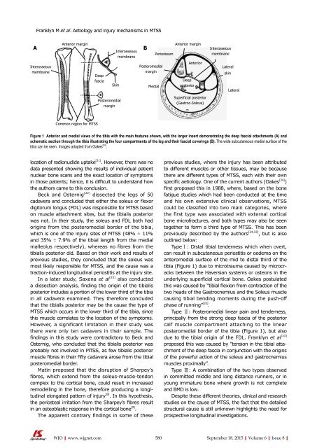
Figure 1 Anterior and medial views <strong>of</strong> the tibia with the main features shown, with the larger insert demonstrating the deep fascial attachments (A) and<br />
schematic section through the tibia illustrating the four compartments <strong>of</strong> the leg and their fascial coverings (B). The wide subcutaneous medial surface <strong>of</strong> the<br />
tibia can be seen. Images adapted from Oakes [24] .<br />
location <strong>of</strong> radionuclide uptake [21] . However, there was no<br />
data presented showing the results <strong>of</strong> individual patient<br />
nuclear bone scans and the exact location <strong>of</strong> symptoms<br />
in those patients; hence, it is difficult to understand how<br />
the authors came to this conclusion.<br />
Beck and Osternig [22] dissected the legs <strong>of</strong> 50<br />
cadavera and concluded that either the soleus or flexor<br />
digitorum longus (FDL) was responsible for MTSS based<br />
on muscle attachment sites, but the tibialis posterior<br />
was not. In their study, the soleus and FDL both had<br />
origins from the posteromedial border <strong>of</strong> the tibia,<br />
which is one <strong>of</strong> the injury sites <strong>of</strong> MTSS (48% ± 11%<br />
and 35% ± 7.9% <strong>of</strong> the tibial length from the medial<br />
malleolus respectively), whereas no fibres from the<br />
tibialis posterior did. Based on their work and results <strong>of</strong><br />
previous studies, they concluded that the soleus was<br />
most likely responsible for MTSS, and the cause was a<br />
traction-induced longitudinal periostitis at the injury site.<br />
In a later study, Saxena et al [23]<br />
also conducted<br />
a dissection analysis, finding the origin <strong>of</strong> the tibialis<br />
posterior includes a portion <strong>of</strong> the lower third <strong>of</strong> the tibia<br />
in all cadavera examined. They therefore concluded<br />
that the tibialis posterior may be the cause the type <strong>of</strong><br />
MTSS which occurs in the lower third <strong>of</strong> the tibia, since<br />
this muscle correlates to the location <strong>of</strong> the symptoms.<br />
However, a significant limitation in their study was<br />
there were only ten cadavers in their sample. The<br />
findings in this study were contradictory to Beck and<br />
Osternig, who concluded that the tibialis posterior was<br />
probably not involved in MTSS, as few tibialis posterior<br />
muscle fibres in their fifty cadavera arose from the tibial<br />
posteromedial border.<br />
Matin proposed that the disruption <strong>of</strong> Sharpey’s<br />
fibres, which extend from the soleus-muscle-tendon<br />
complex to the cortical bone, could result in increased<br />
remodelling in the bone, therefore producing a longitudinal<br />
elongated pattern <strong>of</strong> injury [8] . In this hypothesis,<br />
the periosteal irritation from the Sharpey’s fibres result<br />
in an osteoblastic response in the cortical bone [9] .<br />
The apparent contrary findings in some <strong>of</strong> these<br />
previous studies, where the injury has been attributed<br />
to different muscles or other tissues, may be because<br />
there are different types <strong>of</strong> MTSS, each with their own<br />
specific aetiology. One <strong>of</strong> the current authors (Oakes [24] )<br />
first proposed this in 1988, where, based on the bone<br />
fatigue studies which had been conducted at the time<br />
and his own extensive clinical observations, MTSS<br />
could be classified into two main categories, where<br />
the first type was associated with external cortical<br />
bone micr<strong>of</strong>ractures, and both types may also be seen<br />
together to form a third type <strong>of</strong> MTSS. This has been<br />
previously described by the authors [24,33] , but is also<br />
outlined below:<br />
Type Ⅰ: Distal tibial tenderness which when overt,<br />
can result in subcutaneous periostitis or oedema on the<br />
anteromedial surface <strong>of</strong> the mid to distal third <strong>of</strong> the<br />
tibia (Figure 1) due to microtrauma caused by microcracks<br />
between the Haversian systems or osteons in the<br />
underlying superficial cortical bone. Oakes postulated<br />
this was caused by “tibial flexion from contraction <strong>of</strong> the<br />
two heads <strong>of</strong> the Gastrocnemius and the Soleus muscle<br />
causing tibial bending moments during the push-<strong>of</strong>f<br />
phase <strong>of</strong> running” [33] .<br />
Type Ⅱ: Posteromedial linear pain and tenderness,<br />
principally from the strong deep fascia <strong>of</strong> the posterior<br />
calf muscle compartment attaching to the linear<br />
posteromedial border <strong>of</strong> the tibia (Figure 1), but also<br />
due to the tibial origin <strong>of</strong> the FDL. Franklyn et al [33]<br />
proposed this was caused by “tension in the tibial attachment<br />
<strong>of</strong> the deep fascia in conjunction with the origins<br />
<strong>of</strong> the powerful action <strong>of</strong> the soleus and gastrocnemius<br />
muscles proximally”.<br />
Type Ⅲ: A combination <strong>of</strong> the two types observed<br />
in committed middle and long distance runners, or in<br />
young immature bone where growth is not complete<br />
and BMD is low.<br />
Despite these different theories, clinical and research<br />
studies on the cause <strong>of</strong> MTSS, the fact that the detailed<br />
structural cause is still unknown highlights the need for<br />
prospective longitudinal investigations.<br />
WJO|www.wjgnet.com 580<br />
September 18, 2015|Volume 6|Issue 8|