Life science special report 161215 IY opt

Create successful ePaper yourself
Turn your PDF publications into a flip-book with our unique Google optimized e-Paper software.
VIRTUAL ABDOMINAL AORTIC ANEURYSM LIFE SCIENCES<br />
KNOWLEDGE-BASED VIRTUAL<br />
PROCESS TO AID ABDOMINAL AORTIC<br />
ANEURYSM DIAGNOSIS<br />
DR. SIMONE BARTESAGHI, PROF. GIORGIO COLOMBO<br />
Politecnico di Milano<br />
INTRODUCTION<br />
A range of diseases and a stressful<br />
lifestyle increase the vulnerability of the<br />
human body. Among such disorders,<br />
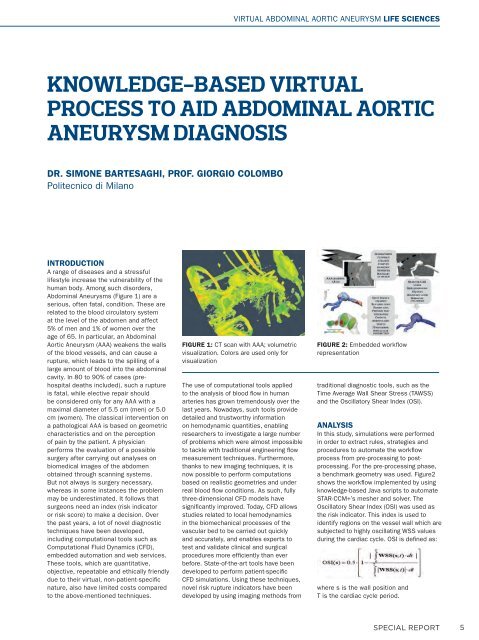
Abdominal Aneurysms (Figure 1) are a<br />
serious, often fatal, condition. These are<br />
related to the blood circulatory system<br />
at the level of the abdomen and affect<br />
5% of men and 1% of women over the<br />
age of 65. In particular, an Abdominal<br />
Aortic Aneurysm (AAA) weakens the walls<br />
of the blood vessels, and can cause a<br />
rupture, which leads to the spilling of a<br />
large amount of blood into the abdominal<br />
cavity. In 80 to 90% of cases (prehospital<br />
deaths included), such a rupture<br />
is fatal, while elective repair should<br />
be considered only for any AAA with a<br />
maximal diameter of 5.5 cm (men) or 5.0<br />
cm (women). The classical intervention on<br />
a pathological AAA is based on geometric<br />
characteristics and on the perception<br />
of pain by the patient. A physician<br />
performs the evaluation of a possible<br />
surgery after carrying out analyses on<br />
biomedical images of the abdomen<br />
obtained through scanning systems.<br />
But not always is surgery necessary,<br />
whereas in some instances the problem<br />
may be underestimated. It follows that<br />
surgeons need an index (risk indicator<br />
or risk score) to make a decision. Over<br />
the past years, a lot of novel diagnostic<br />
techniques have been developed,<br />
including computational tools such as<br />
Computational Fluid Dynamics (CFD),<br />
embedded automation and web services.<br />
These tools, which are quantitative,<br />
objective, repeatable and ethically friendly<br />
due to their virtual, non-patient-specifi c<br />
nature, also have limited costs compared<br />
to the above-mentioned techniques.<br />
FIGURE 1: CT scan with AAA; volumetric<br />
visualization. Colors are used only for<br />
visualization<br />
The use of computational tools applied<br />
to the analysis of blood fl ow in human<br />
arteries has grown tremendously over the<br />
last years. Nowadays, such tools provide<br />
detailed and trustworthy information<br />
on hemodynamic quantities, enabling<br />
researchers to investigate a large number<br />
of problems which were almost impossible<br />
to tackle with traditional engineering fl ow<br />
measurement techniques. Furthermore,<br />
thanks to new imaging techniques, it is<br />
now possible to perform computations<br />
based on realistic geometries and under<br />
real blood fl ow conditions. As such, fully<br />
three-dimensional CFD models have<br />
signifi cantly improved. Today, CFD allows<br />
studies related to local hemodynamics<br />
in the biomechanical processes of the<br />
vascular bed to be carried out quickly<br />
and accurately, and enables experts to<br />
test and validate clinical and surgical<br />
procedures more effi ciently than ever<br />
before. State-of-the-art tools have been<br />
developed to perform patient-specifi c<br />
CFD simulations. Using these techniques,<br />
novel risk rupture indicators have been<br />
developed by using imaging methods from<br />
FIGURE 2: Embedded workfl ow<br />
representation<br />
traditional diagnostic tools, such as the<br />
Time Average Wall Shear Stress (TAWSS)<br />
and the Oscillatory Shear Index (OSI).<br />
ANALYSIS<br />
In this study, simulations were performed<br />
in order to extract rules, strategies and<br />
procedures to automate the workfl ow<br />
process from pre-processing to postprocessing.<br />
For the pre-processing phase,<br />
a benchmark geometry was used. Figure2<br />
shows the workfl ow implemented by using<br />
knowledge-based Java scripts to automate<br />
STAR-CCM+’s mesher and solver. The<br />
Oscillatory Shear Index (OSI) was used as<br />
the risk indicator. This index is used to<br />
identify regions on the vessel wall which are<br />
subjected to highly oscillating WSS values<br />
during the cardiac cycle. OSI is defi ned as:<br />
where s is the wall position and<br />
T is the cardiac cycle period.<br />
SPECIAL REPORT<br />
5