Interventionelle Therapieansätze bei der akuten Lungenembolie
- No tags were found...

You also want an ePaper? Increase the reach of your titles
YUMPU automatically turns print PDFs into web optimized ePapers that Google loves.
JOURNAL OF THE AMERICAN COLLEGE OF CARDIOLOGY VOL. 67, NO. 8, 2016<br />
ª 2016 BY THE AMERICAN COLLEGE OF CARDIOLOGY FOUNDATION ISSN 0735-1097/$36.00<br />
PUBLISHED BY ELSEVIER<br />
http://dx.doi.org/10.1016/j.jacc.2015.12.024<br />
COUNCIL PERSPECTIVES<br />
Acute Pulmonary Embolism<br />
With an Emphasis on an Interventional Approach<br />
Wissam A. Jaber, MD, a Pete P. Fong, MD, b Giora Weisz, MD, c Omar Lattouf, MD, d James Jenkins, MD, e<br />
Kenneth Rosenfield, MD, MHCDS, f Tanveer Rab, MD, a Stephen Ramee, MD g<br />
ABSTRACT<br />
Compared with recent advances in treatment of serious cardiovascular diseases, such as myocardial infarction and<br />
stroke, the treatment and outcome of acute pulmonary embolism (PE) have remained relatively unchanged over the<br />
last few decades. This has prompted several experts to call for the formation of multidisciplinary PE response teams<br />
with a more proactive approach to the treatment of PE. In the current document, we discuss the formation of such<br />
teams and describe the available treatment options beyond anticoagulation, with a focus on the interventional<br />
approach. Acknowledging the paucity of data to support widespread adoption of such techniques, we call for the<br />
collection of outcomes data in multicenter registries and support for randomized trials to evaluate interventional<br />
treatments in patients with high-risk PE. (J Am Coll Cardiol 2016;67:991–1002) © 2016 by the American College of<br />
Cardiology Foundation.<br />
The American Heart Association classifies<br />
pulmonary embolism (PE) into low-risk,<br />
intermediate-risk (submassive), and highrisk<br />
(massive) categories (1). Whereas massive PE is<br />
defined by the presence of persistent hypotension,<br />
submassive PE is defined as occurring in normotensive<br />
patients with evidence of right ventricular (RV)<br />
strain by echocardiogram, computed tomography<br />
(CT) scan, or cardiac biomarkers. The most recent<br />
European Society of Cardiology guidelines further<br />
divide the intermediate-risk group into intermediatelow–<br />
and intermediate-high–risk subgroups, with<br />
the latter defined by the presence of both RV dysfunction<br />
and increased cardiac biomarkers (2). Despitea<br />
high case fatality rate, most patients with massive<br />
andsubmassivePEcontinuetobetreatedconservatively<br />
with anticoagulation alone. This has prompted<br />
alternate, intensive treatment options, including<br />
systemic fibrinolysis, catheter-based therapy, and<br />
surgical embolectomy. Because adequate studies<br />
evaluating these therapies are scarce, and given the<br />
difficulty in managing PE patients, multiple centers<br />
have formed multidisciplinary pulmonary embolism<br />
response teams (PERT, a trademark of the National<br />
PERT Consortium) to engage specialists from different<br />
backgrounds to discuss treatment options and provide<br />
immediate advice and therapy for patients in<br />
the massive and submassive categories (3,4).<br />
Listen to this manuscript’s<br />
audio summary by<br />
JACC Editor-in-Chief<br />
Dr. Valentin Fuster.<br />
The views expressed in this manuscript by the American College of Cardiology (ACC’s) Interventional Council do not necessarily<br />
reflect the views of the Journal of the American College of Cardiology or the ACC.<br />
From the a Division of Cardiology, Emory University School of Medicine, Atlanta, Georgia; b Van<strong>der</strong>bilt Heart and Vascular Institute,<br />
Nashville, Tennessee; c Division of Cardiology, Shari Zadek Medical Center, Jerusalem, Israel; d Division of Cardiothoracic Surgery,<br />
Emory University School of Medicine, Atlanta, Georgia; e Ochsner Medical Center, New Orleans, Louisiana; f Section of Vascular<br />
Medicine, & Intervention, Division of Cardiology, Massachusetts General Hospital, Boston, Massachusetts; and the g Structural and<br />
Valvular Heart Disease Program, Ochsner Medical Center, New Orleans, Louisiana. Dr. Rosenfield has served as a consultant to<br />
Abbott, Cardinal Health, Inari Medical, Surmodics, Volcano, Capture Vascular, and Shockwave; has served as a board member for<br />
VIVA Physicians, a 501c3 organization; has received research support from the National Institutes of Health, Atrium, Lutonix-Bard,<br />
Abbott Vascular, and Gore; and has personal equity in CardioMems, Embolitech, MD Insi<strong>der</strong>, Primacea, and Vortex. All other<br />
authors have reported that they have no relationships relevant to the contents of this paper to disclose.<br />
Manuscript received September 28, 2015; revised manuscript received November 17, 2015, accepted December 14, 2015.
992<br />
Jaber et al. JACC VOL. 67, NO. 8, 2016<br />
Pulmonary Embolism MARCH 1, 2016:991– 1002<br />
ABBREVIATIONS<br />
This document summarizes current invasive<br />
treatment options beyond anti-<br />
AND ACRONYMS<br />
coagulation available to intermediate- and<br />
CDF = catheter-directed<br />
high-risk PE patients, with the un<strong>der</strong>standing<br />
that data supporting any of them is<br />
fibrinolysis<br />
CDT = catheter-directed<br />
therapy<br />
either inconclusive or lacking, and therefore<br />
CT = computed tomography<br />
not covered by American and European<br />
guidelines. Most PE patients only require<br />
IV = intravenous<br />
simple anticoagulation; patients with highrisk<br />
features deserve consi<strong>der</strong>ation for an<br />
PA = pulmonary artery<br />
PE = pulmonary embolism<br />
invasive treatment approach.<br />
PERT = pulmonary embolism<br />
response team<br />
BUILDING AN ACUTE PE TEAM AND<br />
RV = right ventricle/ventricular MANAGEMENT PATHWAY<br />
t-PA = tissue-type<br />
plasminogen activator<br />
Intensive management of acute PE begins<br />
with formation of a PERT. Assembling a team of<br />
specialists and coordinating care through a system<br />
similar to the management of ST-segment elevation<br />
myocardial infarction (STEMI) has been described at<br />
several institutions (3,5,6). A PERT may consist of<br />
specialists from vascular medicine, pulmonary critical<br />
care, emergency medicine, interventional cardiology/<br />
radiology, hematology, vascular surgery, and cardiothoracic<br />
surgery. Not every hospital system will<br />
engage all of these subspecialists, but we recommend,<br />
at a minimum, representatives from medicine, interventional<br />
cardiology/radiology, and surgery, as the<br />
decisions to be made require an un<strong>der</strong>standing of<br />
the risks and benefits of all treatment modalities.<br />
The PERT’s responsibilityistoassesseachcaseina<br />
timely manner, examine the patient, review the<br />
available data, perform any additional testing, and<br />
then (in conjunction with the patient, family members,<br />
and care team) develop a consensus regarding<br />
the optimal treatment plan. In certain patients with<br />
massive PE and rapid deterioration, the decision to<br />
give fibrinolytic agents, go to the interventional laboratory,<br />
or proceed to the operating room will need to<br />
be made urgently by a limited number of PERT<br />
members. In such cases, prior experience assessing<br />
multiple patients with PERT colleagues will lend<br />
advantage to the on-call team members and inform<br />
their decision making. As a foundation, we recommend<br />
that the local PERT review current published<br />
reports and society guidelines (1,2,7) and establish an<br />
institutional acute PE protocol, as in the Central<br />
Illustration (6). The key to team management is activation<br />
of the PERT with a single phone call. Team<br />
members should have an easily accessible online<br />
system allowing all members to review the available<br />
medical information, including computed tomography<br />
(CT) scans, echocardiograms, electrocardiograms,<br />
and laboratory data. PE teams should leverage<br />
existing systems, such as hospital electronic medical<br />
records, imaging systems, virtual meeting rooms,<br />
and STEMI or acute stroke activation protocols.<br />
Furthermore, teams should identify the admission<br />
location best able to manage sick patients with submassive<br />
and massive PE. A defined area enables<br />
consistency and further development of expertise in<br />
management of complex PE patients. A cardiovascular<br />
intensive care unit where the surgeon, cardiovascular<br />
specialist, and critical care specialist round<br />
together is a reasonable choice. This type of team<br />
approach is a well-established Class I recommendation<br />
for management of ischemic heart disease (8).<br />
In May 2015, the National PERT Consortium<br />
launch meeting, sponsored by the Massachusetts<br />
GeneralHospitalPERT,washeldinBoston,Massachusetts,<br />
and was attended by approximately 40<br />
hospital-based PE teams. There was an important<br />
call to action to gather and share data on patients<br />
with PE. We encourage PERTs to establish institutional<br />
review board–approved databases that can be<br />
shared among like-minded institutions for further<br />
advancement of PE management, such as Research<br />
Electronic Data Capture (RedCap). Participation in a<br />
multicenter registry, such as that un<strong>der</strong> development<br />
by the National PERT Consortium, will enable systematic,<br />
broad-based assessment of outcomes and<br />
further our knowledge regarding optimal care and<br />
best practices.<br />
PRE-INTERVENTION<br />
Unless contraindicated, anticoagulation should be<br />
initiated when PE is suspected, prior to additional<br />
work-up. Intravenous heparin is a good initial choice<br />
while alternative options (such as invasive therapies)<br />
are <strong>bei</strong>ng evaluated. After confirmation, the first<br />
question is whether the PE is low risk versus submassive<br />
to massive. Massive PE is currently defined<br />
by hypotension with a systolic blood pressure<br />
(SBP) 15 min or the requirement of<br />
inotropic support to maintain SBP >90 mm Hg. Submassive<br />
PE is defined by SBP >90 mm Hg with evidence<br />
of right heart dysfunction, as noted by a<br />
dilated RV with an RV to left ventricle ratio >0.9 in<br />
the 4-chamber view by CT or echocardiogram, or<br />
elevated biomarkers, such as troponin or B-type<br />
natriuretic peptide. The echocardiogram has the<br />
advantage of demonstrating depressed RV function<br />
and providing an estimate of pulmonary arterial<br />
systolic pressure. Other high-risk markers of submassive<br />
PE include tachycardia, tachypnea, and<br />
hypoxia, which may be less specific, and thus less<br />
helpful in management selection. Special attention
JACC VOL. 67, NO. 8, 2016<br />
MARCH 1, 2016:991– 1002<br />
Jaber et al.<br />
Pulmonary Embolism<br />
993<br />
should be given to patients who had syncope, which<br />
may represent transient hemodynamic instability<br />
and the potential for higher risk (9). Hemodynamically<br />
stable patients without evidence of RV strain<br />
are consi<strong>der</strong>ed to be at low risk, may not require<br />
PERT activation, and can be treated with anticoagulation<br />
alone. That said, the PERT might help the<br />
clinical team define the level of risk and optimal<br />
therapy for each individual patient.<br />
Once the PERT has been activated, members typically<br />
meet, either at the patient’s bedside or virtually,<br />
and review all patient-related data. The most important<br />
data include the presenting history, with a focus<br />
on symptoms and signs of hemodynamic instability,<br />
vital signs, CT, echocardiogram, and laboratory data.<br />
Team members should discuss indications and contraindications<br />
to fibrinolytic therapy, catheter-based<br />
intervention, and surgical embolectomy, followed<br />
by discussion with the patient and family members,<br />
including the risks and benefits of each therapy proposed.<br />
A sample algorithm to help in triaging patients<br />
presenting to the emergency room of a hospital with a<br />
PERT is provided (Figure 1). The PERT should<br />
consi<strong>der</strong> the degree of hemodynamic compromise<br />
and the existence of variable contraindications. The<br />
simplified PE severity index may be a useful aid when<br />
further consi<strong>der</strong>ing the risks and benefits of interventional<br />
therapies (10).<br />
SYSTEMIC FIBRINOLYSIS<br />
Traditionally, intravenous (IV) fibrinolysis has been<br />
consi<strong>der</strong>ed the primary intensive therapy option in<br />
patients with high-risk PE, although the data supporting<br />
its use in massive PE is poor. Most trials that<br />
randomized patients with PE to fibrinolytics versus<br />
standard anticoagulation included submassive PE<br />
only. A meta-analysis of trials including patients with<br />
massive PE showed a reduction in the composite of<br />
recurrent PE and death with use of IV fibrinolytic<br />
agents, but not in death alone (11). Univariateanalysis<br />
of a large inpatient sample found that among unstable<br />
patients with PE, use of IV fibrinolytic therapy was<br />
associated with a lower mortality rate (12), butonly<br />
30% of unstable patients received such therapy.<br />
Patients with submassive PE were better represented<br />
in randomized trials. The MAPPET (Management,<br />
Strategies and Prognosis of Pulmonary<br />
Embolism)-3 trial (13) randomized 256 patients with<br />
PE and pulmonary hypertension or RV dysfunction to<br />
100 mg of IV alteplase or placebo infused over 2 h plus<br />
anticoagulation. IV alteplase was associated with a<br />
lower risk of further need to escalate the treatment and<br />
with a similar risk of death. Mortality was lower than<br />
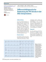
CENTRAL ILLUSTRATION<br />
Medical therapy<br />
PERT Protocol<br />
Patient with suspected pulmonary embolism (PE)<br />
Anticoagulation initiated, unless contraindicated<br />
Acute PE confirmed by Computed Tomography (CT) scan<br />
Multidisciplinary PE response team (PERT) alerted:<br />
Interventionalist, cardiac surgeon,<br />
radiology, pulmonary/critical care medicine<br />
PERT members review the available medical information<br />
and develop optimal treatment plan<br />
Catheter<br />
directed therapy<br />
Jaber, W.A. et al. J Am Coll Cardiol. 2016; 67(8):991–1002.<br />
expected in both groups (3.4% in the alteplase and<br />
2.2% in the placebo group; p ¼ 0.71). More recently,<br />
the PEITHO (Pulmonary Embolism Thrombolysis) trial<br />
(14) randomized 1,006 patients with submassive<br />
PE (normal blood pressure, RV enlargement, and<br />
increased troponin level) to tenecteplase or placebo.<br />
The PEITHO trial showed a reduction in the primary<br />
endpoint of hemodynamic collapse at 7 days with<br />
tenecteplase, but a significant increase in hemorrhagic<br />
stroke (most in patients ol<strong>der</strong> than 75 years of age),<br />
with similar mortality in both groups. The smaller<br />
MOPETT (Mo<strong>der</strong>ate Pulmonary Embolism Treated<br />
With Thrombolysis) trial (15) randomized 121 patients<br />
with mo<strong>der</strong>ate-risk PE to half-dose alteplase<br />
(maximum 50 mg over 2 h) with anticoagulation versus<br />
anticoagulation alone. Low-dose alteplase was associated<br />
with lower pulmonary pressure at 28 months<br />
and no major bleeding. A 1,700 patient meta-analysis<br />
Surgical<br />
embolectomy<br />
Example of an intensive PE management pathway utilizing a single phone call to the PE<br />
team lea<strong>der</strong>. Further review with PERT members can occur while the patient is transferred<br />
to the critical care unit, interventional laboratory, or operating room. Adapted with<br />
permission from Bloomer et al. (6). CT¼ computed tomography; PE ¼ pulmonary<br />
embolism; PERT ¼ pulmonary embolism response team.
994<br />
Jaber et al. JACC VOL. 67, NO. 8, 2016<br />
Pulmonary Embolism MARCH 1, 2016:991– 1002<br />
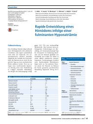
FIGURE 1<br />
ER PE Protocol Utilizing PERT Consultation and sPESI Score<br />
PE confirmed:<br />
Anticoagulate<br />
Stable patient<br />
Unstable patient<br />
Massive PE<br />
(SBP < 90)<br />
PERT consult<br />
Low risk PE pt<br />
sPESI* 0<br />
Submassive PE<br />
suspected<br />
sPESI ≥ 1<br />
1. Discuss IV lytics/<br />
catheter/surgery<br />
with PERT lea<strong>der</strong><br />
2. If lytics,<br />
consi<strong>der</strong><br />
initiation in ER<br />
Echo<br />
+<br />
Troponin<br />
Echo and troponin<br />
(-) for RV<br />
dysfunction<br />
Echo or troponin (+)<br />
for RV dysfunction<br />
PERT consult<br />
Anticoagulate,<br />
admit to medicine<br />
floor<br />
Admit to critical<br />
care unit<br />
*Simplified pulmonary embolism severity index (sPESI) score ¼ 1 point for age >80 years, cancer, chronic heart failure or chronic pulmonary<br />
disease, heart rate >110 beats/min, SBP
JACC VOL. 67, NO. 8, 2016<br />
MARCH 1, 2016:991– 1002<br />
Jaber et al.<br />
Pulmonary Embolism<br />
995<br />
acute PE, giving advanced therapies a limited<br />
recommendation because of a lack of randomized<br />
control data (1,7).<br />
Multiple percutaneous techniques have been<br />
described for treatment of unstable patients with PE<br />
(Table 1). They aim at relieving the obstruction in the<br />
pulmonary circulation, thus immediately improving<br />
the patient’s hemodynamics and potentially reducing<br />
the long-term risk of pulmonary hypertension (17).<br />
These techniques have been poorly studied, and they<br />
facethechallengeoftryingtoremovelarge,sometimes<br />
organized thrombi from a large space with<br />
numerous 3-dimensional branches and multiple<br />
angles. The simplest and most commonly performed<br />
catheter-based therapy is a local, slow infusion of a<br />
fibrinolytic agent through low-profile catheters<br />
placed in the obstructed pulmonary artery (PA). CDF<br />
is best suited for more stable patients or those who<br />
have been hemodynamically stabilized, as thrombus<br />
resolution may take several hours.<br />
For unstable patients who require immediate<br />
intervention and/or those with contraindication to<br />
fibrinolysis, mechanical thrombus fragmentation,<br />
debulking, or aspiration of occlusive thrombi may be<br />
attempted.<br />
Potential complications of any catheter-based<br />
therapy may include pulmonary arterial injury, pericardial<br />
tamponade, major bleeding, hemodynamic<br />
deterioration, distal embolization and “no-reflow”<br />
phenomenon, and access site bleeding. A metaanalysis<br />
of CDT using #10-F low-profile devices<br />
reported minor and major procedural complications<br />
of 7.9% and 2.4%, respectively (18). Minor complications<br />
included: groin hematomas not requiring transfusion,<br />
transient bradyarrhythmia, heart block,<br />
hemoglobinuria, mild hemoptysis, temporary renal<br />
insufficiency, embolus dislocation (n ¼ 1), and PA<br />
dissection (n ¼ 1). Major complications included: groin<br />
hematomas requiring transfusion, massive hemoptysis<br />
requiring transfusion, renal failure requiring<br />
hemodialysis, cardiac tamponade (n ¼ 1), and death<br />
(n ¼ 5; 1 each from bradyarrhythmia and apnea,<br />
distal embolization, and cerebral vascular hemorrhage,<br />
plus 2 procedure-related deaths not otherwise<br />
specified) (18).<br />
FRAGMENTATION AND ASPIRATION. Fragmentation<br />
and aspiration of PE may be helpful in stabilizing<br />
patients with massive PE, especially when systemic<br />
fibrinolysis is contraindicated or has failed. Multiple<br />
techniques have been tried with some success (19). By<br />
rotating a pigtail catheter in the PA, the PE can be<br />
fragmented (20). The aim is to reduce the load on the<br />
RV by partially relieving the obstruction in the main<br />
TABLE 1<br />
Catheter-Based Therapies<br />
Device Size Mechanism of Action<br />
Pigtail catheter 6- to 8-F Fragmentation<br />
Peripheral balloon 5 to 10 mm Fragmentation<br />
Catheter-directed<br />
fibrinolysis<br />
4- to 6-F Direct infusion of fibrinolytic agent<br />
Ultrasound-accelerated<br />
thrombolysis<br />
6-F Direct infusion of fibrinolytic agent<br />
plus ultrasound for clot separation.<br />
Currently the only catheter-based<br />
therapy FDA-approved for PE treatment.<br />
Guide catheter 6- to 10-F Manual aspiration<br />
Pronto Xl catheter 6- to 14-F Manual aspiration<br />
Penumbra Indigo system 6- to 8-F Suction pump aspiration<br />
Inari FlowTriever 22-F sheath Disruption, retraction, and aspiration of clot<br />
AngioVac<br />
26-F sheath<br />
and 18-F cannula<br />
Large-volume aspiration with return of<br />
filtered blood utilizing a centrifugal pump<br />
FDA ¼ Food and Drug Administration; PE ¼ pulmonary embolism.<br />
PA branches. Fragmentation alone may cause distal<br />
embolization and potentially worsen distal branch<br />
obstruction (21). Fragmentation is frequently combined<br />
with local infusion of small-dose fibrinolytic<br />
agents (e.g., 4 to 10 mg of tissue-type plasminogen<br />
activator [t-PA]), delivered either at the time of the<br />
procedure or subsequently via an infusion catheter<br />
left in place. Concomitant aspiration can reduce the<br />
risk of worsening obstruction (21). Fragmentation can<br />
also be performed by inflation of an angioplasty<br />
balloon, with care to keep inflationinlargervessels<br />
and to choose a balloon smaller than the artery<br />
diameter. Injection through a diagnostic catheter<br />
(e.g., a multipurpose catheter) placed distal to the<br />
intended inflation site can help determine the size of<br />
the distal artery before selecting a balloon catheter.<br />
Aspiration can be attempted using regular 8-F<br />
guide catheters or specialized catheters. One of the<br />
first aspiration catheters was the Greenfield embolectomy<br />
catheter (22), which consisted of a suction<br />
cup at the tip of a straight catheter. Its complexity<br />
and the requirement for a surgical cutdown for access<br />
prevented it from <strong>bei</strong>ng widely adopted. Other<br />
specialized devices used to treat peripheral thrombi<br />
have also been used off-label to treat PE. These<br />
include the 10-F Aspirex thrombectomy catheter<br />
(Straub Medical, Wangs, Switzerland), currently unavailable<br />
in the United States, which combines rotational<br />
thrombus fragmentation with aspiration (23);<br />
the 7-F Helix Clot Buster (ev3, Plymouth, Minnesota),<br />
approved for dialysis graft thrombosis treatment (24);<br />
and8-to14-FProntoXLmanualextractioncatheters<br />
(Vascular Solutions, Minneapolis, Minnesota).<br />
The Angiojet Rheolytic Thrombectomy System<br />
(Possis, Minneapolis, Minnesota) deserves special<br />
mention. This 8-F peripheral catheter utilizes the
996<br />
Jaber et al. JACC VOL. 67, NO. 8, 2016<br />
Pulmonary Embolism MARCH 1, 2016:991– 1002<br />
FIGURE 2<br />
B<br />
A<br />
Venturi-Bernoulli effect, using multiple highvelocity<br />
saline jets introduced through the distal<br />
tip, creating a low-pressure vacuum through small<br />
slits in the catheter that can entrain and fragment<br />
thrombi. A meta-analysis reported higher mortality<br />
and morbidity, including massive hemoptysis, renal<br />
failure, and death from bradycardia and apnea or<br />
from widespread distal embolization (18), which<br />
resulted in a black-box warning from the Food and<br />
Drug Administration (FDA) for use of Angiojet in<br />
acute PE.<br />
Additional embolectomy devices are discussed in<br />
the following sections.<br />
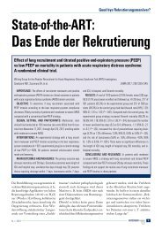
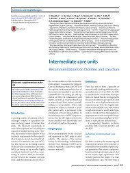
AngioVac thrombectomy device. The AngioVac<br />
Cannula (Angiodynamics, Latham, New York), a 22-F<br />
venous catheter that can remove soft thrombi utilizing<br />
the centrifugal pump and venous reinfusion<br />
cannula used in cardiopulmonary bypass (Figure 2), is<br />
FDA approved for the removal of undesirable<br />
AngioVac Device<br />
Filter<br />
Centrifugal Pump Console<br />
Saline Bag<br />
Reinfusion<br />
Cannula<br />
AngioVac<br />
Circuit<br />
C<br />
AngioVac Cannula<br />
(A) AngioVac cannula. (B) Diagram of AngioVac insertion and reinfusion circuit. The cannula<br />
has been inserted into the right internal jugular vein. Blood and thrombus is aspirated<br />
through the filter canister, allowing clot capture utilizing a centrifugal pump canister,<br />
prior to return of blood to the patient via the reinfusion cannula placed into the femoral<br />
vein. (C) Example of thrombus captured in the filter canister. Images from Angiodynamics.<br />
intravascular material, including fresh, soft thrombi<br />
or emboli. The AngioVac catheter consists of a<br />
balloon-expandable, funnel-shaped distal tip, which<br />
improves removal of large clots en masse. Patients are<br />
prepped in 2 body locations that will allow for large<br />
venous sheath placements (common femoral or internal<br />
jugular veins). A 26-F sheath is placed in 1 vein<br />
and an 18-F reinfusion cannula is placed in another<br />
vein. The AngioVac cannula is then attached to the<br />
inflow tubing of the centrifuge pump and the outflow<br />
tubing connected to the 18-F reinfusion cannula,<br />
creating a “veno-veno” bypass circuit. The cannula is<br />
inserted into the 26-F sheath and is advanced to the<br />
thrombus, which is suctioned out and captured by a<br />
filtration canister inserted proximal to the centrifuge<br />
pump; filtered blood is returned continuously via the<br />
reinfusion cannula. Limitations of this device include<br />
the large dual sheaths required for access, leading to a<br />
higher likelihood of bleeding complications, and the<br />
relatively stiff suction catheter, which is difficult to<br />
maneuver into the RV and PA. Furthermore, the<br />
active participation of an experienced perfusionist is<br />
required for AngioVac setup and operation, as there is<br />
a learning curve for its use. AngioVac has been utilized<br />
in PE, although it is more commonly used to<br />
retrieve thrombi from the vena cava and right atrium<br />
(25). The rapidity of initiation may limit its use in<br />
massive PE situations; future iterations may ren<strong>der</strong> it<br />
more useful for PE.<br />
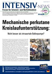
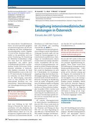
FlowTriever device. The FlowTriever catheter<br />
(Inari Medical, Irvine, California) is a recently<br />
released device that has FDA 510(k) approval for<br />
removal of emboli and thrombi from blood vessels<br />
(Figure 3). The FlowTriever Infusion Aspiration System<br />
requires a 22-F venous sheath and consists of<br />
3 parts: the Flow Restoration Catheter, which is made<br />
up of 3 self-expanding nitinol disks; the Aspiration<br />
Guide Catheter; and the Retraction Aspirator Device.<br />
The FlowTriever device is advanced over the wire and<br />
into the thrombus, where the expandable disks are<br />
deployed using a pin and pull method. The disks and<br />
disrupted thrombus are then retracted and removed<br />
through the aspiration catheter. Set-up is rapid, and<br />
there is a modest learning curve for device utilization.<br />
Limitations include the large size requirement of the<br />
access sheath, and manipulation of the large-bore<br />
catheter into the PA.<br />
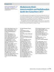
Penumbra Indigo thrombectomy system. The<br />
Indigo mechanical thrombectomy system (Penumbra,<br />
Inc., Alameda, California) consists of a pump, 6- to<br />
8-F straight or angled catheters, and a Separator device<br />
(Figure 4). It is approved for thrombus removal<br />
in both peripheral arterial and venous systems.<br />
An advantage is that it only requires an 8-F venous
JACC VOL. 67, NO. 8, 2016<br />
MARCH 1, 2016:991– 1002<br />
Jaber et al.<br />
Pulmonary Embolism<br />
997<br />
sheath and can be placed into the PA system quickly,<br />
in an over-the-wire technique. Once placed proximal<br />
to the clot, the thrombectomy catheter is advanced<br />
while suction is supplied with the ACER pump. The<br />
provided Separator wire is used to clear the system of<br />
thrombus as the catheter is manipulated inside the<br />
artery.<br />
A distinct limitation of these last 3 devices is the<br />
absence of published data on their overall success and<br />
safety.<br />
FIGURE 3<br />
A<br />
FlowTriever Device<br />
CATHETER-DIRECTED FIBRINOLYSIS. General<br />
consi<strong>der</strong>ations. Given that full-dose systemic fibrinolysis<br />
is helpful in stabilizing high-risk PE patients<br />
and reducing pulmonary pressure, but at the cost of<br />
increased systemic bleeding, interest has risen in local<br />
delivery of low-dose fibrinolytics close to or into<br />
the PA thrombus. Unfortunately, data supporting<br />
such therapy is limited and mostly from small<br />
case series (18,26–28). One small trial randomized<br />
34 patients with angiographically large PE to IV- or<br />
catheter-based infusion of t-PA at a dose of 50 mg over<br />
2h(29), and showed similar safety and angiographic<br />
and hemodynamic results by both techniques. However,<br />
the local fibrinolytic dose used in this ol<strong>der</strong> trial<br />
was much higher than what is currently used. In a<br />
more recent prospective registry of 101 massive and<br />
submassive PE patients treated with catheter-based<br />
therapy (mostly local fibrinolysis), there was a significant<br />
decrease in PA pressure and improvement in<br />
RV function, with no reported major complications,<br />
major bleeding, or strokes (26). Giventhelowriskfor<br />
major complications, it is reasonable to consi<strong>der</strong> CDF<br />
in patients with already stabilized massive PE who<br />
have contraindications to systemic fibrinolysis and in<br />
patients with intermediate-high–risk PE (those with<br />
RV dysfunction and increased biomarkers), particularly<br />
those deemed at increased bleeding risk with<br />
full-dose systemic fibrinolysis. In a series of 52 PE<br />
patients treated with CDF, a more prominent hemodynamic<br />
benefit was obtained in patients with symptom<br />
duration
998<br />
Jaber et al. JACC VOL. 67, NO. 8, 2016<br />
Pulmonary Embolism MARCH 1, 2016:991– 1002<br />
FIGURE 4<br />
Penumbra Indigo Aspiration System<br />
fibrinolytic agent, hence theoretically achieving a<br />
faster thrombus breakdown (30). The technique<br />
used for in vivo insertion is similar to other infusion<br />
catheters, as described earlier. Once the<br />
catheter is in position over the 0.035-inch guidewire,<br />
the guidewire is replaced with the microsonic<br />
cable, which is then locked into place. The catheter<br />
has 2 infusion ports: 1 for the fibrinolytic<br />
infusion, and the other for a coolant solution<br />
(normal saline at $35 ml/h).<br />
Limited evidence supports the use of ultrasoundaccelerated<br />
thrombolysis (discussed in detail by<br />
Konstantinides et al. [31]). It is uncertain whether<br />
this treatment is suitable for patients who are<br />
hemodynamically unstable and need faster resolution<br />
of the PE or if there is long-term benefit ofthe<br />
prolonged treatment in prevention of future pulmonary<br />
hypertension, un<strong>der</strong>scoring the need for more<br />
evidence.<br />
(A) The 6- to 8-F straight or angled aspiration catheter (CAT6 or CAT8, respectively)<br />
is advanced to the thrombus and aspiration performed with the (B) ACER pump. Separator<br />
wires may be inserted into the catheter and utilized in a gentle back-and-forth motion to<br />
clear the catheter of thrombus. Images from Penumbra, Inc.<br />
contralateral PA through a second venous sheath, if<br />
needed, using the same technique. Alternatively, a<br />
10- to 12-F sheath may be placed at the beginning of<br />
the procedure and both catheters placed through the<br />
larger sheath. A commonly used t-PA dose is 0.5 to 1.0<br />
mg/h per catheter. The total t-PA dose is typically<br />
between 12 and 24 mg, delivered over 6 to 24 h. Lowdose,<br />
weight-adjusted heparin infusion is usually<br />
continued during t-PA infusion, with a target partial<br />
thromboplastin time on the low end of the therapeuticrange(e.g.,40to60s).<br />
Commonly available infusion catheters used offlabel<br />
for PE include the Cragg-McNamera catheter<br />
(ev3 Endovascular Inc.), the Fountain catheter (Merit<br />
Medical, South Jordan, Utah), and the Unifuse catheter<br />
(Angiodynamics). The EkoSonic catheter, (EKOS<br />
Corp., Brothell, Washington), discussed later, is<br />
currently the only catheter specifically approved by<br />
FDA for the treatment of high-risk PE.<br />
The risk of serious complications, including major<br />
hemorrhage, using CDT has been low in published<br />
studies. The risk of intracranial hemorrhage is
JACC VOL. 67, NO. 8, 2016<br />
MARCH 1, 2016:991– 1002<br />
Jaber et al.<br />
Pulmonary Embolism<br />
999<br />
FIGURE 5<br />
ExampleofaPETreatedWithCDF<br />
(A and B) CT angiogram of a patient with acute submassive PE, with thrombi seen in bilateral main PAs extending into the lower branches<br />
(yellow arrows). (C) Pulmonary angiography of the same patient, demonstrating hand injection of contrast into the lower left PA branches,<br />
with thrombus noted by the orange arrow. (D) Infusion catheters noted in both PAs. Manual injection of contrast agent performed through the<br />
left catheter (orange arrows), documenting that the catheter is imbedded in the clot. Note EkoSonic catheter markers in the right PA (red<br />
arrow). CDF ¼ catheter-directed fibrinolysis; CT ¼ computed tomography; PA ¼ pulmonary artery; PE ¼ pulmonary embolism.<br />
in-hospital mortality and a 10-year actuarial survival<br />
rate of 93%; both late mortalities were unrelated to<br />
PE or related therapy (39).<br />
SURGICAL TECHNIQUE. After median sternotomy,<br />
patients are anticoagulated with heparin and placed<br />
on cardiopulmonary bypass. Dual venous cannulation<br />
allows excellent venous drainage and full access<br />
to the right heart. The PA is typically opened longitudinally,<br />
distal to the pulmonic valve, to a length of<br />
approximately 5 cm. Sponge forceps are used to grasp<br />
and remove visible clots. Small clot fragments may be<br />
extracted using targeted gentle suction. The aorta<br />
may be circumferentially freed and gently retracted<br />
to allow “deeper” visualization of the right PA.<br />
Occasionally, a secondary distal incision of the right<br />
PA is made to allow even more distal access. Clots are<br />
typically not adhered to the artery wall, and thus are<br />
easily removable in acute PE cases.<br />
VENA CAVA FILTER<br />
Placement of an inferior vena cava (IVC) filter is<br />
indicated in patients with acute PE who have absolute<br />
contraindications to anticoagulation or in patients<br />
who have recurrent PE, despite adequate anticoagulation<br />
(1,2). The position of the filter below or<br />
above the renal veins depends on the absence or<br />
presence of renal vein thrombus, respectively.<br />
Retrievable filters are preferable because they are<br />
associated with lower complication rates (40). Both<br />
the American and the European guidelines do not<br />
recommend routine use of IVC filters in patients<br />
with PE (1,2). These recommendations are supported
1000<br />
Jaber et al. JACC VOL. 67, NO. 8, 2016<br />
Pulmonary Embolism MARCH 1, 2016:991– 1002<br />
FIGURE 6 EkoSonic Endovascular Device<br />
The 5.2-F infusion catheter (A), which contains 3 lumens: 1 each for the inner ultrasound<br />
cable, drug infusion, and normal saline as a coolant. The inner cable (B) is shown with<br />
ultrasound crystals (arrows). Ultrasound energy separates fibrin strands, allowing for<br />
enhanced thrombus penetration of fibrinolytic agent. Images from EKOS Corporation.<br />
anticoagulants, including rivaroxaban, dabigatran,<br />
apixaban, and edoxaban, can be used (45–48). However,<br />
no guidelines indicate when or how these<br />
agents should be initiated post-CDT, especially if<br />
fibrinolytic agents have been administered. If an<br />
alternative anticoagulant agent is utilized, we suggest<br />
heparin alone for the first 24 to 48 h post-intervention<br />
and then discontinuation of the heparin at the time of<br />
the first alternative anticoagulant agent dosing. This<br />
strategy does not include dabigatran or edoxaban<br />
usage, which require at least 5 days of parenteral<br />
therapy before initiation.<br />
Appropriate transition of the patient from the<br />
inpatient to the outpatient setting is important. This<br />
includes assessment of adequacy of anticoagulation<br />
or affordability of novel anticoagulant agents, if prescribed.<br />
Outpatient follow-up with a medical provi<strong>der</strong><br />
familiar with PE care is imperative; several institutions<br />
incorporate a PE follow-up clinic as part of<br />
thePERTprogram.Tobeaddressedatfollow-upare:<br />
monitoring of anticoagulation; assessment of length<br />
of anticoagulation and bleeding risk; retrieval of IVC<br />
filter, if appropriate; screening for the development<br />
of chronic pulmonary hypertension in patients at risk;<br />
and completion of a hypercoagulable profile, when<br />
indicated.<br />
by the PREPIC2 (Prevention of Recurrent Pulmonary<br />
Embolism by Vena Cava Interruption 2) study,<br />
recently conducted in intermediate- and low-risk<br />
patients (41). However, 3 large analyses, including a<br />
U.S. nationwide hospital sample (42) and a study from<br />
Japan (43), suggestthatIVCfilters may result in better<br />
outcomes in patients with massive or intermediatehigh–risk<br />
PE. In the International Cooperative Pulmonary<br />
Embolism registry, IVC filter use in patients<br />
with massive PE was associated with reduced rates of<br />
recurrent PE and mortality at 90 days (44).<br />
POST-INTERVENTION<br />
Maintenance of anticoagulation post-intervention is<br />
critical to prevent recurrent clot formation. However,<br />
patients who have had a recent catheter-based intervention<br />
are at risk of access site bleeding. One strategy<br />
to potentially reduce bleeding risk is to hold the heparin<br />
drip for 1 to 2 h after sheath removal, then restart<br />
without a bolus. Warfarin is administered on the night<br />
of the procedure, and parenteral anticoagulation and<br />
warfarin are overlapped until the international<br />
normalized ratio is 2 to 3 for at least 24 h, as per<br />
American College of Chest Physicians guidelines (7).<br />
Low molecular weight heparin can be utilized in<br />
lieu of IV heparin. Alternatively, novel oral<br />
CONCLUSIONS<br />
At this time, there is not enough evidence to strongly<br />
support routine utilization of any of the previously<br />
discussed techniques in the management of submassive<br />
or massive PE, beyond anticoagulation. Most<br />
PE patients should continue to be treated conservatively,<br />
with aggressive treatment options reserved for<br />
those at high- or intermediate-high–risk without<br />
contraindications. Several studies have shown benefit<br />
from systemic fibrinolysis in this patient population,<br />
at the expense of an increased bleeding risk.<br />
Currently, CDF with use of the EKOS catheter is the<br />
only FDA-approved catheter-based therapy for use in<br />
treatment of acute PE, although adequate comparative<br />
studies are lacking. Other catheter-based therapies<br />
focus on direct thrombus removal without use of<br />
fibrinolytic agents and may be an option for patients<br />
who either cannot receive fibrinolysis or cannot wait<br />
for CDF to take effect. Although some centers have<br />
reported favorable outcomes withsurgicalembolectomy<br />
as a first-line management of intermediatehigh–<br />
and high-risk PE, it is reasonable to reserve it<br />
for patients with massive PE and shock, who have<br />
contraindications to fibrinolysis, who have failed<br />
other treatments, or who have concomitant intracardiac<br />
thrombus or paradoxical embolus.
JACC VOL. 67, NO. 8, 2016<br />
MARCH 1, 2016:991– 1002<br />
Jaber et al.<br />
Pulmonary Embolism<br />
1001<br />
Until appropriate studies fill knowledge gaps, we<br />
recommend utilization of multidisciplinary PERTs<br />
and collection and sharing of data in registries or<br />
formal studies. We have provided sample algorithms<br />
and pathways to coordinate response to PE and<br />
encourage multidisciplinary decision making. Similar<br />
to a “Code Stroke” or “Code STEMI,” PE should<br />
be consi<strong>der</strong>ed as a “lung attack,” and appropriate<br />
resources utilized. Formation of hospital-based<br />
PERT programs and collaboration across multiple institutions<br />
through the National PERT Consortium can<br />
provide the foundation for global prospective registries<br />
and much-needed randomized trials.<br />
REPRINT REQUESTS AND CORRESPONDENCE: Dr.<br />
Tanveer Rab, Division of Cardiology, Emory University<br />
Hospital, 1364 Clifton Road Northeast, F-606,<br />
Atlanta, Georgia 30322. E-mail: srab@emory.edu.<br />
REFERENCES<br />
1. Jaff MR, McMurtry MS, Archer SL, et al., for the<br />
American Heart Association Council on Cardiopulmonary,<br />
Critical Care, Perioperative and Resuscitation,<br />
Council on Peripheral Vascular Disease,<br />
and Council on Arteriosclerosis, Thrombosis and<br />
Vascular Biology. Management of massive and<br />
submassive pulmonary embolism, iliofemoral deep<br />
vein thrombosis, and chronic thromboembolic<br />
pulmonary hypertension: a scientific statement<br />
from the American Heart Association. Circulation<br />
2011;123:1788–830.<br />
2. Konstantinides SV, Torbicki A, Agnelli G, et al.,<br />
for the Task Force for the Diagnosis and Management<br />
of Acute Pulmonary Embolism of the<br />
European Society of Cardiology (ESC). 2014 ESC<br />
guidelines on the diagnosis and management of<br />
acute pulmonary embolism. Eur Heart J 2014;35:<br />
3033–69, 3069a–k.<br />
3. Kabrhel C, Jaff MR, Channick RN, et al.<br />
A multidisciplinary pulmonary embolism response<br />
team. Chest 2013;144:1738–9.<br />
4. Provias T, Dudzinski DM, Jaff MR, et al. The<br />
Massachusetts General Hospital Pulmonary<br />
Embolism Response Team (MGH PERT): creation<br />
of a multidisciplinary program to improve care<br />
of patients with massive and submassive pulmonary<br />
embolism. Hosp Pract (1995) 2014;42:<br />
31–7.<br />
5. Sista AK, Friedman OA, Horowitz JM, et al.<br />
Building a pulmonary embolism lysis practice.<br />
Endovascular Today 2013;12:61–4.<br />
6. Bloomer TL, Thomassee EJ, Fong PP. Acute<br />
pulmonary embolism network and multidisciplinary<br />
response team approach to treatment. Crit<br />
Pathw Cardiol 2015;14:90–6.<br />
7. Kearon C, Akl EA, Comerota AJ, et al. Antithrombotic<br />
therapy for VTE disease: antithrombotic<br />
therapy and prevention of thrombosis, 9th ed:<br />
American College of Chest Physicians evidencebased<br />
clinical practice guidelines. Chest 2012;141:<br />
e419S–94S.<br />
8. Fihn SD, Blankenship JC, Alexan<strong>der</strong> KP, et al.<br />
2014 ACC/AHA/AATS/PCNA/SCAI/STS focused<br />
update of the guideline for the diagnosis and<br />
management of patients with stable ischemic<br />
heart disease: a report of the American College<br />
of Cardiology/American Heart Association Task<br />
Force on Practice Guidelines, and the American<br />
Association for Thoracic Surgery, Preventive<br />
Cardiovascular Nurses Association, Society for<br />
Cardiovascular Angiography and Interventions,<br />
and Society of Thoracic Surgeons. J Am Coll Cardiol<br />
2014;64:1929–49.<br />
9. Kabrhel C, Rempell JS, Avery LL, et al. Case<br />
records of the Massachusetts General Hospital.<br />
Case 29-2014. A 60-year-old woman with syncope.<br />
N Engl J Med 2014;371:1143–50.<br />
10. Jiménez D, Aujesky D, Moores L, et al., for<br />
the RIETE Investigators. Simplification of the<br />
pulmonary embolism severity index for prognostication<br />
in patients with acute symptomatic<br />
pulmonary embolism. Arch Intern Med 2010;170:<br />
1383–9.<br />
11. Wan S, Quinlan DJ, Agnelli G, et al. Thrombolysis<br />
compared with heparin for the initial<br />
treatment of pulmonary embolism: a metaanalysis<br />
of the randomized controlled trials. Circulation<br />
2004;110:744–9.<br />
12. Stein PD, Matta F. Thrombolytic therapy in<br />
unstable patients with acute pulmonary embolism:<br />
saves lives but un<strong>der</strong>used. Am J Med 2012;125:<br />
465–70.<br />
13. Konstantinides S, Geibel A, Heusel G, et al., for<br />
the Management Strategies and Prognosis of<br />
Pulmonary Embolism-3 Trial Investigators. Heparin<br />
plus alteplase compared with heparin alone in<br />
patients with submassive pulmonary embolism.<br />
N Engl J Med 2002;347:1143–50.<br />
14. Meyer G, Vicaut E, Danays T, et al., for the<br />
PEITHO Investigators. Fibrinolysis for patients<br />
with intermediate-risk pulmonary embolism.<br />
N Engl J Med 2014;370:1402–11.<br />
15. Sharifi M, Bay C, Skrocki L, et al., for the<br />
“MOPETT” Investigators. Mo<strong>der</strong>ate pulmonary<br />
embolism treated with thrombolysis (from the<br />
“MOPETT” Trial). Am J Cardiol 2013;111:273–7.<br />
16. Chatterjee S, Chakraborty A, Weinberg I, et al.<br />
Thrombolysis for pulmonary embolism and risk of<br />
all-cause mortality, major bleeding, and intracranial<br />
hemorrhage: a meta-analysis. JAMA 2014;311:<br />
2414–21.<br />
17. Todoran TM, Sobieszczyk P. Catheter-based<br />
therapies for massive pulmonary embolism. Prog<br />
Cardiovasc Dis 2010;52:429–37.<br />
18. Kuo WT, Gould MK, Louie JD, et al. Catheterdirected<br />
therapy for the treatment of massive<br />
pulmonary embolism: systematic review and<br />
meta-analysis of mo<strong>der</strong>n techniques. J Vasc Interv<br />
Radiol 2009;20:1431–40.<br />
19. Kuo WT. Endovascular therapy for acute pulmonary<br />
embolism. J Vasc Interv Radiol 2012;23:<br />
167–79.e4, quiz 179.<br />
20. Schmitz-Rode T, Janssens U, Duda SH, et al.<br />
Massive pulmonary embolism: percutaneous<br />
emergency treatment by pigtail rotation catheter.<br />
J Am Coll Cardiol 2000;36:375–80.<br />
21. Nakazawa K, Tajima H, Murata S, et al. Catheter<br />
fragmentation of acute massive pulmonary<br />
thromboembolism: distal embolisation and pulmonary<br />
arterial pressure elevation. Br J Radiol<br />
2008;81:848–54.<br />
22. Greenfield LJ, Proctor MC, Williams DM, et al.<br />
Long-term experience with transvenous catheter<br />
pulmonary embolectomy. J Vasc Surg 1993;18:<br />
450–7, discussion 457–8.<br />
23. Kucher N, Windecker S, Banz Y, et al. Percutaneous<br />
catheter thrombectomy device for acute<br />
pulmonary embolism: in vitro and in vivo testing.<br />
Radiology 2005;236:852–8.<br />
24. Uflacker R, Strange C, Vujic I. Massive pulmonary<br />
embolism: preliminary results of treatment<br />
with the Amplatz thrombectomy device.<br />
J Vasc Interv Radiol 1996;7:519–28.<br />
25. Donaldson CW, Baker JN, Narayan RL, et al.<br />
Thrombectomy using suction filtration and venovenous<br />
bypass: single center experience with a<br />
novel device. Catheter Cardiovasc Interv 2015;86:<br />
E81–7.<br />
26. Kuo WT, Banerjee A, Kim PS, et al. Pulmonary<br />
Embolism Response to Fragmentation, Embolectomy,<br />
and Catheter Thrombolysis (PERFECT):<br />
initial results from a prospective multicenter registry.<br />
Chest 2015;148:667–73.<br />
27. Piazza G, Hohlfel<strong>der</strong> B, Jaff MR, et al., for the<br />
SEATTLE II Investigators. A prospective, singlearm,<br />
multicenter trial of ultrasound-facilitated,<br />
catheter-directed, low-dose fibrinolysis for acute<br />
massive and submassive pulmonary embolism: The<br />
SEATTLE II Study. J Am Coll Cardiol Intv 2015;8:<br />
1382–92.<br />
28. Engelberger RP, Moschovitis A, Fahrni J, et al.<br />
Fixed low-dose ultrasound-assisted catheterdirected<br />
thrombolysis for intermediate and high-risk<br />
pulmonary embolism. Eur Heart J 2015;36:597–604.<br />
29. Verstraete M, Miller GA, Bounameaux H, et al.<br />
Intravenous and intrapulmonary recombinant<br />
tissue-type plasminogen activator in the treatment<br />
of acute massive pulmonary embolism. Circulation<br />
1988;77:353–60.<br />
30. Braaten JV, Goss RA, Francis CW. Ultrasound<br />
reversibly disaggregates fibrin fibers. Thromb<br />
Haemost 1997;78:1063–8.<br />
31. Konstantinides SV, Barco S, Lankeit M,<br />
Meyer G. Management of pulmonary embolism: an<br />
update. J Am Coll Cardiol 2016;67:976–90.
1002<br />
Jaber et al. JACC VOL. 67, NO. 8, 2016<br />
Pulmonary Embolism MARCH 1, 2016:991– 1002<br />
32. Misawa Y. Extracorporeal membrane oxygenation<br />
support for acute pulmonary embolism. Circulation<br />
2004;109:e229.<br />
33. Carroll BJ, Shah RV, Murthy V, et al. Clinical<br />
features and outcomes in adults with cardiogenic<br />
shock supported by extracorporeal membrane<br />
oxygenation. Am J Cardiol 2015;116:1624–30.<br />
34. Cross FS, Mowlem A. A survey of the current<br />
status of pulmonary embolectomy for massive<br />
pulmonary embolism. Circulation 1967;35:I86–91.<br />
35. Stulz P, Schläpfer R, Feer R, et al. Decision<br />
making in the surgical treatment of massive pulmonary<br />
embolism. Eur J Cardiothorac Surg 1994;<br />
8:188–93.<br />
36. Yalamanchili K, Fleisher AG, Lehrman SG, et al.<br />
Open pulmonary embolectomy for treatment of<br />
major pulmonary embolism. Ann Thorac Surg<br />
2004;77:819–23.<br />
37. Digonnet A, Moya-Plana A, Aubert S, et al. Acute<br />
pulmonary embolism: a current surgical approach.<br />
Interact Cardiovasc Thorac Surg 2007;6:27–9.<br />
38. Leacche M, Unic D, Goldhaber SZ, et al.<br />
Mo<strong>der</strong>n surgical treatment of massive pulmonary<br />
embolism: results in 47 consecutive patients after<br />
rapid diagnosis and aggressive surgical approach.<br />
J Thorac Cardiovasc Surg 2005;129:1018–23.<br />
39. Lattouf O, Leeper K, Kelli H. Life-threatening<br />
pulmonary embolism: a multidisciplinary approach<br />
to diagnosis and management. World J Cardiovasc<br />
Surg 2013;3:190–7.<br />
40. Weinberg I, Kaufman J, Jaff MR. Inferior vena<br />
cava filters. J Am Coll Cardiol Intv 2013;6:539–47.<br />
41. Mismetti P, Laporte S, Pellerin O, et al., for the<br />
PREPIC2 Study Group. Effect of a retrievable<br />
inferior vena cava filter plus anticoagulation vs<br />
anticoagulation alone on risk of recurrent pulmonary<br />
embolism: a randomized clinical trial. JAMA<br />
2015;313:1627–35.<br />
42. Stein PD, Matta F, Keyes DC, et al. Impact of<br />
vena cava filters on in-hospital case fatality rate<br />
from pulmonary embolism. Am J Med 2012;125:<br />
478–84.<br />
43. Isogai T, Yasunaga H, Matsui H, et al. Effectiveness<br />
of inferior vena cava filters on mortality<br />
as an adjuvant to antithrombotic therapy. Am J<br />
Med 2015;128:312.e23–31.<br />
44. Kucher N, Rossi E, De Rosa M, et al. Massive<br />
pulmonary embolism. Circulation 2006;113:<br />
577–82.<br />
45. Schulman S, Kearon C, Kakkar AK, et al., for<br />
the RE-COVER Study Group. Dabigatran versus<br />
warfarin in the treatment of acute venous thromboembolism.<br />
N Engl J Med 2009;361:2342–52.<br />
46. EINSTEIN–PE Investigators. Oral rivaroxaban<br />
for the treatment of symptomatic pulmonary<br />
embolism. N Engl J Med 2012;366:1287–97.<br />
47. Agnelli G, Buller HR, Cohen A, et al., for the<br />
AMPLIFY Investigators. Oral apixaban for the<br />
treatment of acute venous thromboembolism.<br />
N Engl J Med 2013;369:799–808.<br />
48. Hokusai-VTE Investigators. Edoxaban versus<br />
warfarin for the treatment of symptomatic venous<br />
thromboembolism. N Engl J Med 2013;369:<br />
1406–15.<br />
KEY WORDS embolectomy, fibrinolysis,<br />
interventional management, pulmonary<br />
artery