Approximation and contact of the maxillary central incisor roots with the incisive canal after maximum retraction with temporary anchorage devices_ Report of 2 patients
artigos ortodontia
artigos ortodontia

Create successful ePaper yourself
Turn your PDF publications into a flip-book with our unique Google optimized e-Paper software.
494 Chung, Choi, <strong>and</strong> Kim<br />
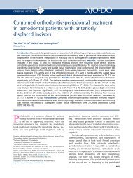
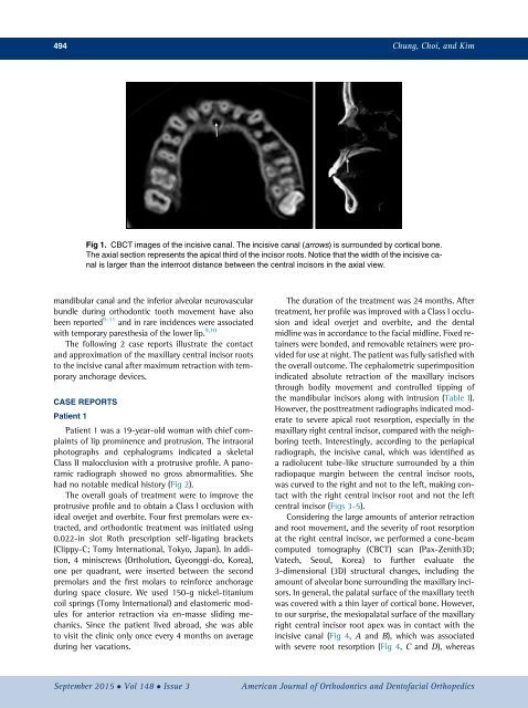
Fig 1. CBCT images <strong>of</strong> <strong>the</strong> <strong>incisive</strong> <strong>canal</strong>. The <strong>incisive</strong> <strong>canal</strong> (arrows) is surrounded by cortical bone.<br />
The axial section represents <strong>the</strong> apical third <strong>of</strong> <strong>the</strong> <strong>incisor</strong> <strong>roots</strong>. Notice that <strong>the</strong> width <strong>of</strong> <strong>the</strong> <strong>incisive</strong> <strong>canal</strong><br />
is larger than <strong>the</strong> interroot distance between <strong>the</strong> <strong>central</strong> <strong>incisor</strong>s in <strong>the</strong> axial view.<br />
m<strong>and</strong>ibular <strong>canal</strong> <strong>and</strong> <strong>the</strong> inferior alveolar neurovascular<br />
bundle during orthodontic tooth movement have also<br />
been reported 9-11 <strong>and</strong> in rare incidences were associated<br />
<strong>with</strong> <strong>temporary</strong> pares<strong>the</strong>sia <strong>of</strong> <strong>the</strong> lower lip. 9,10<br />
The following 2 case reports illustrate <strong>the</strong> <strong>contact</strong><br />
<strong>and</strong> approximation <strong>of</strong> <strong>the</strong> <strong>maxillary</strong> <strong>central</strong> <strong>incisor</strong> <strong>roots</strong><br />
to <strong>the</strong> <strong>incisive</strong> <strong>canal</strong> <strong>after</strong> <strong>maximum</strong> <strong>retraction</strong> <strong>with</strong> <strong>temporary</strong><br />
<strong>anchorage</strong> <strong>devices</strong>.<br />
CASE REPORTS<br />
Patient 1<br />
Patient 1 was a 19-year-old woman <strong>with</strong> chief complaints<br />
<strong>of</strong> lip prominence <strong>and</strong> protrusion. The intraoral<br />
photographs <strong>and</strong> cephalograms indicated a skeletal<br />
Class II malocclusion <strong>with</strong> a protrusive pr<strong>of</strong>ile. A panoramic<br />
radiograph showed no gross abnormalities. She<br />
had no notable medical history (Fig 2).<br />
The overall goals <strong>of</strong> treatment were to improve <strong>the</strong><br />
protrusive pr<strong>of</strong>ile <strong>and</strong> to obtain a Class I occlusion <strong>with</strong><br />
ideal overjet <strong>and</strong> overbite. Four first premolars were extracted,<br />
<strong>and</strong> orthodontic treatment was initiated using<br />
0.022-in slot Roth prescription self-ligating brackets<br />
(Clippy-C; Tomy International, Tokyo, Japan). In addition,<br />
4 miniscrews (Ortholution, Gyeonggi-do, Korea),<br />
one per quadrant, were inserted between <strong>the</strong> second<br />
premolars <strong>and</strong> <strong>the</strong> first molars to reinforce <strong>anchorage</strong><br />
during space closure. We used 150-g nickel-titanium<br />
coil springs (Tomy International) <strong>and</strong> elastomeric modules<br />
for anterior <strong>retraction</strong> via en-masse sliding mechanics.<br />
Since <strong>the</strong> patient lived abroad, she was able<br />
to visit <strong>the</strong> clinic only once every 4 months on average<br />
during her vacations.<br />
The duration <strong>of</strong> <strong>the</strong> treatment was 24 months. After<br />
treatment, her pr<strong>of</strong>ile was improved <strong>with</strong> a Class I occlusion<br />
<strong>and</strong> ideal overjet <strong>and</strong> overbite, <strong>and</strong> <strong>the</strong> dental<br />
midline was in accordance to <strong>the</strong> facial midline. Fixed retainers<br />
were bonded, <strong>and</strong> removable retainers were provided<br />
for use at night. The patient was fully satisfied <strong>with</strong><br />
<strong>the</strong> overall outcome. The cephalometric superimposition<br />
indicated absolute <strong>retraction</strong> <strong>of</strong> <strong>the</strong> <strong>maxillary</strong> <strong>incisor</strong>s<br />
through bodily movement <strong>and</strong> controlled tipping <strong>of</strong><br />
<strong>the</strong> m<strong>and</strong>ibular <strong>incisor</strong>s along <strong>with</strong> intrusion (Table I).<br />
However, <strong>the</strong> posttreatment radiographs indicated moderate<br />
to severe apical root resorption, especially in <strong>the</strong><br />
<strong>maxillary</strong> right <strong>central</strong> <strong>incisor</strong>, compared <strong>with</strong> <strong>the</strong> neighboring<br />
teeth. Interestingly, according to <strong>the</strong> periapical<br />
radiograph, <strong>the</strong> <strong>incisive</strong> <strong>canal</strong>, which was identified as<br />
a radiolucent tube-like structure surrounded by a thin<br />
radiopaque margin between <strong>the</strong> <strong>central</strong> <strong>incisor</strong> <strong>roots</strong>,<br />
was curved to <strong>the</strong> right <strong>and</strong> not to <strong>the</strong> left, making <strong>contact</strong><br />
<strong>with</strong> <strong>the</strong> right <strong>central</strong> <strong>incisor</strong> root <strong>and</strong> not <strong>the</strong> left<br />
<strong>central</strong> <strong>incisor</strong> (Figs 3-5).<br />
Considering <strong>the</strong> large amounts <strong>of</strong> anterior <strong>retraction</strong><br />
<strong>and</strong> root movement, <strong>and</strong> <strong>the</strong> severity <strong>of</strong> root resorption<br />
at <strong>the</strong> right <strong>central</strong> <strong>incisor</strong>, we performed a cone-beam<br />
computed tomography (CBCT) scan (Pax-Zenith3D;<br />
Vatech, Seoul, Korea) to fur<strong>the</strong>r evaluate <strong>the</strong><br />
3-dimensional (3D) structural changes, including <strong>the</strong><br />
amount <strong>of</strong> alveolar bone surrounding <strong>the</strong> <strong>maxillary</strong> <strong>incisor</strong>s.<br />
In general, <strong>the</strong> palatal surface <strong>of</strong> <strong>the</strong> <strong>maxillary</strong> teeth<br />
was covered <strong>with</strong> a thin layer <strong>of</strong> cortical bone. However,<br />
to our surprise, <strong>the</strong> mesiopalatal surface <strong>of</strong> <strong>the</strong> <strong>maxillary</strong><br />
right <strong>central</strong> <strong>incisor</strong> root apex was in <strong>contact</strong> <strong>with</strong> <strong>the</strong><br />
<strong>incisive</strong> <strong>canal</strong> (Fig 4, A <strong>and</strong> B), which was associated<br />
<strong>with</strong> severe root resorption (Fig 4, C <strong>and</strong> D), whereas<br />
September 2015 Vol 148 Issue 3<br />
American Journal <strong>of</strong> Orthodontics <strong>and</strong> Dent<strong>of</strong>acial Orthopedics