Approximation and contact of the maxillary central incisor roots with the incisive canal after maximum retraction with temporary anchorage devices_ Report of 2 patients
artigos ortodontia
artigos ortodontia

Create successful ePaper yourself
Turn your PDF publications into a flip-book with our unique Google optimized e-Paper software.
500 Chung, Choi, <strong>and</strong> Kim<br />
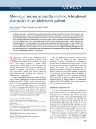
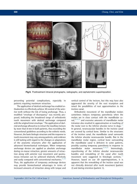
Fig 8. Posttreatment intraoral photographs, radiographs, <strong>and</strong> cephalometric superimposition.<br />
preventing potential complications, especially in<br />
<strong>patients</strong> requiring <strong>maximum</strong> <strong>retraction</strong>.<br />
The application <strong>of</strong> skeletal <strong>anchorage</strong> has enabled orthodontists<br />
to effectively achieve 3D control <strong>of</strong> <strong>the</strong> anterior<br />
teeth <strong>with</strong>out <strong>the</strong> risk <strong>of</strong> losing <strong>anchorage</strong>. Thus, a<br />
modified “envelope <strong>of</strong> discrepancy” was recently proposed,<br />
indicating <strong>the</strong> broadened range <strong>of</strong> orthodontic<br />
tooth movement <strong>with</strong> skeletal <strong>anchorage</strong> compared<br />
<strong>with</strong> <strong>the</strong> original inner envelope. 2 The application <strong>of</strong> skeletal<br />
<strong>anchorage</strong> allowed us to retract <strong>the</strong> <strong>maxillary</strong> <strong>incisor</strong>s<br />
by more than 8 mm in both <strong>patients</strong>, thus exceeding <strong>the</strong><br />
conventional guidelines according to <strong>the</strong> es<strong>the</strong>tic needs.<br />
However, <strong>the</strong> basic biologic structure <strong>and</strong> <strong>the</strong> response to<br />
tooth movement may vary among <strong>patients</strong>, <strong>and</strong> evidence<br />
is still lacking <strong>with</strong> regard to <strong>the</strong> changes or adaptations<br />
<strong>of</strong> <strong>the</strong> anatomic structures <strong>after</strong> <strong>the</strong> application <strong>of</strong><br />
advanced biomechanical techniques. When <strong>temporary</strong><br />
<strong>anchorage</strong> <strong>devices</strong> are applied as absolute <strong>anchorage</strong><br />
during en-masse <strong>retraction</strong>, greater amounts <strong>of</strong> <strong>retraction</strong><br />
along <strong>with</strong> anterior root movement <strong>and</strong> spontaneous<br />
intrusion can be achieved relatively effectively<br />
<strong>and</strong> easily compared <strong>with</strong> conventional mechanics. 21,22<br />
Thus, <strong>the</strong> application <strong>of</strong> <strong>temporary</strong> <strong>anchorage</strong> <strong>devices</strong><br />
may provide biomechanical advantages in terms <strong>of</strong><br />
increased amounts <strong>of</strong> <strong>retraction</strong> along <strong>with</strong> torque <strong>and</strong><br />
vertical control <strong>of</strong> <strong>the</strong> <strong>incisor</strong>s, but this may have also<br />
aggravated <strong>the</strong> severity <strong>of</strong> <strong>the</strong> root resorption <strong>and</strong><br />
raised <strong>the</strong> possibilities <strong>of</strong> root approximation to <strong>the</strong><br />
<strong>incisive</strong> <strong>canal</strong>.<br />
Orthodontic movement <strong>of</strong> <strong>the</strong> m<strong>and</strong>ibular molars<br />
sometimes induces <strong>temporary</strong> pares<strong>the</strong>sia when <strong>the</strong><br />
molars are in close <strong>contact</strong> <strong>with</strong> <strong>the</strong> m<strong>and</strong>ibular <strong>canal</strong>,<br />
9,10,23 <strong>and</strong> excessive amounts <strong>of</strong> m<strong>and</strong>ibular molar<br />
intrusion also resulted in approximation or touching <strong>of</strong><br />
<strong>the</strong> molar root to <strong>the</strong> inferior alveolar nerve in dogs. 11<br />
In general, neurovascular bundles in <strong>the</strong> <strong>incisive</strong> <strong>canal</strong><br />
are covered by cortical bone. Similar to <strong>the</strong> structures<br />
<strong>of</strong> <strong>the</strong> <strong>incisive</strong> <strong>canal</strong>, <strong>the</strong> m<strong>and</strong>ibular <strong>canal</strong> surrounds<br />
<strong>the</strong> inferior alveolar neurovascular bundle. But in <strong>the</strong><br />
m<strong>and</strong>ibular molar region, cortical bone surrounding<br />
<strong>the</strong> m<strong>and</strong>ibular <strong>canal</strong> is deficient in some <strong>patients</strong>,<br />
possibly causing <strong>temporary</strong> pares<strong>the</strong>sia in response to<br />
m<strong>and</strong>ibular molar movement. 9,24,25 Interestingly,<br />
repositioning <strong>of</strong> <strong>the</strong> inferior alveolar neurovascular<br />
bundles <strong>and</strong> remodeling <strong>of</strong> <strong>the</strong> <strong>canal</strong> <strong>after</strong> tooth<br />
movement were suggested in histologic sections. 11<br />
However, based on our 3D superimpositions, it is<br />
apparent that <strong>the</strong> remodeling <strong>of</strong> <strong>the</strong> <strong>incisive</strong> <strong>canal</strong> was<br />
not clinically detected <strong>after</strong> tooth movement (patient<br />
2) <strong>and</strong> during retention (patient 1).<br />
September 2015 Vol 148 Issue 3<br />
American Journal <strong>of</strong> Orthodontics <strong>and</strong> Dent<strong>of</strong>acial Orthopedics