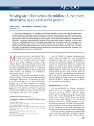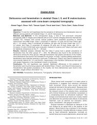
Approximation and contact of the maxillary central incisor roots with the incisive canal after maximum retraction with temporary anchorage devices_ Report of 2 patients
artigos ortodontia
artigos ortodontia

Create successful ePaper yourself
Turn your PDF publications into a flip-book with our unique Google optimized e-Paper software.
Chung, Choi, <strong>and</strong> Kim 499<br />
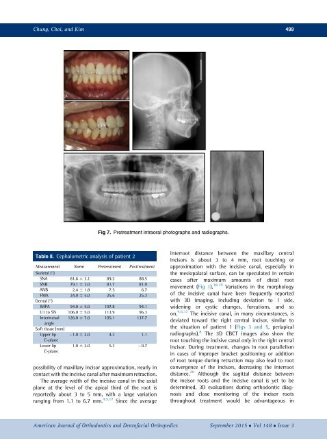
Fig 7. Pretreatment intraoral photographs <strong>and</strong> radiographs.<br />
Table II. Cephalometric analysis <strong>of</strong> patient 2<br />
Measurement Norm Pretreatment Posttreatment<br />
Skeletal ( )<br />
SNA 81.6 6 3.1 89.2 88.5<br />
SNB 79.1 6 3.0 81.7 81.9<br />
ANB 2.4 6 1.8 7.5 6.7<br />
FMA 24.0 6 5.0 25.6 25.3<br />
Dental ( )<br />
IMPA 94.0 6 5.0 107.8 94.1<br />
U1 to SN 106.0 6 5.0 113.9 96.3<br />
Interincisal 126.0 6 7.0 105.1 137.7<br />
angle<br />
S<strong>of</strong>t tissue (mm)<br />
Upper lip 1.0 6 2.0 4.3 1.1<br />
E-plane<br />
Lower lip<br />
E-plane<br />
1.0 6 2.0 5.3 0.7<br />
possibility <strong>of</strong> <strong>maxillary</strong> <strong>incisor</strong> approximation, nearly in<br />
<strong>contact</strong> <strong>with</strong> <strong>the</strong> <strong>incisive</strong> <strong>canal</strong> <strong>after</strong> <strong>maximum</strong> <strong>retraction</strong>.<br />
The average width <strong>of</strong> <strong>the</strong> <strong>incisive</strong> <strong>canal</strong> in <strong>the</strong> axial<br />
plane at <strong>the</strong> level <strong>of</strong> <strong>the</strong> apical third <strong>of</strong> <strong>the</strong> root is<br />
reportedly about 3 to 5 mm, <strong>with</strong> a large variation<br />
ranging from 1.1 to 6.7 mm. 4,5,17 Since <strong>the</strong> average<br />
interroot distance between <strong>the</strong> <strong>maxillary</strong> <strong>central</strong><br />
<strong>incisor</strong>s is about 3 to 4 mm, root touching or<br />
approximation <strong>with</strong> <strong>the</strong> <strong>incisive</strong> <strong>canal</strong>, especially in<br />
<strong>the</strong> mesiopalatal surface, can be speculated in certain<br />
cases <strong>after</strong> <strong>maximum</strong> amounts <strong>of</strong> distal root<br />
movement (Fig 1). 18,19 Variations in <strong>the</strong> morphology<br />
<strong>of</strong> <strong>the</strong> <strong>incisive</strong> <strong>canal</strong> have been frequently reported<br />
<strong>with</strong> 3D imaging, including deviation to 1 side,<br />
widening or cystic changes, furcations, <strong>and</strong> so<br />
on. 4,5,17 The <strong>incisive</strong> <strong>canal</strong>, in many circumstances, is<br />
deviated toward <strong>the</strong> right <strong>central</strong> <strong>incisor</strong>, similar to<br />
<strong>the</strong> situation <strong>of</strong> patient 1 (Figs 3 <strong>and</strong> 5, periapical<br />
radiographs). 8 The 3D CBCT images also show <strong>the</strong><br />
root touching <strong>the</strong> <strong>incisive</strong> <strong>canal</strong> only in <strong>the</strong> right <strong>central</strong><br />
<strong>incisor</strong>. During treatment, changes in root parallelism<br />
in cases <strong>of</strong> improper bracket positioning or addition<br />
<strong>of</strong> root torque during <strong>retraction</strong> may also lead to root<br />
convergence <strong>of</strong> <strong>the</strong> <strong>incisor</strong>s, decreasing <strong>the</strong> interroot<br />
distance. 20 Although <strong>the</strong> sagittal distance between<br />
<strong>the</strong> <strong>incisor</strong> <strong>roots</strong> <strong>and</strong> <strong>the</strong> <strong>incisive</strong> <strong>canal</strong> is yet to be<br />
determined, 3D evaluations during orthodontic diagnosis<br />
<strong>and</strong> close monitoring <strong>of</strong> <strong>the</strong> <strong>incisor</strong> <strong>roots</strong><br />
throughout treatment would be advantageous in<br />
American Journal <strong>of</strong> Orthodontics <strong>and</strong> Dent<strong>of</strong>acial Orthopedics September 2015 Vol 148 Issue 3